This website uses cookies to ensure you get the best experience on our website.
- Table of Contents


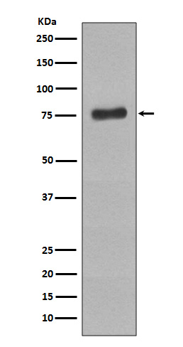
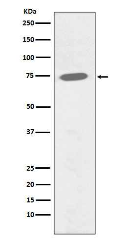
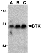
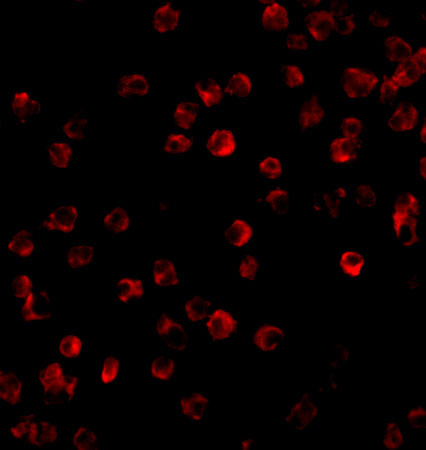
Facts about Tyrosine-protein kinase BTK.

Following BCR engagement and activation at the plasma membrane, phosphorylates PLCG2 at many sites, sparking the downstream signaling pathway through calcium mobilization, followed by activation of the protein kinase C (PKC) family members. PLCG2 phosphorylation is done in close collaboration with the adapter protein B-cell linker protein BLNK.
| Human | |
|---|---|
| Gene Name: | BTK |
| Uniprot: | Q06187 |
| Entrez: | 695 |

| Belongs to: |
|---|
| protein kinase superfamily |

Agammaglobulinaemia tyrosine kinase; AGMX1; AGMX1MGC126262; AT; ATKIMD1; B-cell progenitor kinase; BPK; Bruton agammaglobulinemia tyrosine kinase; Bruton tyrosine kinase; BTK; dominant-negative kinase-deficient Brutons tyrosine kinase; EC 2.7.10; EC 2.7.10.2; IMD1; PSCTK1; tyrosine-protein kinase BTK; XLA; XLAMGC126261
Mass (kDA):
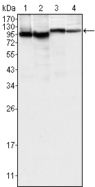
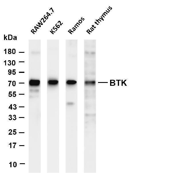
76.281 kDA

| Human | |
|---|---|
| Location: | Xq22.1 |
| Sequence: | X; NC_000023.11 (101349447..101390796, complement) |
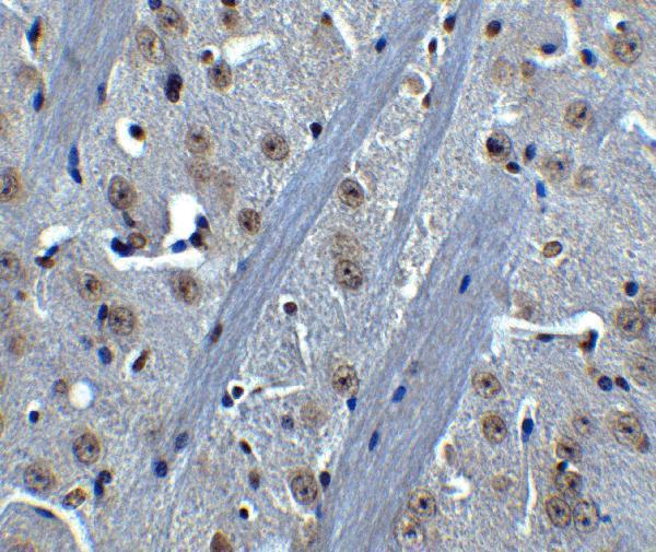
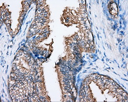
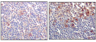
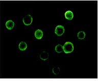
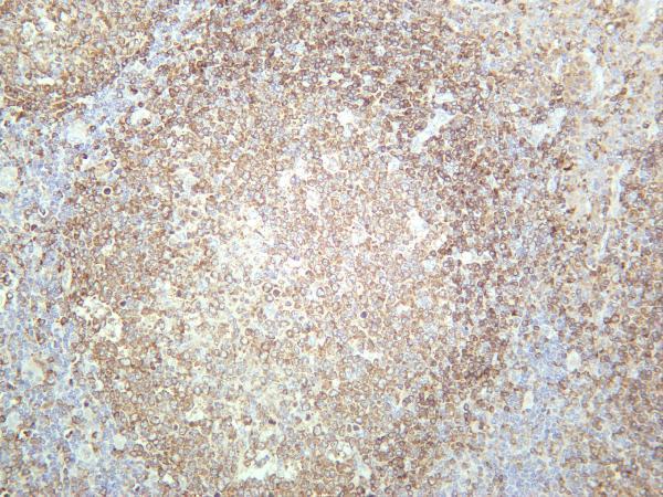
Predominantly expressed in B-lymphocytes.
Cytoplasm. Cell membrane; Peripheral membrane protein. Nucleus. In steady state, BTK is predominantly cytosolic. Following B-cell receptor (BCR) engagement by antigen, translocates to the plasma membrane through its PH domain. Plasma membrane localization is a critical step in the activation of BTK. A fraction of BTK also shuttles between the nucleus and the cytoplasm, and nuclear export is mediated by the nuclear export receptor CRM1.


PMID: 8380905 by Vetrie D., et al. The gene involved in X-linked agammaglobulinaemia is a member of the src family of protein-tyrosine kinases.
PMID: 8090769 by Ohta Y., et al. Genomic organization and structure of Bruton agammaglobulinemia tyrosine kinase: localization of mutations associated with varied clinical presentations and course in X chromosome-linked agammaglobulinemia.