This website uses cookies to ensure you get the best experience on our website.


- Table of Contents
Plan chromogenic IHC for BUD31 in paraffin sections using the nuclear and cytoplasmic tissue pattern reported by HPA (HPA tissue IHC). This guide covers fixation, retrieval, controls and scoring, with the catalog antibody’s 1:50–1:200 IHC dilution range (datasheet: 1:50–1:200).

Expected localisation, validated protocols, controls and antibodies — the at-a-glance facts below, then the full design guide.
| Expected localisation | Nuclear (UniProt); nuclear and cytoplasmic in tissue (HPA tissue IHC) | |
| Staining pattern | Glandular and other cells show nuclear and cytoplasmic staining (HPA tissue IHC) | |
| Antigen retrieval | Tris-EDTA pH 9.0 HIER, 95–98 °C, 20 min (rule: nuclear antigen) | |
| Positive control | Adipose tissue+4 more · see all | |
| Negative control | Adrenal gland+3 more · see all |
| Fixation | Keep fixation consistent across paraffin sections. (standard IHC practice; not target-specific) | |
| Caveat | Staining and RNA show medium consistency (HPA tissue IHC) | |
| Regulation | Low tissue RNA specificity (HPA tissue RNA) | |
| Isoform / epitope | 2 isoforms; epitope coverage is unestablished (UniProt) |
Compare the catalog antibody’s IHC-P protocol (datasheet) with published BUD31 protocols for FFPE sections (PMC10094631; PMC9587234).
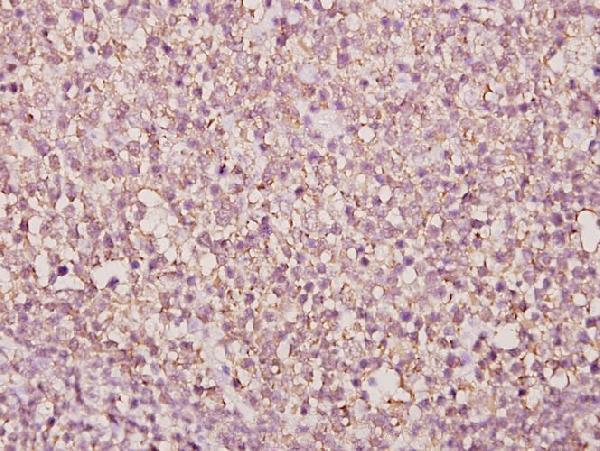
| Sample | Paraffin-embedded human tonsil carcinoma tissue; fixative not specified (datasheet A11662) |
| Fixation | Image fixative and duration unreported (datasheet A11662); verify before use. |
| Sectioning | 4–5 µm sections on charged slides (standard) |
| Deparaffinisation | Xylene, graded ethanol series to water (standard) |
| Antigen retrieval | Heat-induced epitope retrieval in Tris-EDTA buffer, pH 9.0, 20 min at 95–98 °C (standard rule: nuclear antigen) |
| Peroxidase block | 3% H2O2, 10 min, room temperature (standard) |
| Blocking | 10% normal serum of the secondary host, 30 min, room temperature (standard) |
| Primary antibody | Rabbit anti-BUD31, 1:50-1:200 (datasheet A11662) |
| Primary incubation | Overnight at 4 °C (standard) |
| Detection | HRP-polymer secondary, DAB chromogen 5–10 min (standard) |
| Counterstain | Hematoxylin, blue, dehydrate and mount (standard) |
| Expected result | BUD31-positive staining in adipocytes of adipose tissue (HPA tissue IHC: Medium). HPA tissue profile: Cytoplasmic and nuclear expression in several tissues. No signal in the no-primary control. |
BUD31 is a nuclear protein with no transmembrane segment (UniProt P41223: localization and topology). In paraffin sections, expect nuclear staining in some epithelial and stromal cells, with possible cytoplasmic staining: HPA reports cytoplasmic and nuclear expression across several tissues, while UniProt reports protein in benign prostate hyperplasia epithelial and stromal cells (HPA tissue IHC; UniProt P41223: tissue specificity). HPA rates its tissue IHC profile Approved, with medium consistency against RNA expression (HPA tissue IHC).
| Nuclear staining in appendix or colon glandular cells, with possible cytoplasmic staining (HPA tissue IHC). | This fits the reported tissue pattern: both cell groups have Medium staining, and HPA describes cytoplasmic and nuclear expression (HPA tissue IHC). Score the compartments separately; cytoplasmic color alone is less persuasive for a protein UniProt places in the nucleus (UniProt P41223: localization). |
| Strong membrane outlining or staining restricted to a compartment other than the nucleus. | A membrane pattern conflicts with the reported nuclear localization and absence of a transmembrane segment (UniProt P41223: localization and topology). Cytoplasmic staining can occur (HPA tissue IHC), so assess whether convincing nuclei accompany it before calling the pattern an artefact. |
| Prominent color in adrenal gland glandular cells or ovarian stroma cells (HPA tissue IHC: Not detected). | Those sampled cell groups were Not detected by HPA; unexpected staining warrants review for cross-reactivity or endogenous chromogenic activity (HPA tissue IHC; general IHC practice). These observations do not prove every specimen from either tissue is BUD31-negative. |
| Diffuse color across tissue, including areas without clear cell boundaries or nuclear detail. | That distribution is difficult to assign to BUD31-positive cells; nonspecific reagent binding or detection background are possible causes (general IHC practice). Compare a matched control slide and judge cell-localized signal after background is controlled (general IHC practice). |
| No staining in appendix or colon glandular cells (HPA tissue IHC: Medium). | These are reasonable positive-control cell groups, though Medium is an observed level, not a guaranteed result in every section (HPA tissue IHC). Check tissue preservation, retrieval, antibody dilution and detection controls before concluding the specimen lacks BUD31 (general IHC practice). |
| Compartment and topology | UniProt places BUD31 in the nucleus and reports no transmembrane segment; HPA tissue IHC also reports cytoplasmic expression (UniProt P41223: localization and topology; HPA tissue IHC). Interpret mixed nuclear and cytoplasmic staining in that context, while investigating a membrane-only pattern. |
| Cell type and reference intensity | HPA reports Medium staining in appendix and colon glandular cells, Low staining in prostate glandular cells, and Not detected in adrenal gland glandular cells (HPA tissue IHC). Compare the same cell type when using these references; Low and Not detected are observed categories, not universal biological absolutes. |
| Strength of IHC evidence | HPA calls tissue IHC Approved but describes only medium consistency between staining and RNA expression; its listed antibody HPA028943 is IHC Approved (HPA tissue IHC; HPA antibodies). This supports a reference pattern without establishing that every positive structure in a new assay is specific. |
| Isoforms and antigen retrieval | UniProt lists 2 isoforms, but the supplied record gives no antibody epitope or isoform-specific IHC behavior (UniProt P41223: isoforms). Retrieval conditions should therefore be checked against the chosen antibody's validated procedure; no BUD31-specific retrieval effect is established here. |
| IF/ICC Q: What pattern should a separate IF/ICC assay show? | A: HPA supports mainly nucleoplasmic localization; additional microtubule and centrosome localizations are uncertain (HPA subcellular ICC-IF). Use that evidence to interpret IF images, while keeping this section's primary readout chromogenic IHC in paraffin sections. |
| Situation | Likely cause | Next action |
|---|---|---|
| Nuclei are unstained in appendix or colon glandular cells (HPA tissue IHC: Medium). | A missed assay step or inadequate signal is possible; the HPA level alone cannot identify the cause (HPA tissue IHC; general IHC practice). | Check section quality, retrieval, antibody dilution and detection reagents using the chosen antibody's validated IHC procedure (general IHC practice). |
| Only cytoplasm stains, with no convincing nuclear signal. | HPA reports cytoplasmic and nuclear tissue staining, but UniProt places BUD31 in the nucleus (HPA tissue IHC; UniProt P41223: localization). | Review nuclear counterstain and cellular boundaries, then compare with a known HPA-positive cell group and a matched control (HPA tissue IHC; general IHC practice). |
| Cell membranes form the dominant staining pattern. | That pattern is unsupported by the nuclear localization and lack of a transmembrane segment (UniProt P41223: localization and topology). | Reassess specificity with appropriate controls and inspect whether precipitate or edge staining is producing apparent membrane outlines (general IHC practice). |
| Adrenal gland glandular cells show prominent color (HPA tissue IHC: Not detected). | Cross-reactivity or endogenous detection activity is possible; the HPA observation cannot distinguish these causes (HPA tissue IHC; general IHC practice). | Compare a control without primary antibody and the chosen detection system's endogenous-activity control, then judge cell-specific staining (general IHC practice). |
| Color spreads over nuclei, cytoplasm and empty spaces alike. | Diffuse reagent background or chromogen deposit can obscure cellular localization (general IHC practice). | Inspect matched controls, washing and chromogen development; score only signal whose cellular compartment remains clear (general IHC practice). |
| A weak prostate glandular-cell result disagrees with stronger staining elsewhere. | HPA reports Low staining in prostate glandular cells but Medium in appendix and colon glandular cells (HPA tissue IHC). | Compare equivalent cell types and assay conditions before treating different tissue intensities as an assay failure (HPA tissue IHC; general IHC practice). |


Comprehensive Human Protein Atlas IHC scoring per tissue (reliability: Approved — Medium consistency between antibody staining and RNA expression data.). Rows are taken directly from the HPA tissue chart — click any row's HPA link to view the source.
| Tissue | Cell type | Level | Evidence | Source |
|---|---|---|---|---|
| Adipose tissue | Adipocytes | Medium | Protein (IHC) | HPA → |
| Appendix | Glandular cells | Medium | Protein (IHC) | HPA → |
| Bronchus | Respiratory epithelial cells | Medium | Protein (IHC) | HPA → |
| Caudate | Glial cells | Medium | Protein (IHC) | HPA → |
| Cerebellum | Cells in molecular layer | Medium | Protein (IHC) | HPA → |
| Tissue | Cell type | Level | Evidence | Source |
|---|---|---|---|---|
| Adrenal gland | Glandular cells | Not detected | Protein (IHC) | HPA → |
| Ovary | Ovarian stroma cells | Not detected | Protein (IHC) | HPA → |
| Soft tissue | Peripheral nerve | Not detected | Protein (IHC) | HPA → |
| Spleen | Cells in red pulp | Not detected | Protein (IHC) | HPA → |
Troubleshooting BUD31 staining in paraffin sections, with one entry for IF/ICC assay design.
A11662 has real BUD31 IHC data from a paraffin-embedded human tissue sample (catalog image caption); the catalog lists Human, Mouse, and Rat reactivity, with no IF data (catalog applications and images).
A11662 will render with an IHC image of paraffin-embedded human tonsil carcinoma tissue at 1:50 (catalog image caption). Its listed applications are IHC and WB, and its listed reactivity is Human, Mouse, and Rat (catalog applications and reactivity).
Which to pick: Choose A11662 for paraffin-section IHC: its own image caption documents staining in paraffin-embedded human tonsil carcinoma tissue at 1:50; the fixative is unreported (catalog image caption). For Mouse or Rat tissue, A11662 has listed reactivity, but its IHC image documents only a human sample (catalog reactivity and image caption). No catalog antibody here is listed for IF/ICC, so there is no supported IF/ICC pick (catalog applications and images).