BZW2 / eIF5-mimic protein 1 · IHC design guide

Design Immunohistochemistry for BZW2

Plan chromogenic IHC on paraffin sections using the catalog antibody at 2–5 μg/ml (datasheet A11832-1). Use tissue staining to select controls and assess cytoplasmic signal, with medium consistency between antibody staining and RNA expression (HPA tissue IHC).

Evidence assembled Oct 2026 · For research use; verify linked source records and product datasheet before use
Immunohistochemistry protocol sheet for BZW2 (IHC for BZW2): expected localisation Variable cytoplasmic staining across tissues (HPA tissue IHC), antibody A11832-1, validated IHC image, and IHC protocol steps
Printable BZW2 IHC protocol sheet — expected localisation Variable cytoplasmic staining across tissues (HPA tissue IHC), antibody A11832-1, controls and protocol steps. Open the full BZW2 IHC guide →

BZW2 Immunohistochemistry Experimental Design Guide

Expected localisation, validated protocols, controls and antibodies — the at-a-glance facts below, then the full design guide.

Must know before staining
Expected localisation Variable cytoplasmic staining across tissues (HPA tissue IHC)
Staining pattern Cytoplasmic staining in glandular and Leydig cells varies by tissue (HPA tissue IHC)
Antigen retrieval EDTA pH 8.0 HIER, heat-mediated (datasheet A11832-1)
Positive control ⓘ Breast+4 more · see all
Negative control ⓘ Appendix+4 more · see all
Important caveats
Reasons your staining may differ from the expected pattern.
Fixation Matched tissue-IHC evidence does not establish the fixation claim. Validate the specimen-specific method before use. (selected-SKU IHC image A11832-1)
Caveat Appendix glands undetected; appendiceal tumor section stained (HPA tissue IHC; datasheet A11832-1)
Regulation No expression regulator annotated (UniProt)
Isoform / epitope 2 isoforms; assess epitope coverage; no extracellular domain (UniProt)
Section 1

Recommended BZW2 IHC & IF Protocols

The catalog antibody’s IHC-P protocol (datasheet A11832-1) is followed by published BZW2 IHC methods for pancreatic, liver and colorectal specimens (PMC9576853; PMC8317536; PMC10197465).

Recommended immunohistochemistry (IHC-P) protocol parameters
SampleParaffin-embedded human appendiceal adenocarcinoma tissue; fixative not specified (datasheet A11832-1)
FixationImage fixative and duration unreported (datasheet A11832-1); verify before use.
Sectioning4–5 µm sections on charged slides (standard)
DeparaffinisationXylene, graded ethanol series to water (standard)
Antigen retrievalHeat retrieval: EDTA pH 8.0 (datasheet A11832-1); 20 min, 95–100 °C (standard)
Peroxidase block3% H2O2, 10 min, room temperature (standard)
Blocking10% goat serum (datasheet A11832-1)
Primary antibodyRabbit anti-BZW2, 2-5 μg/ml (datasheet A11832-1)
Primary incubationOvernight at 4 °C (datasheet A11832-1)
DetectionStreptavidin-biotin complex (SABC), DAB chromogen (datasheet A11832-1)
CounterstainHematoxylin, blue, dehydrate and mount (standard)
Expected resultBZW2-positive staining in glandular cells of breast (HPA tissue IHC: High). HPA tissue profile: Cytoplasmic expression with varying intensity in most tissues. No signal in the no-primary control.
💡Decision noteStart with heat-mediated EDTA retrieval at pH 8.0 (datasheet A11832-1); the colorectal study reports heated retrieval without specifying a buffer (PMC10197465).
Section 2

What Is the Expected BZW2 Staining Pattern?

In paraffin-section IHC, expect predominantly cytoplasmic BZW2 staining with variable intensity across tissues (HPA: cytoplasmic expression in most tissues). Breast and gallbladder glandular cells and testicular Leydig cells are reported as high-staining examples (HPA: tissue IHC). BZW2 is cytoplasmic and has no transmembrane segment (UniProt Q9Y6E2). HPA rates the tissue profile Enhanced, with medium staining–RNA consistency and external verification pending (HPA: tissue IHC reliability).

What am I looking at on my slide?
Cytoplasmic chromogen is strong in breast or gallbladder glandular cells, or testicular Leydig cells (HPA: High in each cell population).This matches the reported compartment and high-staining cell populations (HPA: tissue IHC; UniProt Q9Y6E2: cytoplasm). Score the named cells and their cytoplasmic signal, rather than treating every cell in the section as equally positive (general IHC practice).
Staining is exclusively nuclear, with no convincing cytoplasmic signal.An exclusively nuclear IHC pattern does not match the reported cytoplasmic tissue profile (HPA: tissue IHC; UniProt Q9Y6E2: cytoplasm). Check the negative control, morphology and detection conditions before interpreting it as BZW2 (general IHC practice).
Strong signal appears in appendix glandular cells or lung alveolar cells (HPA: Not detected in those cell populations).Those particular cells are useful contrasts to the reported high-staining populations; their unexpected signal raises cross-reactivity or endogenous chromogenic activity as possibilities (HPA: tissue IHC; general IHC practice). A tissue label alone cannot classify every cell in that tissue as negative.
Broad chromogen covers tissue and empty areas without a clear cell boundary.Diffuse background is not the reported cell-associated cytoplasmic pattern (HPA: tissue IHC). Compare a reagent control and inspect blocking, washing and detection conditions; background by itself does not establish BZW2 expression (general IHC practice).
No signal appears in the expected cells of a breast, gallbladder or testis positive-control section (HPA: High in the specified cells).First assess whether the control's glandular or Leydig cells are present and whether the staining run worked (HPA: tissue IHC; general IHC practice). A blank run cannot establish absence of BZW2 in the test specimen.
💡Expected BZW2 appearanceCall a result positive when cytoplasmic staining is identifiable in the relevant cells, particularly the high-staining breast or gallbladder glandular cells or testicular Leydig cells; exclusively nuclear signal or diffuse cell-free background is suspect (HPA: tissue IHC; UniProt Q9Y6E2: cytoplasm; general IHC practice).
How each factor affects the staining
Cell population and tissue choiceHPA reports High staining in breast and gallbladder glandular cells and testicular Leydig cells, Medium staining in several other named populations, and Not detected in others (HPA: tissue IHC). Choose and score the documented cell population; RNA enrichment in heart muscle, skeletal muscle and tongue is not an IHC intensity claim (HPA: RNA specificity).
Antibody validationHPA022813 is IHC Approved; HPA026709 is IHC Enhanced (HPA: antibodies). HPA describes the tissue profile as Enhanced but notes medium agreement with RNA and pending external verification (HPA: tissue IHC reliability). Interpret a new staining pattern against controls rather than treating validation status as proof for every specimen.
Intracellular location and processingBZW2 is annotated cytoplasmic, with no transmembrane segment or signal peptide; its recorded chain spans residues 1–419 (UniProt Q9Y6E2: subcellular location, topology and processing). These annotations support a cytoplasmic IHC expectation, but do not identify an antibody epitope or establish fixation sensitivity.
Isoforms and antigen retrievalTwo isoforms are listed (UniProt Q9Y6E2: isoforms). Their relative tissue staining and antibody epitope coverage are not supplied. Antigen retrieval is a general paraffin-IHC optimization step; no BZW2-specific retrieval condition or fixation effect is established by the supplied sources (general IHC practice).
IF/ICC Q: what pattern is reported?A: HPA reports approved plasma membrane, actin-filament and cytosol locations in ICC-IF images from A-431, U-251MG and U2OS (HPA: subcellular ICC-IF). That cellular imaging result adds context; the paraffin-tissue IHC profile is reported as cytoplasmic (HPA: tissue IHC).
Why is my staining missing, weak or wrong?
SituationLikely causeNext action
A documented high-staining control cell population is blank (HPA: breast or gallbladder glandular cells; testis Leydig cells).The expected cells may be absent from the section, or the IHC run may have failed; a blank control alone does not distinguish these causes (general IHC practice).Confirm control-cell morphology, then check the catalog antibody's IHC-P instructions, retrieval, dilution and detection controls for the run (general IHC practice). No BZW2-specific settings are supplied here.
Signal is strong only in nuclei.That distribution conflicts with the reported cytoplasmic IHC pattern (HPA: tissue IHC; UniProt Q9Y6E2: cytoplasm). Artefact or nonspecific detection is possible, not proven.Review tissue morphology and reagent controls, then repeat or corroborate the finding with an independently validated antibody if available (general IHC practice; HPA: antibody validation).
Appendix glandular or lung alveolar cells show strong chromogen (HPA: Not detected in those cells).The result conflicts with HPA's cell-specific observations; cross-reactivity or endogenous detection activity are possible explanations (HPA: tissue IHC; general IHC practice).Compare the named cells with a documented high-staining control and run the appropriate detection control; investigate persistent cell-specific staining before calling it BZW2 (general IHC practice).
The section has widespread haze or precipitate.Diffuse material outside recognizable cytoplasm is inconsistent with HPA's cell-associated pattern; detection background or deposited reagent is possible (HPA: tissue IHC; general IHC practice).Inspect a reagent control and review blocking, wash steps, reagent handling and chromogen development; score only interpretable cellular staining (general IHC practice).
A low-staining or Not detected population is being used to judge whether the run worked (HPA: tissue IHC).A weak or absent result in that population may agree with the reported profile and therefore cannot serve as a reliable positive-run check (HPA: tissue IHC).Use a documented high-staining population containing the specified cells as the positive comparison, and record the exact population assessed (HPA: tissue IHC; general IHC practice).

Sample controls for BZW2 IHC & IF

🧪Run breast first and expect staining in glandular cells (HPA: High in breast glandular cells); run normal appendix glandular cells as the negative tissue (HPA: Not detected in appendix glandular cells). On the breast slide, use adjacent nonglandular cells as internal background comparators only if they lack specific staining; the supplied HPA call applies to glandular cells (HPA: High in breast glandular cells).
Positive control tissue: Breast (Glandular cells, HPA High)
Negative control tissue: Appendix (HPA Not detected)
ICC-IF cell lines (HPA subcellular resource): HPA ICC-IF images show BZW2 in A-431, U-251MG, U2OS, with annotated localisation: Plasma membrane (approved), Actin filaments (approved), Cytosol (approved) (HPA subcellular).
Technical controls: Include a no-primary, secondary-only control, a concentration-matched rabbit IgG isotype control, and BZW2-knockout tissue or cells as a biological negative (selected-SKU caption: rabbit primary antibody). For chromogenic detection, quench endogenous peroxidase and check for endogenous biotin background when using the caption’s biotinylated secondary and streptavidin–biotin system (selected-SKU caption: SABC with DAB).
⚠️Feasibility: A target-specific fixation window and fixation effect are unreported, and the selected-SKU paraffin-section caption does not state a fixative (selected-SKU caption: fixative not stated). Heat retrieval in EDTA at pH 8.0 produced the captioned IHC result, but a strict retrieval dependency is unreported (selected-SKU caption: heat-mediated EDTA retrieval). No supplied comparison establishes frozen sections or IF as easier; interpret the caption’s appendiceal adenocarcinoma staining separately from the normal appendix negative control (selected-SKU caption: appendiceal adenocarcinoma; HPA: Not detected in appendix glandular cells).

HPA tissue IHC evidence for BZW2

Comprehensive Human Protein Atlas IHC scoring per tissue (reliability: Enhanced — Medium consistency between antibody staining and RNA expression data. Pending external verification.). Rows are taken directly from the HPA tissue chart — click any row's HPA link to view the source.

Positive expression · recommended positive controls

TissueCell typeLevelEvidenceSource
Breast Glandular cells High Protein (IHC) HPA →
Gallbladder Glandular cells High Protein (IHC) HPA →
Testis Leydig cells High Protein (IHC) HPA →
Adrenal gland Glandular cells Medium Protein (IHC) HPA →
Bone marrow Hematopoietic cells Medium Protein (IHC) HPA →

Undetected expression · recommended negative controls

TissueCell typeLevelEvidenceSource
Appendix Glandular cells Not detected Protein (IHC) HPA →
Caudate Glial cells Not detected Protein (IHC) HPA →
Cerebellum Cells in granular layer Not detected Protein (IHC) HPA →
Epididymis Glandular cells Not detected Protein (IHC) HPA →
Hippocampus Glial cells Not detected Protein (IHC) HPA →
Section 3

Advanced BZW2 IHC Tips

Troubleshoot BZW2 staining by checking retrieval, cell compartment, controls and scoring against the reported tissue patterns (HPA tissue IHC; HPA subcellular).

How should I retrieve BZW2 antigen in paraffin sections with weak cytoplasmic staining?
Start with heat-mediated retrieval in EDTA pH 8.0 for paraffin sections (datasheet A11832-1). The selected antibody detected BZW2 in a paraffin-embedded human appendiceal adenocarcinoma section after that retrieval step (caption A11832-1). If staining is weak, check that sections received consistent heating and cooling, then compare a longer retrieval period on serial sections while holding detection conditions constant (standard IHC practice). Use a known positive tissue compartment and a no-primary control in the comparison, because excessive retrieval can increase tissue damage and background (standard IHC practice). Record retrieval duration and assess cytoplasmic signal alongside tissue morphology rather than selecting the darkest slide alone (UniProt: cytoplasm; standard IHC practice).
Could fixation explain weak BZW2 staining in my paraffin sections?
BZW2-specific sensitivity to fixative type or fixation duration is unknown from the supplied evidence; the selected paraffin-section caption does not state its fixative (caption A11832-1). For a fixation comparison, process matched samples with documented fixative and duration, then hold section thickness, retrieval and detection conditions constant (standard IHC practice). Start the staining comparison with EDTA pH 8.0 retrieval and the reported 2 μg/ml primary concentration (datasheet A11832-1; caption A11832-1). Examine morphology and cytoplasmic staining together, since damaged or unevenly fixed regions can mislead assessment (UniProt: cytoplasm; standard IHC practice). Do not treat a change in signal as BZW2-specific fixation sensitivity until matched samples reproduce it (standard IHC practice).
Which staining compartment should I accept as BZW2 positive in tissue IHC?
Prioritise cytoplasmic staining in intact cells: UniProt places BZW2 in the cytoplasm, and HPA reports variable cytoplasmic expression across most tissues (UniProt: cytoplasm; HPA tissue IHC). In ICC/IF, HPA also reports cytosol, plasma membrane and actin filaments, but that observation should be checked in the tissue and detection method being scored (HPA subcellular; standard IHC practice). BZW2 has no annotated transmembrane segment, so an exclusively sharp membrane rim warrants scrutiny rather than automatic acceptance (UniProt: topology; standard IHC practice). Compare the suspected signal with nearby intact cells and a no-primary slide at the same exposure or microscope settings (standard IHC practice). Record mixed compartments separately if reproducible, including whether cytoplasmic signal remains present (standard IHC practice).
Can isoforms or modified residues explain inconsistent BZW2 IHC staining?
BZW2 has 2 annotated isoforms, while the supplied antibody caption does not identify its epitope or establish isoform selectivity (UniProt: isoforms; caption A11832-1). The record places the W2 domain at residues 248–415 and lists modifications including acetyllysine 117 and phosphoserines 412 and 414 (UniProt: domains; UniProt: modified residues). Those annotations alone cannot show that an isoform or modification changes recognition by this antibody (UniProt: record; standard IHC interpretation). Keep retrieval and antibody concentration constant when comparing serial sections, and document which cell compartment changes (standard IHC practice). Seek an epitope map or independent specificity evidence before assigning differential staining to an isoform or modified residue (standard IHC practice).
How should I adapt the BZW2 question to multiplex IF?
Treat IF as a separate optimisation: the selected antibody evidence describes chromogenic staining of a paraffin section, whereas HPA reports BZW2 locations from ICC/IF images (caption A11832-1; HPA subcellular). Multiplex BZW2 with a marker that identifies the cell population under study, then assess whether signal lies in those cells and matches the expected cytosolic pattern (HPA: cytosol; standard IF practice). Choose fluorophores after inspecting unstained tissue autofluorescence, and place the weaker signal in a cleaner spectral channel when possible (standard IF practice). Because BZW2 has no transmembrane segment and is reported in cytosol, optimise permeabilisation for intracellular epitope access without assuming an exposed surface epitope (UniProt: topology; HPA subcellular; standard IF practice). Include single-label and no-primary controls to assess bleed-through and background (standard IF practice).
How can I distinguish BZW2 staining from chromogenic background?
First compare the test section with a no-primary control and inspect whether colour follows intact cellular cytoplasm or damaged tissue edges (UniProt: cytoplasm; standard IHC practice). The selected caption used 10% goat serum blocking, 2 μg/ml primary antibody overnight at 4°C, and a biotinylated secondary followed by SABC and DAB (caption A11832-1). If diffuse colour persists, assess blocking, washing and primary concentration on matched sections without changing every variable together (standard IHC practice). Include the appropriate endogenous peroxidase block for chromogenic detection, and consider endogenous biotin when using a biotin-based system (standard IHC practice; caption A11832-1). Score staining only where morphology is preserved and the control supports specific signal (standard IHC practice).
What is a defensible way to quantify BZW2 in chromogenic IHC? ⚠ ANSWER MARKED FOR VERIFICATION
Define the cell population and cytoplasmic compartment before scoring, since HPA describes BZW2 as variably cytoplasmic across tissues (HPA tissue IHC; standard IHC practice). For a semiquantitative result, record staining intensity and the percentage of positive target cells, then calculate an H-score using one prespecified intensity scale across slides (standard IHC practice). If cell counts matter more than intensity, report positive cells per mm² of viable tissue and normalise to the number or area of eligible cells (standard IHC practice). Exclude necrosis, folds and tissue edges using the same written rules for every section (standard IHC practice). Review representative fields against no-primary controls and keep acquisition and threshold settings consistent (standard IHC practice).
When is unexpected BZW2 staining a true positive rather than artefact?
A plausible positive shows reproducible staining in intact cellular cytoplasm, consistent with UniProt and HPA tissue IHC localisation (UniProt: cytoplasm; HPA tissue IHC). Cell identity matters: HPA reports high staining in breast glandular cells and testis Leydig cells, but does not detect staining in normal appendix glandular cells (HPA tissue IHC). The selected antibody nevertheless stained human appendiceal adenocarcinoma, so do not equate that tumour observation with normal appendix glandular cells (caption A11832-1; HPA tissue IHC). Treat isolated nuclear colour, edge effects, necrotic debris or colour reproduced on a no-primary slide as potential artefact (standard IHC practice). Check endogenous peroxidase activity and the biotin-based detection controls before assigning such colour to BZW2 (standard IHC practice; caption A11832-1).
Boster reagents

Best BZW2 / eIF5-mimic protein 1 IHC Antibodies

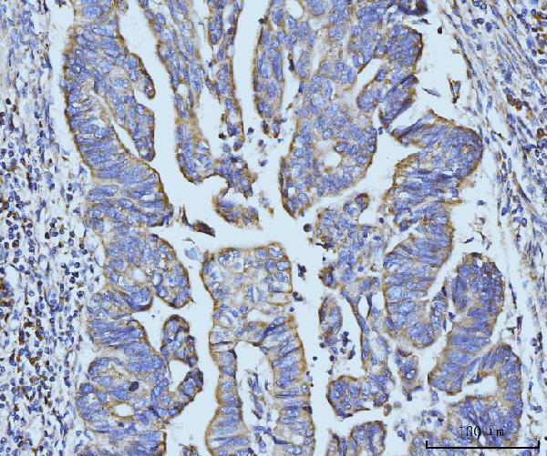
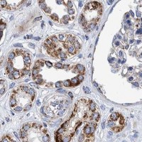
A11832-1 has IHC images of paraffin sections from human appendiceal adenocarcinoma, gastric adenocarcinoma and placenta, plus mouse colon (catalog image captions). No IF data are supplied (catalog applications and images).

Real IHC data IHC analysis of BZW2 using anti-BZW2 antibody (A11832-1). BZW2 was detected in a paraffin-embedded section of human appendiceal adenocarcinoma tissue. Heat mediated antigen retrieval was performed in EDTA buffer (pH 8.0, epitope retrieval solution). The tissue section was blocked with 10% goat serum. The tissue section was then incubated with 2 μg/ml rabbit anti-BZW2 Antibody (A11832-1) overnight at 4°C. Biotinylated goat anti-rabbit IgG was used as secondary antibody and incubated for 30 minutes at 37°C. The tissue section was developed using Strepavidin-Biotin-Complex (SABC) (Catalog # SA1022) with DAB as the chromogen.
Anti-BZW2 Antibody ®
Cat # A11832-1

A11832-1 is listed for IHC and shows BZW2 staining in the four paraffin-section sample types named above (catalog applications and image captions). Human, mouse and rat reactivity is listed, but the supplied IHC images cover human and mouse only (catalog reactivity and image captions).

Which to pick: Choose A11832-1 for paraffin-section IHC: its rabbit antibody was used at 2 μg/ml after EDTA retrieval at pH 8.0, with biotin-based DAB detection (catalog image captions); the fixative is unreported (catalog image captions). For IF/ICC, no SKU has a listed application or image, so an IF choice is unsupported (catalog applications and images). For work across species, A11832-1 lists human, mouse and rat reactivity, with IHC images for human and mouse; rat IHC performance and clonality are unreported (catalog reactivity, image captions and clone field).

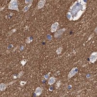
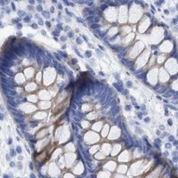
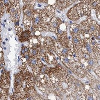
Each figure is that product's own IHC / IF validation image from its datasheet.

References

  1. UniProt Consortium. UniProt entry Q9Y6E2 (5MP1_HUMAN, eIF5-mimic protein 1).
  2. Human Protein Atlas. BZW2 tissue IHC expression (reliability: Enhanced).
  3. Human Protein Atlas. BZW2 subcellular location (ICC-IF): Localized to the plasma membrane, cytosol and actin filaments..
  4. Human Protein Atlas. BZW2 antibody validation summary (2 antibodies).
  5. Expression, oncological and immunological characterizations of BZW1/2 in pancreatic adenocarcinoma. Frontiers in genetics 2022 — PMC9576853.
  6. BZW2 promotes malignant progression in lung adenocarcinoma through enhancing the ubiquitination and degradation of GSK3β. Cell death discovery 2024 — PMC10904796.
  7. BZW2/5MP1 acts as a promising target in hepatocellular carcinoma. Journal of Cancer 2021 — PMC8317536.
  8. Polyamine metabolism patterns characterized tumor microenvironment, prognosis, and response to immunotherapy in colorectal cancer. Cancer cell international 2023 — PMC10197465.
  9. PubMed PMID:11042152 — UniProt-cited evidence.
  10. PubMed PMID:14702039 — UniProt-cited evidence.
  11. PubMed PMID:12690205 — UniProt-cited evidence.