This website uses cookies to ensure you get the best experience on our website.


- Table of Contents
Start C1QTNF6 chromogenic IHC-P with the catalog antibody at 10 μg/mL (datasheet: A12414-1). Compare placental or endometrial staining with an HPA tissue showing no detected signal, and interpret the cytoplasmic pattern in light of the protein’s secretion (HPA tissue IHC; UniProt).

Expected localisation, validated protocols, controls and antibodies — the at-a-glance facts below, then the full design guide.
| Expected localisation | Cytoplasmic in trophoblasts and endometrial stroma (HPA tissue IHC) | |
| Staining pattern | Cytoplasmic trophoblast and endometrial stromal staining (HPA tissue IHC) | |
| Antigen retrieval | Citrate pH 6.0 HIER, 95–98 °C, 20 min (rule: cytoplasmic / membrane antigen) | |
| Positive control | Placenta+1 more · see all | |
| Negative control | Adipose tissue+4 more · see all |
| Fixation | Matched tissue-IHC evidence does not establish the fixation claim. Validate the specimen-specific method before use. (selected-SKU IHC image A12414-1) | |
| Caveat | Secretion may separate tissue RNA and protein patterns (HPA tissue IHC) | |
| Regulation | No expression regulator annotated (UniProt) | |
| Isoform / epitope | 3 isoforms; signal 1–46 is cleaved; epitope map unknown (UniProt) |
The catalog antibody’s IHC-P protocol is accompanied by four published C1QTNF6 IHC workflows for bladder, lung and oral cancer specimens (PMC7585664; PMC7805025; PMC8670214; PMC9310408).
| Sample | Tissue sections; selected-image fixative not specified (standard IHC workflow) |
| Fixation | Image fixative and duration unreported (datasheet A12414-1); verify before use. |
| Sectioning | 4–5 µm sections on charged slides (standard) |
| Deparaffinisation | Xylene, graded ethanol series to water (standard) |
| Antigen retrieval | Heat-induced epitope retrieval in citrate buffer, pH 6.0, 20 min at 95–98 °C (standard rule: cytoplasmic / membrane antigen) |
| Peroxidase block | 3% H2O2, 10 min, room temperature (standard) |
| Blocking | 10% normal serum of the secondary host, 30 min, room temperature (standard) |
| Primary antibody | Rabbit anti-C1QTNF6, 10 μg/mL (datasheet A12414-1) |
| Primary incubation | Overnight at 4 °C (standard) |
| Detection | HRP-polymer secondary, DAB chromogen 5–10 min (standard) |
| Counterstain | Hematoxylin, blue, dehydrate and mount (standard) |
| Expected result | C1QTNF6-positive staining in endothelial cells of placenta (HPA tissue IHC: High). HPA tissue profile: Cytoplasmic expression in placental trophoblasts and endometrial stroma. No signal in the no-primary control. |
C1QTNF6 is secreted and has no transmembrane segment (UniProt Q9BXI9 topology). In tissue IHC, expect cytoplasmic staining in placental trophoblasts and endometrial stromal cells (HPA: tissue profile). HPA also records high staining in placental endothelial cells and medium staining in endometrial stromal cells (HPA: tissue IHC). Its tissue IHC reliability is Enhanced, with a caveat that secretion can separate the locations of RNA and protein (HPA: reliability description).
| Cytoplasmic staining appears in placental trophoblasts or endometrial stromal cells. | This matches the reported tissue pattern (HPA: tissue profile). Compare cell identity and signal with a negative control before scoring; a cytoplasmic stain alone does not establish specificity (general IHC practice). |
| Placental endothelial cells stain strongly; endometrial stromal cells stain less strongly. | HPA reports High staining in placental endothelial cells and Medium staining in endometrial stromal cells (HPA: tissue IHC). These are observations in different tissue and cell contexts, not a required intensity ratio for every specimen (HPA: tissue IHC). |
| Prominent nuclear staining dominates a paraffin tissue section. | Treat this as discordant with the reported cytoplasmic tissue pattern and investigate artefact or off-target staining (HPA: tissue profile; general IHC practice). HPA separately reports approved nucleoplasmic ICC-IF localisation; that observation does not redefine the expected tissue IHC pattern (HPA: subcellular ICC-IF). |
| Unexpected cell types stain, including adipocytes in adipose tissue. | HPA reports adipocytes as Not detected in adipose tissue (HPA: tissue IHC). Check for cross-reactivity or endogenous detection activity with an appropriate negative control; neither cause can be assigned from appearance alone (general IHC practice). |
| Colour is diffuse across tissue, or a known-positive compartment has no signal. | Diffuse colour limits cell-specific scoring and calls for a background control (general IHC practice). If placental endothelial cells or endometrial stromal cells are blank, first verify that the staining run worked before interpreting a biological absence (HPA: tissue IHC; general IHC practice). |
| Secreted protein and tissue context | C1QTNF6 has a signal peptide at residues 1–46 and a chain at 47–278, with no transmembrane segment (UniProt Q9BXI9 topology and processing). Secreted protein can make protein staining diverge from local RNA expression; HPA explicitly flags this limitation (HPA: reliability description). |
| Structural features and epitope uncertainty | UniProt lists a glycosylation site at residue 91, a collagen-like region at 97–138, and a C1q domain at 139–259 (UniProt Q9BXI9). The supplied record gives no antibody epitope, so these features cannot determine an antigen retrieval condition or predict loss of staining after fixation. |
| Isoforms and antibody validation | UniProt lists three isoforms (UniProt Q9BXI9). HPA lists HPA002042 as a rabbit polyclonal antibody with Enhanced IHC validation (HPA: antibodies). Without an epitope map or isoform-specific staining data, do not infer which isoform accounts for a positive cell. |
| IF/ICC Q&A: Should the IF result be judged by the tissue IHC pattern? | No. HPA reports approved nucleoplasmic localisation in ICC-IF and lists images from A-431, U-251MG, and U2OS (HPA: subcellular ICC-IF). Use that as an assay-specific observation; the reported tissue IHC pattern is cytoplasmic (HPA: tissue profile). |
| Situation | Likely cause | Next action |
|---|---|---|
| No staining in a placental or endometrial section. | A run failure or weak detection is possible; HPA reports positive placental endothelial and endometrial stromal staining (HPA: tissue IHC). | Check a known-positive section in the same run, then review antibody dilution, retrieval, detection reagents, and counterstain as general IHC controls (general IHC practice). No target-specific retrieval condition is supplied. |
| The whole section has diffuse chromogenic colour. | Nonspecific binding or background from detection reagents can obscure cell-specific staining (general IHC practice). | Compare a no-primary control, review blocking and washes, and adjust detection or antibody concentration if the controls identify background (general IHC practice). Score only interpretable cellular signal. |
| Strong nuclear colour appears in tissue IHC. | This conflicts with HPA's cytoplasmic tissue profile; nucleoplasmic ICC-IF approval belongs to a separate assay (HPA: tissue profile; HPA: subcellular ICC-IF). | Check the negative control and staining distribution, then confirm cell identity before treating nuclear tissue colour as C1QTNF6 (general IHC practice). |
| Adipocytes or other unexpected cells stain. | Adipocytes are Not detected in HPA adipose tissue IHC; cross-reactivity or endogenous detection activity is possible (HPA: tissue IHC; general IHC practice). | Review a no-primary control and compare the suspect cells with a reported positive population; investigate the detection system if the control also stains (general IHC practice). |
| Placental and endometrial cell intensities differ. | HPA records High placental endothelial staining and Medium endometrial stromal staining, while secretion complicates correspondence between RNA and protein location (HPA: tissue IHC and reliability description). | Score each identified cell population against its own tissue context and the run controls; do not require equal intensity across the two tissues (HPA: tissue IHC; general IHC practice). |
| The investigator wants to change retrieval because the target is glycosylated. | The listed glycosylation site does not establish fixation sensitivity or a preferred retrieval method (UniProt Q9BXI9; supplied evidence scope). | Use a validated IHC-P condition if available and evaluate any retrieval change against positive and negative controls (general IHC practice). Target-specific fixation sensitivity is unreported in the supplied sources. |
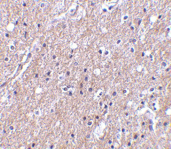
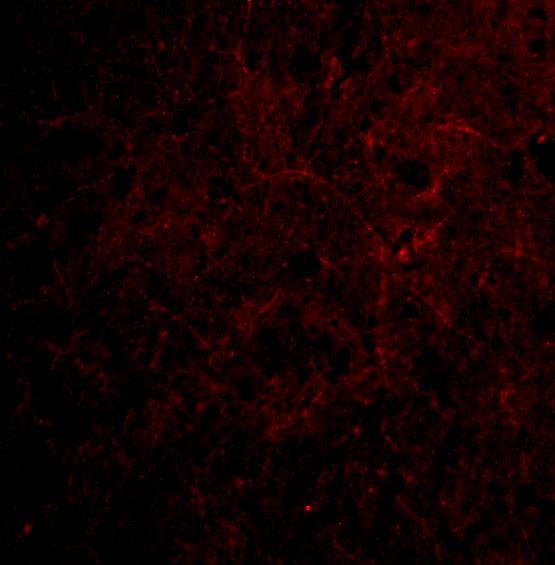
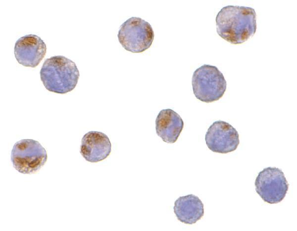

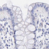


Comprehensive Human Protein Atlas IHC scoring per tissue (reliability: Enhanced — High consistency between antibody staining and RNA expression data. At least one protein variant secreted, tissue location of RNA and protein might differ and correlation is complex.). Rows are taken directly from the HPA tissue chart — click any row's HPA link to view the source.
| Tissue | Cell type | Level | Evidence | Source |
|---|---|---|---|---|
| Placenta | Endothelial cells | High | Protein (IHC) | HPA → |
| Endometrium | Cells in endometrial stroma | Medium | Protein (IHC) | HPA → |
| Tissue | Cell type | Level | Evidence | Source |
|---|---|---|---|---|
| Adipose tissue | Adipocytes | Not detected | Protein (IHC) | HPA → |
| Adrenal gland | Glandular cells | Not detected | Protein (IHC) | HPA → |
| Appendix | Endocrine cells | Not detected | Protein (IHC) | HPA → |
| Bone marrow | Hematopoietic cells | Not detected | Protein (IHC) | HPA → |
| Breast | Adipocytes | Not detected | Protein (IHC) | HPA → |
Troubleshoot CTRP6 staining in paraffin section IHC, with a separate note on IF multiplexing and interpretation.
The catalog includes a paraffin-section IHC antibody with a human brain tissue image (A12414-1 image caption) and IF/ICC antibodies with human cell images (catalog image captions). Listed reactivity spans human, mouse and rat (catalog reactivity).
A12414-1 lists IHC-P and IF and shows IHC in human brain tissue and IF in human brain cells (catalog applications; A12414-1 image captions). A12414 lists IF and ICC and shows both in HeLa cells (catalog applications; A12414 image captions).
Which to pick: Choose A12414-1 for paraffin-section IHC: it lists IHC-P and shows staining in human brain tissue at 10 μg/mL; the fixative is unreported (catalog applications; A12414-1 IHC image caption). Choose A12414 for cell IF/ICC, supported by its HeLa cell images; for the broadest listed species reactivity, choose A12414-1, which lists human, mouse and rat, while A12414 lists human and mouse (A12414 image captions; catalog reactivity). Both antibodies list rabbit as host, and clonality is unreported (catalog host and clone fields).