This website uses cookies to ensure you get the best experience on our website.
- Table of Contents
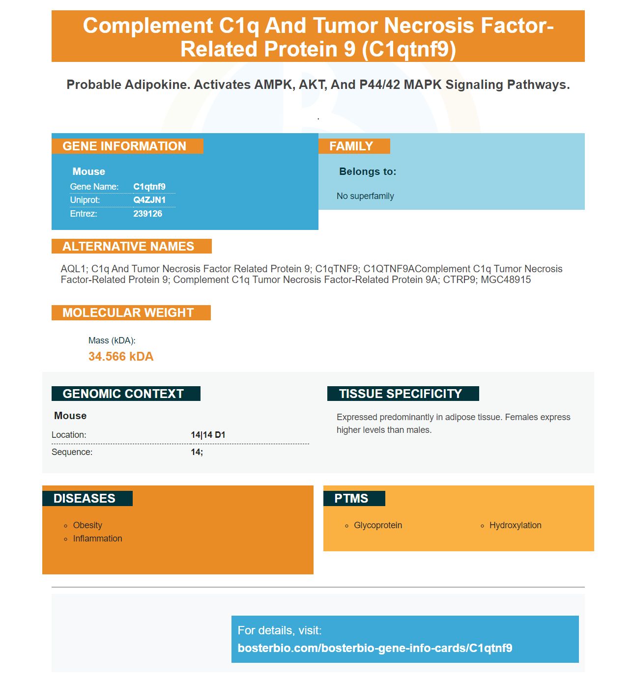
Facts about Complement C1q and tumor necrosis factor-related protein 9.
.
Mouse | |
---|---|
Gene Name: | C1qtnf9 |
Uniprot: | Q4ZJN1 |
Entrez: | 239126 |
Belongs to: |
---|
No superfamily |
AQL1; C1q and tumor necrosis factor related protein 9; C1qTNF9; C1QTNF9AComplement C1q tumor necrosis factor-related protein 9; complement C1q tumor necrosis factor-related protein 9A; CTRP9; MGC48915
Mass (kDA):
34.566 kDA
Mouse | |
---|---|
Location: | 14|14 D1 |
Sequence: | 14; |
Expressed predominantly in adipose tissue. Females express higher levels than males.
If you're planning on using the C1QTNF9 Marker in your research, you'll find it useful to read this article first. This article will explain the difference between Boster Bio's High-affinity primary antibodies and the ECL chemiluminescent detection system. Then you can decide whether the C1QTNF9Marker is right to use in your experiments.
Boster Biotechnology is a biotechnology company that develops antibodies that detect C1QTNF9 from a variety of animal samples. Boster Bio uses rabbit and mouse samples to develop antibodies against this marker. Its products include CTRP9 which is a novel secreted glycoprotein, which reduces serum glucose levels in mice and forms heterotrimers of adiponectin.
NGS has changed antibody repertoire analysis by extending sample depth. Next-generation sequencing allows for the determination of paired H and L chains at large scales, which provides a genetic record of evolutionary events. Computational tools permit reconstruction of antibody-clonal lineages. Recent research has provided new insights into the mechanism behind affinity maturation. The molecular biology of antibody molecules is progressing at an incredible pace, and NGS is making great strides in this area.
The affinity-matured antibodies were first examined against a protein and not a hapten. The affinity-matured antibody that recognized hen egg white Lysozyme represented different stages of affinity maturation. Although these antibodies had higher affinity for somatic mutations than others, the increased affinity was not due to formation of additional hydrogen bonds, salt bridges or total buried area. The VH–HEL interface had a higher shape complementarity and the affinity increased.
Next-generation sequencing of immune system has allowed reconstruction and inference of germline progenitor sequences. The derived sequences may not be identical to the unmutated sequences from the true ancestors. However, there is high confidence in mutations in the VL or VH gene segments. However, the original VLJL/VHDJH junctional sequencings remain elusive. It is difficult for us to know the exact sequence of insertions and deletions that occur during affinity maturation.
These peptides are very useful in confirmation. Researchers can confirm that the primary antibody is binding with the target antigen by using these antibodies. It is also useful to confirm whether the antibody is binding to non-specific antigens. This allows high-affinity primary antibodies to be simultaneously screened for multiple antigens.
Boster Bio's C1QTNF9 protein, a 33.7 kDa oligopeptide containing 324 amin acids fused to one peptide, is 33.7 kDa. Once prepared, the C1QTNF9 protein should be stored at -20degC. After reconstitution of the protein, aliquot it and keep at 4degC until use. This method is applicable to researchers and scientists worldwide.
The secondary antibodies that are bound to the western blot anti-body detection methods are the basis of the detection. There are two main types: ECL chemiluminescent and DAB chromogenic. The Boster ECL-chemiluminescent detection method relies on exposure of the secondary antibody and incubation using a substrate.
Chemiluminescence, also known as fluorescent immunoblotting or fluorescent immunoblotting is the use of secondary antibodies that are attached to a fluorescent protein molecule. Fluorophore light emits light that is proportional to the amount of protein present on the membrane. In contrast, Bradford protein assay uses a Coomassie Brilliant Blue dye. The concentration of the protein determines the intensity of this dye.
Nitrocellulose Membrane is a popular protein blotting material. Its high affinity for proteins makes it compatible with a variety of detection methods. Boster's Nitrocellulose Membrane is compatible with a variety protein molecularweights and nucleic substances. With Boster's Nitrocellulose Membrane, you can detect proteins with very low molecular weight.
The Boster Bio C1QTNF9 Chemiluminescent-Detection System can detect C1QTNF9. The system uses monoclonal and polyclonal antibodies to target CTRP9. The antibody binds directly to the protein, forming heterotrimers with Adiponectin. These antibodies are very specific and have a low background.
Boster Bio C1QTNF9 Chemiluminescent-Detection System can detect horseradish oxidase in western blood blots. There are enough reagents to cover 800 cm2 membrane. Apply the substrate solution evenly to the membrane. The edge should be gently blotted to remove any excess. The membrane should then be covered with a clear preservative film. To prevent air bubbles, use transparent glass paper to blot the membrane.
This colorimetric tUNEL system measured the apoptosis rate of colonic mucosal epithelial cell cells. To identify apoptotic cells, samples were incubated in a DAB chromogenic solution for three to six minutes. After that, the samples were counterstained by hematoxylin. FHC cells were planted in 96-well plates. The survival rates were measured using optical density.
PMID: 15231994 by Wong G.W., et al. A family of Acrp30/adiponectin structural and functional paralogs.
PMID: 18787108 by Wong G.W., et al. Identification and characterization of CTRP9, a novel secreted glycoprotein, from adipose tissue that reduces serum glucose in mice and forms heterotrimers with adiponectin.