This website uses cookies to ensure you get the best experience on our website.


- Table of Contents
Plan C3AR1 staining in paraffin sections using an IHC-validated antibody at 1:100–1:300 (datasheet). Use cytoplasmic staining in immune cells as the tissue reference (HPA tissue IHC), while considering the receptor’s membrane topology when interpreting the pattern (UniProt).

Expected localisation, validated protocols, controls and antibodies — the at-a-glance facts below, then the full design guide.
| Expected localisation | Immune-cell cytoplasm (HPA tissue IHC); cell membrane (UniProt) | |
| Staining pattern | Mainly cytoplasmic staining in immune cells (HPA tissue IHC) | |
| Antigen retrieval | Citrate pH 6.0 HIER, 95–98 °C, 20 min (rule: cytoplasmic / membrane antigen) | |
| Positive control | Appendix+4 more · see all | |
| Negative control | Adipose tissue+4 more · see all |
| Fixation | Keep fixation consistent across sections (standard IHC practice; not target-specific) | |
| Caveat | Phosphorylation-associated internalization may shift staining (UniProt) | |
| Regulation | Expression regulation not specified (UniProt) | |
| Isoform / epitope | No isoforms or processing noted; epitope side matters (UniProt) |
The catalog antibody’s IHC-P protocol is accompanied by published protocols for placental, ovarian, and carotid tissues (PMC11780793; PMC10067206; PMC11061441).
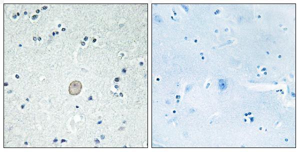
| Sample | Paraffin-embedded human brain tissue; fixative not specified (datasheet A06350) |
| Fixation | Image fixative and duration unreported (datasheet A06350); verify before use. |
| Sectioning | 4–5 µm sections on charged slides (standard) |
| Deparaffinisation | Xylene, graded ethanol series to water (standard) |
| Antigen retrieval | Heat-induced epitope retrieval in citrate buffer, pH 6.0, 20 min at 95–98 °C (standard rule: cytoplasmic / membrane antigen) |
| Peroxidase block | 3% H2O2, 10 min, room temperature (standard) |
| Blocking | 10% normal serum of the secondary host, 30 min, room temperature (standard) |
| Primary antibody | Rabbit anti-C3AR1, 1:100 - 1:300 (datasheet A06350) |
| Primary incubation | Overnight at 4 °C (standard) |
| Detection | HRP-polymer secondary, DAB chromogen 5–10 min (standard) |
| Counterstain | Hematoxylin, blue, dehydrate and mount (standard) |
| Expected result | C3AR1-positive staining in lymphoid tissue of appendix (HPA tissue IHC: Medium). HPA tissue profile: Mainly cytoplasmic expression in immune cells. No signal in the no-primary control. |
C3AR1 is a 7-transmembrane cell-membrane receptor (UniProt Q16581 topology). In tissue IHC, expect mainly cytoplasmic staining in immune cells, with medium staining reported in lymphoid tissue and non-germinal-center cells (HPA: tissue IHC profile and levels). HPA rates its tissue staining “Enhanced”: external characterization supports staining, but internal RNA data are unavailable for correlation (HPA: reliability description).
| Discrete staining in immune cells, including non-germinal-center cells in lymph node or tonsil (HPA: tissue IHC). | This fits the reported mainly cytoplasmic immune-cell profile; both named cell populations show medium staining (HPA: tissue IHC). A membrane rim may fit the receptor's topology, but HPA's tissue images do not require a crisp rim in every positive cell (UniProt Q16581 topology; HPA: tissue IHC profile). |
| Predominantly nuclear staining, without a convincing immune-cell pattern. | A nuclear-only result conflicts with the membrane receptor annotation and HPA's mainly cytoplasmic IHC profile (UniProt Q16581 subcellular location; HPA: tissue IHC). Treat it as suspect, then compare the positive control and detection controls before assigning a cause (general IHC practice). |
| Strong staining in a cell population reported as undetected, such as adipocytes. | Adipocytes are “Not detected” in the supplied HPA tissue IHC data (HPA: adipose tissue). Consider cross-reactivity or endogenous detection activity; the discrepancy alone cannot distinguish them (general IHC practice). Do not classify every nonimmune cell as false positive: HPA also reports medium staining in several named nonimmune populations (HPA: tissue IHC). |
| Uniform colour across tissue, stroma, or section edges, without cell-specific contrast. | A diffuse pattern does not resemble HPA's mainly cytoplasmic immune-cell profile (HPA: tissue IHC). Background from detection chemistry or nonspecific binding is plausible; check a control lacking primary antibody and inspect the staining distribution before scoring cells (general IHC practice). |
| No staining in lymph node or tonsil non-germinal-center cells. | Both populations are reported at medium intensity, so a blank result raises concern about the run or specimen (HPA: tissue IHC). Confirm that expected cells are present, then review the catalog antibody's IHC-P conditions and the run controls; one negative section does not establish C3AR1 absence (general IHC practice). |
| Compartment and receptor topology | UniProt places C3AR1 at the cell membrane with 7 transmembrane segments, while HPA describes tissue staining as mainly cytoplasmic (UniProt Q16581 topology; HPA: tissue IHC profile). Interpret both annotations together; cytoplasmic IHC colour alone is not proof of mislocalization or receptor internalization. |
| Choice of positive and comparison tissue | Lymph node and tonsil non-germinal-center cells, and appendix lymphoid tissue, are reported at medium staining (HPA: tissue IHC). Adipocytes are reported “Not detected,” making that named population a useful comparison; this does not certify the whole adipose section as C3AR1-free (HPA: adipose tissue). |
| Strength of the tissue evidence | HPA assigns “Enhanced” reliability to tissue staining and lists antibody CAB009405 as IHC Enhanced (HPA: reliability and antibody validation). Its reliability description also says internal RNA data are unavailable for correlation; use morphology and controls when evaluating an unexpected cell type (HPA: reliability description; general IHC practice). |
| Epitope and molecular modification limits | UniProt lists extracellular and cytoplasmic regions and 3 glycosylation sites (UniProt Q16581 topology and glycosylation). The supplied record does not locate the catalog antibody's epitope, so it cannot predict whether retrieval or permeabilisation will expose that epitope (UniProt Q16581 topology; supplied antibody data). |
| Situation | Likely cause | Next action |
|---|---|---|
| No signal in a named positive tissue. | The expected cell population may be absent, or the IHC run may have failed; the result alone cannot identify which (HPA: tissue IHC; general IHC practice). | Find non-germinal-center cells in lymph node or tonsil, review run controls, and check the catalog antibody's IHC-P instructions before changing conditions (HPA: tissue IHC; general IHC practice). |
| Colour is diffuse or appears on nearly every structure. | Nonspecific staining or detection background may obscure the reported mainly cytoplasmic immune-cell pattern (HPA: tissue IHC profile; general IHC practice). | Compare a control lacking primary antibody, examine section edges and tissue architecture, and adjust blocking or detection only in response to the control result (general IHC practice). |
| Nuclei dominate the staining. | A nuclear-only pattern conflicts with the reported membrane location and mainly cytoplasmic IHC profile; its mechanism is unresolved (UniProt Q16581 subcellular location; HPA: tissue IHC). | Check the counterstain and control without primary antibody, then judge whether any identifiable immune cells retain the expected cytoplasmic pattern (general IHC practice; HPA: tissue IHC profile). |
| An unexpected cell population stains strongly. | Cross-reactivity or endogenous detection activity is possible, but tissue context matters: HPA reports some nonimmune populations at medium intensity (HPA: tissue IHC; general IHC practice). | Name the stained cell type before scoring it. Compare its HPA entry, nearby expected cells, and detection controls; avoid calling all nonimmune staining erroneous (HPA: tissue IHC; general IHC practice). |
| Adipocytes stain despite the HPA comparison. | HPA reports adipocytes as “Not detected”; staining could reflect technical background or a sample difference, which one section cannot resolve (HPA: adipose tissue; general IHC practice). | Verify adipocyte identity and check a control lacking primary antibody; use a named positive cell population from the same run to assess whether the assay behaved as expected (HPA: tissue IHC; general IHC practice). |
| Q: Should IF/ICC show a clear membrane rim? | A: HPA summarizes subcellular location as “Membrane” but supplies no ICC-IF image cell lines or main-location detail (HPA: subcellular record). | Use the separate IF/ICC guide for that application. Here, treat a membrane signal as compatible with UniProt topology, without requiring an IF pattern that the supplied HPA record has not documented (UniProt Q16581 topology; HPA: subcellular record). |


Comprehensive Human Protein Atlas IHC scoring per tissue (reliability: Enhanced — External characterization data supports antibody staining but no internal RNA data available for correlation. Antibody staining in cells/structures not annotated, view images.). Rows are taken directly from the HPA tissue chart — click any row's HPA link to view the source.
| Tissue | Cell type | Level | Evidence | Source |
|---|---|---|---|---|
| Appendix | Lymphoid tissue | Medium | Protein (IHC) | HPA → |
| Endometrium | Cells in endometrial stroma | Medium | Protein (IHC) | HPA → |
| Lung | Alveolar cells type I | Medium | Protein (IHC) | HPA → |
| Lymph node | Non-germinal center cells | Medium | Protein (IHC) | HPA → |
| Nasopharynx | Respiratory epithelial cells | Medium | Protein (IHC) | HPA → |
| Tissue | Cell type | Level | Evidence | Source |
|---|---|---|---|---|
| Adipose tissue | Adipocytes | Not detected | Protein (IHC) | HPA → |
| Adrenal gland | Glandular cells | Not detected | Protein (IHC) | HPA → |
| Caudate | Glial cells | Not detected | Protein (IHC) | HPA → |
| Cerebellum | Cells in granular layer | Not detected | Protein (IHC) | HPA → |
| Cerebral cortex | Endothelial cells | Not detected | Protein (IHC) | HPA → |
Troubleshoot C3AR1 staining in paraffin sections by checking retrieval, cellular distribution, controls and scoring against the available tissue evidence.
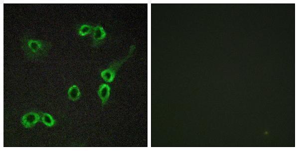
A06350 offers IHC data from paraffin-embedded human brain and IF data from COS7 cells (catalog image captions); its listed reactivity is human, mouse and rat (catalog reactivity).
A06350 will render with its IHC image of paraffin-embedded human brain and a peptide-blocked comparison (catalog IHC caption). The same SKU lists IHC, IF and ICC applications and human, mouse and rat reactivity; its separate IF image shows COS7 cells (catalog applications, reactivity and IF caption).
Which to pick: Choose A06350 for paraffin-section IHC because its own IHC caption shows human brain tissue processed that way (catalog IHC caption); the fixative is unreported (catalog IHC caption). A06350 is also the listed IF/ICC option, supported by an IF image of COS7 cells and an ICC application listing (catalog IF caption and applications). For human, mouse or rat samples, A06350 is the listed cross-species choice, with rabbit polyclonal host and human, mouse and rat reactivity (catalog host, dilution_raw and reactivity).