This website uses cookies to ensure you get the best experience on our website.


- Table of Contents
Plan chromogenic IHC for C5AR1 using high-staining bone marrow hematopoietic cells or spleen red pulp cells as reference tissues (HPA tissue IHC). This guide covers expected staining, antibody selection and interpretation for paraffin sections.

Expected localisation, validated protocols, controls and antibodies — the at-a-glance facts below, then the full design guide.
| Expected localisation | Membranous and cytoplasmic staining in immune-cell subsets (HPA tissue IHC) | |
| Staining pattern | Subset immune cells show membranous and cytoplasmic staining (HPA tissue IHC) | |
| Antigen retrieval | EDTA pH 8.0 HIER, heat-mediated (datasheet A01898-3) | |
| Positive control | Bone marrow+4 more · see all | |
| Negative control | Breast+4 more · see all |
| Fixation | Keep paraffin-section fixation consistent across samples. (standard IHC practice; not target-specific) | |
| Caveat | Phosphorylated receptor can internalize into vesicles (UniProt) | |
| Regulation | Phosphorylation-linked internalization (UniProt) | |
| Isoform / epitope | No annotated isoforms; extracellular versus cytoplasmic epitopes matter (UniProt) |
The catalog antibody protocol uses heat-mediated EDTA retrieval at pH 8.0 (datasheet: A01898-3). One published mouse paw IHC protocol is included (PMC12202429).
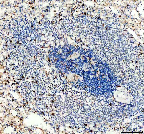
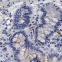
| Sample | Paraffin-embedded human spleen tissue; fixative not specified (datasheet A01898-3) |
| Fixation | Image fixative and duration unreported (datasheet A01898-3); verify before use. |
| Sectioning | 4–5 µm sections on charged slides (standard) |
| Deparaffinisation | Xylene, graded ethanol series to water (standard) |
| Antigen retrieval | Heat retrieval: EDTA pH 8.0 (datasheet A01898-3); 20 min, 95–100 °C (standard) |
| Peroxidase block | 3% H2O2, 10 min, room temperature (standard) |
| Blocking | 10% goat serum (datasheet A01898-3) |
| Primary antibody | Rabbit anti-C5AR1, 1:50 recommended; image 1:200 (datasheet A01898-3) |
| Primary incubation | Overnight at 4 °C (datasheet A01898-3) |
| Detection | HRP-conjugated secondary, DAB chromogen (datasheet A01898-3) |
| Counterstain | Hematoxylin, blue, dehydrate and mount (standard) |
| Expected result | C5AR1-positive staining in hematopoietic cells of bone marrow (HPA tissue IHC: High). HPA tissue profile: Cytoplasmic and membranous expression in subsets of immune cells in several tissues. No signal in the no-primary control. |
C5AR1 is a seven-transmembrane receptor at the cell membrane and in cytoplasmic vesicles (UniProt P21730 topology and subcellular location). In paraffin-section IHC, expect cytoplasmic and membranous staining in subsets of immune cells across several tissues (HPA tissue IHC). HPA rates this tissue pattern Approved, with medium consistency between staining and RNA expression; interpret individual stained cells in their tissue context (HPA tissue IHC).
| Membranous staining with cytoplasmic signal in selected immune cells, including lung macrophages. | This fits the reported tissue pattern and receptor locations (HPA: medium in lung macrophages; UniProt P21730: cell membrane and cytoplasmic vesicles). Score the relevant cells and compartments, since HPA describes staining in subsets rather than every cell in a section (HPA tissue IHC). |
| Predominantly nuclear staining, without a credible membranous or cytoplasmic pattern. | Nuclear localisation is outside the supplied C5AR1 locations (UniProt P21730 subcellular location; HPA tissue IHC). Treat it as suspect staining; review the negative control and detection steps before assigning it to C5AR1 (general IHC practice). |
| Strong staining in a cell population reported as not detected, such as breast adipocytes. | This conflicts with the reported cell-specific result (HPA: breast adipocytes not detected). Cross-reactivity or endogenous detection activity is possible; check a negative control and whether the signal follows the expected cellular pattern (general IHC practice). |
| Diffuse colour over tissue and empty areas, obscuring cell borders. | A field-wide haze cannot establish the selected-cell membranous and cytoplasmic pattern (HPA tissue IHC). Consider excess antibody or detection background, and compare with a negative control processed alongside the section (general IHC practice). |
| No convincing signal in hematopoietic cells of bone marrow or cells in splenic red pulp. | Those populations are reported High and provide useful comparison material (HPA: bone marrow hematopoietic cells High; spleen red-pulp cells High). Confirm that the relevant cells are present, then assess the control and staining run before interpreting a negative specimen (general IHC practice). |
| Cell population and tissue | HPA reports High staining in bone-marrow hematopoietic cells and splenic red-pulp cells, Medium in lung macrophages, and no detection in breast adipocytes (HPA tissue IHC). Compare the same cell population when judging intensity; an unrelated cell in a positive tissue is not an equivalent control (general IHC practice). |
| Receptor location and trafficking | Seven transmembrane segments support a membrane-associated pattern (UniProt P21730 topology). Cytoplasmic vesicular signal can also fit C5AR1: phosphorylated receptor colocalizes with ARRB1 and ARRB2 in vesicles, and their interaction is associated with internalisation (UniProt P21730 subcellular location and subunit). |
| Evidence strength | The tissue pattern is Approved but has medium consistency with RNA expression, so use it as an expected pattern rather than a guarantee for every specimen (HPA tissue IHC). HPA lists IHC Approved for HPA014520 and CAB002720; those statuses do not establish the performance of another antibody (HPA antibodies). |
| Epitope position | C5AR1 has extracellular, transmembrane and cytoplasmic regions (UniProt P21730 topology). An antibody's epitope position would affect which region its staining assesses, but no epitope position is supplied here; do not infer a required retrieval method from topology alone (UniProt P21730 topology; general IHC practice). |
| IF/ICC Q: Should Golgi staining match paraffin-section IHC? | A: HPA reports mainly Golgi and additional vesicular localisation in ICC-IF, whereas its tissue IHC profile is cytoplasmic and membranous in immune-cell subsets (HPA subcellular ICC-IF; HPA tissue IHC). Compare each result with its own preparation and cell context; the ICC-IF finding does not establish a paraffin IHC pattern. |
| Situation | Likely cause | Next action |
|---|---|---|
| Expected positive cells show no stain. | The relevant cells may be absent from the section, or the staining run may have failed; HPA reports High signal specifically in bone-marrow hematopoietic cells and splenic red-pulp cells (HPA tissue IHC). | Verify those cells on the section and assess a concurrently stained known-positive section and detection controls before calling the sample negative (general IHC practice). |
| The whole section has weak or diffuse colour. | General detection background or excessive primary-antibody signal may obscure the selected-cell pattern (general IHC practice; HPA tissue IHC pattern). | Compare with a primary-antibody omission control, review blocking and washes, and optimise primary-antibody dilution using the antibody's IHC instructions (general IHC practice). |
| Nuclei dominate the signal. | That compartment does not match the supplied cell-membrane or cytoplasmic-vesicle locations (UniProt P21730 subcellular location). Nonspecific binding or detection artefact is possible (general IHC practice). | Check the omission control and whether any membranous or cytoplasmic signal remains in HPA-reported positive cells; withhold a C5AR1 call if it does not (HPA tissue IHC; general IHC practice). |
| A reportedly negative cell type stains strongly. | For example, HPA reports no detection in breast adipocytes; apparent signal there may reflect cross-reactivity or endogenous detection activity (HPA tissue IHC; general IHC practice). | Check the same cell type in an appropriate control section and inspect the omission control. Address endogenous detection activity as appropriate to the chromogenic system (general IHC practice). |
| Only cytoplasmic puncta are visible. | Vesicular C5AR1 is plausible, including receptor associated with ARRB1 and ARRB2 after phosphorylation (UniProt P21730 subcellular location and subunit). Puncta alone do not establish antibody specificity (general IHC practice). | Assess whether puncta occur in the expected cells, compare the negative control, and review any membranous component against the HPA tissue profile (HPA tissue IHC; general IHC practice). |
| A medium- or low-staining tissue appears negative. | HPA levels vary by tissue and cell: lung macrophages are Medium, stomach glandular cells Low, and several listed cell populations are not detected (HPA tissue IHC). | Check the specified cell population and a High reference population before treating the finding as a staining failure; report the examined cells and control result (HPA tissue IHC; general IHC practice). |


Comprehensive Human Protein Atlas IHC scoring per tissue (reliability: Approved — Medium consistency between antibody staining and RNA expression data.). Rows are taken directly from the HPA tissue chart — click any row's HPA link to view the source.
| Tissue | Cell type | Level | Evidence | Source |
|---|---|---|---|---|
| Bone marrow | Hematopoietic cells | High | Protein (IHC) | HPA → |
| Spleen | Cells in red pulp | High | Protein (IHC) | HPA → |
| Adrenal gland | Glandular cells | Medium | Protein (IHC) | HPA → |
| Appendix | Lymphoid tissue | Medium | Protein (IHC) | HPA → |
| Cerebral cortex | Neuropil | Medium | Protein (IHC) | HPA → |
| Tissue | Cell type | Level | Evidence | Source |
|---|---|---|---|---|
| Breast | Adipocytes | Not detected | Protein (IHC) | HPA → |
| Bronchus | Basal cells | Not detected | Protein (IHC) | HPA → |
| Cerebellum | Cells in granular layer | Not detected | Protein (IHC) | HPA → |
| Cervix | Glandular cells | Not detected | Protein (IHC) | HPA → |
| Duodenum | Glandular cells | Not detected | Protein (IHC) | HPA → |
Troubleshoot C5AR1 staining in paraffin sections by checking retrieval, cell identity, subcellular pattern, and controls before interpreting signal.
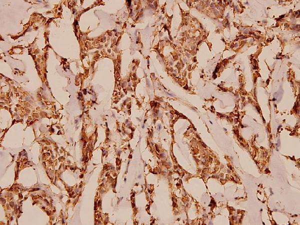
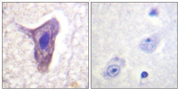
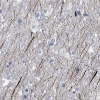
Anti-C5AR1 antibodies have IHC images from paraffin-embedded human spleen, breast carcinoma and brain (catalog image captions); all three list IF, and two list mouse and rat reactivity (catalog applications/reactivity).
A01898-3 shows IHC in human spleen paraffin sections, and A01898S334 shows IHC in human breast carcinoma paraffin sections (respective image captions). A01898S338 shows phospho-Ser338 IHC in human brain paraffin sections with a phosphopeptide blocking comparison (A01898S338 image caption).
Which to pick: For tissue IHC, A01898-3 has a human spleen paraffin example using EDTA retrieval and 1:200 primary antibody; its listed IHC dilution is 1:50 (A01898-3 image caption; catalog dilution). For IF/ICC, choose A01898-3 because this polyclonal antibody lists both applications and human reactivity; for human, mouse or rat IHC/IF, consider A01898S334 or the phospho-Ser338 antibody A01898S338, whose IHC images are from human breast carcinoma and human brain, respectively (A01898-3 datasheet/applications/reactivity; A01898S334 and A01898S338 applications/reactivity and image captions). The fixative is unreported in all three paraffin IHC captions (catalog image captions).