This website uses cookies to ensure you get the best experience on our website.
- Table of Contents
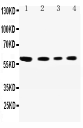
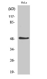
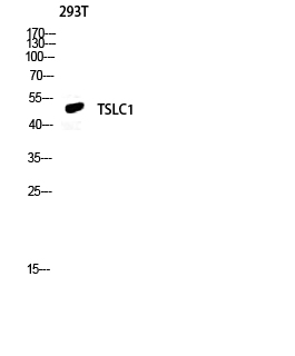
Facts about Cell adhesion molecule 1.
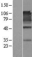
Acts as a tumor suppressor in non-small-cell lung cancer (NSCLC) cells. Interaction with CRTAM promotes natural killer (NK) cell cytotoxicity and interferon-gamma (IFN-gamma) secretion by CD8+ cells in vitro as well as NK cell-mediated rejection of tumors expressing CADM3 in vivo.
Human | |
---|---|
Gene Name: | CADM1 |
Uniprot: | Q9BY67 |
Entrez: | 23705 |
Belongs to: |
---|
nectin family |
BL2; Cadm1; cell adhesion molecule 1; IgSF4; IGSF4A; IGSF4ADKFZp686F1789; IGSF4immunoglobulin superfamily, member 4D variant 2; Immunoglobulin superfamily member 4; immunoglobulin superfamily, member 4; Necl-2; NECL2MGC149785; nectin-like 2; Nectin-like protein 2; RA175; sgIGSF; Spermatogenic immunoglobulin superfamily; ST17; sTSLC-1; Synaptic cell adhesion molecule; SynCAM; SynCAM1; SYNCAMMGC51880; TSLC1; TSLC-1; TSLC1/Nectin-like 2/IGSF4; Tumor suppressor in lung cancer 1SYNCAM1
Mass (kDA):
48.509 kDA
Human | |
---|---|
Location: | 11q23.3 |
Sequence: | 11; NC_000011.10 (115169236..115504428, complement) |
Cell membrane; Single-pass type I membrane protein. Cell junction, synapse. Localized to the basolateral plasma membrane of epithelial cells in gall bladder.
If you're looking for an innovative antibody or are looking for an diagnostic test, the CADM1 Marker is an excellent alternative. The CADM1 protein is used for a variety purposes for example, HTLV-1 associated myelopathy/tropical Spastic Paraplegia, ATLL or other disorders. This article will provide an overview of clinical uses and benefits of the CADM1 marker.
Human T lymphotropic virus type 1 (HTLV-1) infection is a major cause of tropical spastic paralysis and myelopathy. The majority of people with HTLV-1 are not afflicted however, a few suffer from clinical disease. HAM/TSP is a progressive neurological disorder that typically affects adults. Although the symptoms of HAM/TSP may be common to many patients, they do vary widely among patients.
The causes of HAM/TSP are not yet understood. Researchers believe that the virus may be linked to a variety of non-neoplastic inflammatory diseases. The most common of these are Sjogren's Syndrome, uveitis arthritis, bronchoalveolitis, and polymyositis. Some HAM/TSP patients have more than one HTLV-1-associated inflammation disorder.
The virus affects the CNS and results in weakness and paralysis of the legs. It is also associated with urinary symptoms and lower back pain and chronic disability. In areas where HTLV-1 is prevalent, HAM/TSP imposes a significant health burden. While research into the causes of the virus and treatment is ongoing significant advancements have been made towards understanding the disease and its development.
Global travel is increasing the risk of HTLV-1-associated myelopathic/tropical spastic paresis in the human population. Although most cases of the disease are found in endemic areas where it is most prevalent but HTLV-1 is not exclusively in those regions. Multiple sclerosis is often misdiagnosed with HAM/TSP. However the primary form of MS is characterized by some of the same clinical characteristics as HAM/TSP. Due to the wide variety of symptoms, it is vital to improve screening guidelines for HTLV-1.
The HTLV-1 infection can affect the immune system by affecting the CTL. The infection can lead to numerous diseases, including a debilitating myelopathy. ATLL has been recognized as a major cause of death in HAM/TSP patients. In this study, the genomic changes of cells infected by patients with HAM/TSP during the transition of ATLL to ATLL were discovered.
HAM/TSP usually develops during adulthood. The disease is slow-growing and has no recovery. Some patients suffer from rapid or mild progression. Patients must undergo multiple tests to determine if the disease is caused by infected HTLV-1.
In the moment, sCADM1 is still the sole biomarker used for ATLL. This is a good thing that sCADM1 is a biomarker for ATLL. The measurement of sCADM1 may help to predict the stage of the disease and progression. This research is funded by Takeda Science Foundation. This study could become the next standard in the diagnosis of ATLL. It is, however, important to understand the goal of the biomarker before this happens.
The sCADM1 protein is that is found in bloodstreams and is crucial for ATLL. It is possible to measure the response to therapy by measuring the levels of sCADM1 in plasma. It may also be used as a test for determining the ability of a transplant to be stable and maintenance therapy. These are just some of the possible uses for the CADM1 biomarker. For now, however, it is only used to detect disease progression or relapse in patients with this disorder.
sCADM1 may be used to monitor the progress of disease in adult patients with T cell leukemia/lymphoma. Researchers have discovered that sCADM1 can be used to monitor the development of ATLL from the indolent to aggressive states. It is not a good indicator for diagnosing ATLL. Instead, it can be beneficial for monitoring treatment response or assessing the burden of leukemia in the patient.
Although plasma sCADM1 can be used to detect aggressive ATLL, it cannot be used to determine the course of the disease. A better marker for ATLL would be sCADM1-related proteinslike T-cell antigens and IL2R. However the sCADM1–sCADM1 indicator can be used to determine the likelihood of relapse in patients suffering from this disease.
Researchers have discovered a brand new marker for lung cancer that is small-cell called CADM1. The study was conducted by Takeshi Ito, Toko Funaki, Goh Tanaka, Takahide Nagase and Yoshinori Murakami. This research has important implications for cancer research and treatment. While the CADM1 marker is not a cancer-specific marker, it could play a part in a particular cell-cell adhesion.
Furthermore, this protein has been associated with laryngeal Squamous Cell carcinoma and a higher expression of CADM1 in these cases is linked to better prognosis. So, CADM1 could be a significant biomarker for the detection of lung cancers called adenocarcinomas. CADM1 has been demonstrated to improve intestinal function in rats suffering from IBS-D.
In addition, CADM1 prevents cell migration and invasion. It also plays a role in EMT. In addition, CADM1 expression in human melanoma cell lines has been associated with lower viability. It is unclear whether CADM1's expression affects the metastatic potential of cancer cells. However, it is believed to prevent the growth of tumor cells in non-adhesive environments. It has been associated with resistance to anoikis.
PCR was used to determine the expression of CADM1. We employed pcDNA3.1 and FuGENE6 vectors. TWIST1 antibody was used to remove DNA from lysates. Anti-V5 epitope antibody was also used. Then, we used DNA Fragment Analyzer to evaluate the relative expression of CADM1 in three independent tests. In the future we might wish to study the interaction between different conditions.
The variant 8/9 is located in the brain as well as the stomach. This variation is more specific to SCLC than it is for other conditions. This small peak could be caused by SCLC precursor cells of neuroendocrine nature. Additionally, CADM1 is expressed in the testis. These data are promising for cancer treatment. Although further research is needed but this marker has shown promising potential for early diagnosis.
While there is no consensus regarding the role of CADM1 in nevi formation It is known to play a role in the suppression of tumors. If CADM1 may play a part in the formation of nevi cells, it could be selected against during clonal evolution. These research suggests that CADM1 could play a role in cancer. However, there are many other conditions that the CADM1 marker is involved.
The CADM1 gene is a membrane-bound cell adhesion molecule that has four regions which include an extracellular region, a transmembrane domain, and an cytoplasmic area. The gene is found in leukemia cells and is linked to cell-cell junctions. CADM1 is associated with neurobiology, and is widely believed to be a tumor suppressor. Both CD34+ and CD34+ cells have the ability to express the CADM1 gene.
CADM1 is a tumor suppressor. It regulates heterophilic cell–cell adhesion. Its decreased expression is associated with poor prognosis and progression of cancer. In vitro studies on tumor cells showed that the expression of CADM1 was elevated in serous adenomas and decreased in LGSC. Patients with SOTs had a negative correlation between CADM1 and STAT3 protein expression.
The CADM1 gene is a tumor suppressor for various kinds of cancers and the deletion of the CADM1 gene in prostate cancer increases its ability to change. TWIST1 expression is also connected with CADM1 which implies that knockdowns of TWIST1 will result in an increase in the expression of CADM1's protein. All cell lines tested showed a shift in the molecular weight of CADM1 when siRNAs were directed at the TWIST1 gene. This change was consistent across all cell types used in this research. In this study, the promoter reporter CADM1 activity was reduced in both cells.
There is growing evidence that CADM1 could play a role in the formation of cell junctions, and also in promoting tumor development. This gene may also play a role in EMT-like processes and cell adhesion within melanoma cells. In addition, CADM1 could be involved in the biology of dedifferentiated cells, such as keratinocytes in hair follicles. CADM1 expression is implicated in the inhibition of wound healing, proliferation, migration and migration of hair follicles' Keratinocytes.
Human sputum expression of CADM1 is highly variable. A single staining of a single cell using a high-intensity fluorescent dye is not enough to determine its sensitivity. A test with low sensitivity is not recommended in this case. Clinical applications of the CADM1 marker
PMID: 15893517 by Zhou Y., et al. Nectin-like molecule 1 is a protein 4.1N associated protein and recruits protein 4.1N from cytoplasm to the plasma membrane.
PMID: 22438059 by Moiseeva E.P., et al. CADM1 isoforms differentially regulate human mast cell survival and homotypic adhesion.