CALCOCO2 / Calcium-binding and coiled-coil domain-containing protein 2 · IHC design guide

Design Immunohistochemistry for CALCOCO2

Plan chromogenic IHC on paraffin sections using CALCOCO2’s variable cytoplasmic tissue staining as the expected readout (HPA tissue IHC). This guide covers cell-type controls, consistent fixation and scoring, with 0.5–1 μg/mL catalog antibody as a starting concentration (datasheet A05876-1).

Evidence assembled Oct 2026 · For research use; verify linked source records and product datasheet before use
Immunohistochemistry protocol sheet for CALCOCO2 (IHC for CALCOCO2): expected localisation Variable cytoplasmic staining (HPA tissue IHC), antibody A05876-1, validated IHC image, and IHC protocol steps
Printable CALCOCO2 IHC protocol sheet — expected localisation Variable cytoplasmic staining (HPA tissue IHC), antibody A05876-1, controls and protocol steps. Open the full CALCOCO2 IHC guide →

CALCOCO2 Immunohistochemistry Experimental Design Guide

Expected localisation, validated protocols, controls and antibodies — the at-a-glance facts below, then the full design guide.

Must know before staining
Expected localisation Variable cytoplasmic staining (HPA tissue IHC)
Staining pattern Variable cytoplasmic staining in glandular and endothelial cells (HPA tissue IHC)
Antigen retrieval EDTA pH 8.0 HIER, heat-mediated (datasheet A05876-1)
Positive control ⓘ Appendix+4 more · see all
Negative control ⓘ Soft tissue+2 more · see all
Important caveats
Reasons your staining may differ from the expected pattern.
Fixation Keep fixation consistent across paraffin sections and controls. (selected-SKU IHC image A05876-1)
Caveat Cell-type variation can confound whole-tissue scores (HPA tissue IHC)
Regulation Tissue-dependent expression (UniProt)
Isoform / epitope 5 isoforms; antibody epitope coverage is unknown (UniProt)
Section 1

Recommended CALCOCO2 IHC & IF Protocols

The catalog antibody protocol uses EDTA retrieval (datasheet A05876-1). One published CALCOCO2 IHC protocol provides a mouse coronary artery comparison (PMC13242735).

Recommended immunohistochemistry (IHC-P) protocol parameters
SampleParaffin-embedded human lung cancer tissue; fixative not specified (datasheet A05876-1)
FixationImage fixative and duration unreported (datasheet A05876-1); verify before use.
Sectioning4–5 µm sections on charged slides (standard)
DeparaffinisationXylene, graded ethanol series to water (standard)
Antigen retrievalHeat retrieval: EDTA pH 8.0 (datasheet A05876-1); 20 min, 95–100 °C (standard)
Peroxidase block3% H2O2, 10 min, room temperature (standard)
Blocking10% goat serum (datasheet A05876-1)
Primary antibodyRabbit anti-CALCOCO2, 0.5-1μg/ml (datasheet A05876-1)
Primary incubationOvernight at 4 °C (datasheet A05876-1)
DetectionStreptavidin-biotin complex (SABC), DAB chromogen (datasheet A05876-1)
CounterstainHematoxylin, blue, dehydrate and mount (standard)
Expected resultCALCOCO2-positive staining in glandular cells of appendix (HPA tissue IHC: Medium). HPA tissue profile: Cytoplasmic expression at variable levels. No signal in the no-primary control.
💡Decision noteStart with heat-mediated EDTA at pH 8.0 for the catalog antibody (datasheet A05876-1). The published citrate protocol used a different antibody (PMC13242735).
Section 2

What Is the Expected CALCOCO2 Staining Pattern?

CALCOCO2 should appear mainly in the cytoplasm, sometimes concentrated near the nucleus or in vesicle-like structures (UniProt Q13137 subcellular location; HPA subcellular). In paraffin-section IHC, expect variable cytoplasmic staining in the cell populations HPA identifies, including glandular, endothelial and hematopoietic cells (HPA tissue IHC). HPA rates the IHC evidence Enhanced, with medium consistency between staining and RNA expression (HPA tissue IHC). CALCOCO2 has no transmembrane segment (UniProt Q13137 topology).

What am I looking at on my slide?
Cytoplasmic stain in appendix or duodenal glandular cells, or in colon endothelial cells (HPA tissue IHC).This fits the reported Medium staining in those specific cell populations (HPA tissue IHC). Score the named cells and their cytoplasm; a tissue-wide score could hide differences between cell types (general IHC scoring practice).
Predominantly nuclear dots, with little cytoplasmic signal.Treat this as an unexpected compartment pattern and check specificity and counterstain interpretation (HPA subcellular; general IHC practice). UniProt notes an older nuclear-dot report but also reports predominantly coarse-grained cytoplasmic localization near the nucleus; HPA supports cytosol as the main location (UniProt Q13137 subcellular location; HPA subcellular).
Strong stain in soft-tissue fibroblasts, splenic red-pulp cells or vaginal squamous cells.These named populations were Not detected in HPA tissue IHC (HPA tissue IHC). Unexpected stain warrants checks for antibody cross-reactivity and endogenous detection activity (general IHC practice). It does not, by itself, establish that every cell in those tissues must be negative.
Uniform haze across cells and extracellular spaces, without recognizable cytoplasmic borders.Diffuse background cannot be scored as CALCOCO2 localization; HPA describes variable cytoplasmic expression (HPA tissue IHC). Compare with a no-primary control and examine blocking, washing and chromogen development as general IHC background checks (general IHC practice).
No stain in the expected glandular cells of appendix or duodenum.Both populations showed Medium staining in HPA tissue IHC (HPA tissue IHC). A blank result calls for a run-control review before a biological absence call; section quality, retrieval, antibody dilution and detection are general IHC variables, with no CALCOCO2-specific fixation sensitivity established by these sources.
💡Expected CALCOCO2 appearanceCall a section positive when the relevant glandular, endothelial or hematopoietic cells show cytoplasmic staining at the variable levels reported by HPA; isolated dominant nuclear dots or staining confined to HPA-undetected cell populations needs verification (HPA tissue IHC; HPA subcellular; UniProt Q13137 subcellular location).
How each factor affects the staining
Cell population and tissueHPA reports Medium stain in appendix and duodenal glandular cells, colon and cerebral-cortex endothelial cells, and bone-marrow hematopoietic cells; endometrial glandular cells are Low (HPA tissue IHC). Interpret the named population rather than treating the entire section as uniformly positive.
Antibody evidenceHPA rates IHC staining Enhanced for HPA022989, HPA023019 and HPA023195, while tissue staining has medium consistency with RNA expression (HPA antibodies; HPA tissue IHC). This supports the reported pattern but leaves individual unexpected signals to be checked.
Localization and processingUniProt lists cytoplasm, perinuclear region, cytoskeleton and autophagosome membrane; it lists no transmembrane segment, signal peptide or propeptide (UniProt Q13137). HPA supports cytosol, with vesicles as an additional ICC-IF location (HPA subcellular). These records do not identify an antibody epitope.
IsoformsUniProt lists 5 isoforms (UniProt Q13137). Their presence alone does not show which forms a given IHC antibody detects; an isoform-specific interpretation requires epitope information absent from the supplied antibody evidence (UniProt Q13137 isoforms; HPA antibodies).
IF/ICC Q: What pattern should I compare?A: HPA supports mainly cytosolic signal, with additional vesicles, in its ICC-IF assessment; images are listed for A-431, U-251MG and U2OS (HPA subcellular). That compartment comparison can inform interpretation, but it does not establish an IHC staining level in those cell lines.
Why is my staining missing, weak or wrong?
SituationLikely causeNext action
The expected positive cells are blank.The stain run may have failed; appendix and duodenal glandular cells showed Medium signal in HPA tissue IHC (HPA tissue IHC).Check a same-run positive control, reagent sequence and detection. Review retrieval and antibody dilution as general IHC steps; no target-specific fixation effect is established here (general IHC practice).
Nuclear dots dominate the section.This conflicts with HPA-supported cytosol and the predominantly cytoplasmic localization noted by UniProt, despite an older nuclear-dot report (HPA subcellular; UniProt Q13137 subcellular location).Recheck the counterstain and compare an independently validated antibody or orthogonal readout where available (general IHC specificity practice).
HPA-undetected cell types stain strongly.Cross-reactivity or endogenous detection activity is possible; HPA reports no detection in the specified soft-tissue fibroblasts, splenic red-pulp cells and vaginal squamous cells (HPA tissue IHC; general IHC practice).Run no-primary and detection-system controls, then compare staining with the expected cytoplasmic pattern (general IHC practice; HPA tissue IHC).
The section shows diffuse brown haze.Nonspecific background can obscure the variable cytoplasmic pattern reported by HPA (general IHC practice; HPA tissue IHC).Compare a no-primary control; review blocking, washing and chromogen development before scoring cells (general IHC practice).
A weakly stained tissue is called negative.HPA reports Low staining in endometrial glandular cells and several other named populations; weak signal may be missed at the chosen scoring threshold (HPA tissue IHC).Inspect the specified cell population alongside a positive run control and record intensity separately from the fraction of stained cells (general IHC scoring practice).
Only some cells in a positive tissue stain.HPA describes variable cytoplasmic expression and assigns levels to particular cell populations (HPA tissue IHC).Identify the stained cell type on the counterstained section and compare it with HPA's cell-level entry before assigning a section-wide result (HPA tissue IHC; general IHC scoring practice).

Sample controls for CALCOCO2 IHC & IF

🧪Run appendix first: its glandular cells show Medium CALCOCO2 staining (HPA: appendix glandular cells, Medium). Run soft tissue fibroblasts as the negative tissue (HPA: soft tissue fibroblasts, Not detected); use non-glandular cells on the appendix slide to assess background, without assuming they are CALCOCO2-negative (HPA: appendix row specifies glandular cells only).
Positive control tissue: Appendix (Glandular cells, HPA Medium)
Negative control tissue: Soft tissue (HPA Not detected)
ICC-IF cell lines (HPA subcellular resource): HPA ICC-IF images show CALCOCO2 in A-431, U-251MG, U2OS, with annotated localisation: Cytosol (supported) (HPA subcellular).
Technical controls: Include a no-primary, secondary-only control and a concentration-matched rabbit IgG isotype control (caption: rabbit primary antibody and biotinylated goat anti-rabbit secondary); use CALCOCO2 knockout material as a biological negative where available (standard IHC practice). Quench endogenous peroxidase and assess endogenous biotin background in appendix when using the caption’s biotinylated secondary, streptavidin-biotin complex and DAB detection (caption: SABC-DAB method; standard IHC practice).
⚠️Feasibility: No target-specific fixation window or fixation effect is reported, and the fixative for the selected A05876-1 paraffin-section caption is unreported (caption: fixative not stated). Heat retrieval in EDTA at pH 8.0 was used for the shown paraffin IHC result, but retrieval dependence is unreported (caption: EDTA heat retrieval). Frozen sections are not shown to be easier; IF/ICC images exist for CALCOCO2, but they do not establish that IF is easier than IHC (HPA: ICC-IF images in A-431, U-251MG and U2OS).

HPA tissue IHC evidence for CALCOCO2

Comprehensive Human Protein Atlas IHC scoring per tissue (reliability: Enhanced — Medium consistency between antibody staining and RNA expression data.). Rows are taken directly from the HPA tissue chart — click any row's HPA link to view the source.

Positive expression · recommended positive controls

TissueCell typeLevelEvidenceSource
Appendix Glandular cells Medium Protein (IHC) HPA →
Bone marrow Hematopoietic cells Medium Protein (IHC) HPA →
Cerebral cortex Endothelial cells Medium Protein (IHC) HPA →
Colon Endothelial cells Medium Protein (IHC) HPA →
Duodenum Glandular cells Medium Protein (IHC) HPA →

Undetected expression · recommended negative controls

TissueCell typeLevelEvidenceSource
Soft tissue Fibroblasts Not detected Protein (IHC) HPA →
Spleen Cells in red pulp Not detected Protein (IHC) HPA →
Vagina Squamous epithelial cells Not detected Protein (IHC) HPA →
Section 3

Advanced CALCOCO2 IHC Tips

Troubleshoot CALCOCO2 staining in paraffin sections by checking retrieval, compartment, cell type and controls before comparing signal intensity (datasheet A05876-1; HPA tissue IHC).

What retrieval should I use when CALCOCO2 staining is weak?
Start with heat-mediated antigen retrieval in EDTA at pH 8.0 (datasheet A05876-1). The catalog antibody produced staining in a paraffin-embedded human lung cancer section after this retrieval, followed by 1 µg/ml primary antibody overnight at 4°C (datasheet A05876-1). If staining remains weak, compare adjacent sections while holding antibody concentration, heating method and detection constant; a different buffer or pH is an exploratory fallback rather than the documented condition (standard IHC practice; datasheet A05876-1). Include a section processed without primary antibody so an apparent gain in signal can be checked against retrieval-related background (standard IHC practice).
How can I assess whether fixation is limiting CALCOCO2 staining?
The selected tissue caption identifies a paraffin-embedded section but gives no fixative, so CALCOCO2-specific fixation sensitivity is unknown (datasheet A05876-1). Record each specimen’s fixative and fixation duration, then compare matched sections using the documented EDTA pH 8.0 retrieval and 1 µg/ml primary antibody condition (datasheet A05876-1; standard IHC practice). Keep section thickness, heating and detection consistent, and compare intact areas before changing antibody concentration (standard IHC practice). Do not infer a fixation effect from tissue staining patterns or from CALCOCO2’s lack of a transmembrane segment or its phosphoserine at residue 445 (HPA tissue IHC; UniProt Q13137 topology and modified residues).
Which staining pattern should I expect in tissue sections?
Expect predominantly cytoplasmic staining, potentially coarse or concentrated near the nucleus, when evaluating CALCOCO2 by chromogenic IHC (HPA tissue IHC; UniProt Q13137 subcellular location). Cytoskeletal and autophagosome-membrane associations are annotated, while supported cell-imaging data place most signal in the cytosol with additional vesicular signal (UniProt Q13137 subcellular location; HPA subcellular). Evaluate staining within identifiable cells rather than treating every brown punctum as a vesicle, and inspect an adjacent negative-control section for deposit or pigment (standard IHC practice). Predominantly nuclear-dot staining needs extra validation because published localisation reports conflict and the cytoplasmic pattern has stronger support in the supplied record (UniProt Q13137 subcellular location; HPA subcellular).
Can this antibody distinguish CALCOCO2 isoforms or modification states?
CALCOCO2 has 5 annotated isoforms, but the supplied tissue caption does not identify the catalog antibody’s epitope or establish isoform selectivity (UniProt Q13137 isoforms; datasheet A05876-1). Therefore, report staining as CALCOCO2 immunoreactivity unless an epitope map or independent isoform-specific validation is available (standard IHC practice). The protein has no annotated transmembrane segment, and phosphoserine at residue 445 is listed among its modifications; neither fact establishes whether this antibody detects a particular isoform or phosphorylation state (UniProt Q13137 topology and modified residues). If interpretation depends on either distinction, validate it with a suitable orthogonal assay and matched tissue controls before assigning that identity to the chromogenic signal (standard IHC practice).
How should I check CALCOCO2 localisation by multiplex IF?
For a separate IF/ICC experiment, pair CALCOCO2 with a marker for the cell population being evaluated, such as glandular cells in appendix or endothelial cells in colon (HPA tissue IHC; standard IF practice). Choose a spectrally separated, preferably far-red detection channel when tissue autofluorescence obscures shorter wavelengths, and acquire single-stain controls before interpreting overlap (standard IF practice). CALCOCO2 is mainly cytosolic, with additional vesicular localisation and no annotated transmembrane segment, so permeabilise sufficiently to expose its intracellular epitope while checking that cellular structure remains intact (HPA subcellular; UniProt Q13137 topology; standard IF practice). Compare puncta with a no-primary control, and avoid inferring physical interaction from fluorescence overlap alone (standard IF practice).
How do I troubleshoot diffuse brown staining or speckles?
First compare the stained section with a no-primary control and inspect tissue edges, folds and damaged areas for signal that follows processing artifacts (standard IHC practice). The documented workflow used 10% goat serum, 1 µg/ml primary antibody overnight at 4°C, a biotinylated secondary and SABC with DAB (datasheet A05876-1). Because that detection system uses biotin and peroxidase chemistry, check endogenous biotin and peroxidase contributions with appropriate controls before assigning diffuse brown deposits to CALCOCO2 (datasheet A05876-1; standard IHC practice). If controls remain clean, compare adjacent sections at a lower primary concentration while preserving the documented EDTA pH 8.0 retrieval as the reference condition (datasheet A05876-1; standard IHC practice).
What is a defensible way to score CALCOCO2 IHC? ⚠ ANSWER MARKED FOR VERIFICATION
Define the cell population and cytoplasmic compartment before scoring, because reported CALCOCO2 staining varies by cell type and is predominantly cytoplasmic (HPA tissue IHC). For a tissue comparison, record the percentage of positive eligible cells and staining intensity, then calculate an H-score using intensity categories 0–3 if that scale is fixed before review (standard IHC practice). Normalise cell counts to the number of eligible cells, or positive-cell density to analysed viable tissue area in mm², rather than to the whole slide (standard IHC practice). Use the same retrieval, imaging thresholds and exclusion rules across sections, and report whether edge, necrotic or control-positive background regions were excluded (standard IHC practice).
When should a positive CALCOCO2 signal be questioned?
A convincing result should localise mainly to cytoplasm within intact cells and agree with the expected cell population, such as appendix glandular cells or colon endothelial cells showing medium staining in the supplied tissue data (HPA tissue IHC; HPA subcellular). Question a signal confined to nuclei, tissue edges or necrotic regions, especially when the same pattern appears without primary antibody (UniProt Q13137 subcellular location; standard IHC practice). Also check endogenous peroxidase or biotin contributions when using the documented biotinylated secondary, SABC and DAB detection sequence (datasheet A05876-1; standard IHC practice). A positive stain supports CALCOCO2 immunoreactivity but does not by itself establish autophagic activity or identify one of its 5 isoforms (UniProt Q13137 function and isoforms; standard IHC practice).
Boster reagents

Best CALCOCO2 / Calcium-binding and coiled-coil domain-containing protein 2 IHC Antibodies

Anti-CALCOCO2 antibodies have real IHC data from human paraffin sections and IF data from HeLa and U20S cells (catalog image captions).

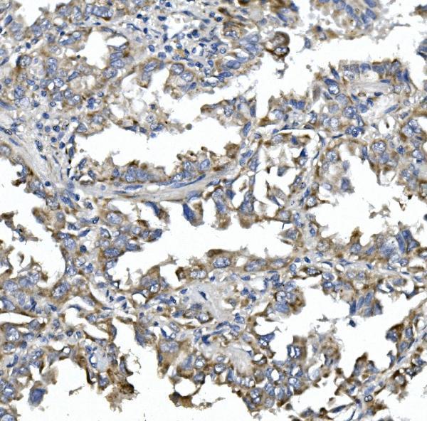
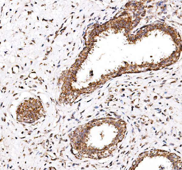
Real IHC data IHC analysis of NDP52/CALCOCO2 using anti-NDP52/CALCOCO2 antibody (A05876-1). NDP52/CALCOCO2 was detected in paraffin-embedded section of human lung cancer tissue. Heat mediated antigen retrieval was performed in EDTA buffer (pH8.0, epitope retrieval solution). The tissue section was blocked with 10% goat serum. The tissue section was then incubated with 1μg/ml rabbit anti-NDP52/CALCOCO2 Antibody (A05876-1) overnight at 4°C. Biotinylated goat anti-rabbit IgG was used as secondary antibody and incubated for 30 minutes at 37°C. The tissue section was developed using Strepavidin-Biotin-Complex (SABC) (Catalog # SA1022) with DAB as the chromogen.
Anti-NDP52/CALCOCO2 Antibody ®
Cat # A05876-1
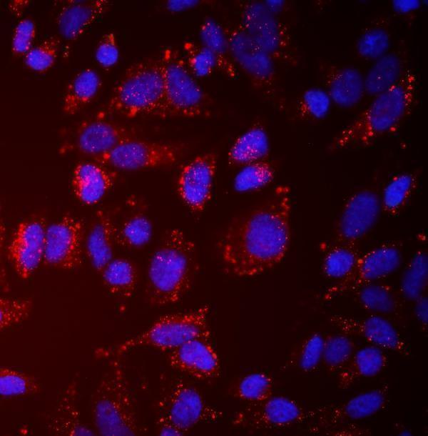
Real IF data IF analysis of NDP52/CALCOCO2 using anti-NDP52/CALCOCO2 antibody (M05876-2). NDP52/CALCOCO2 was detected in an immunocytochemical section of U20S cells. Enzyme antigen retrieval was performed using IHC enzyme antigen retrieval reagent (AR0022) for 15 mins. The cells were blocked with 10% goat serum. And then incubated with 5 μg/mL mouse anti-NDP52/CALCOCO2 Antibody (M05876-2) overnight at 4°C. Cy3 Conjugated Goat Anti-Mouse IgG (BA1031) was used as secondary antibody at 1:100 dilution and incubated for 30 minutes at 37°C. The section was counterstained with DAPI. Visualize using a fluorescence microscope and filter sets appropriate for the label used.
Anti-NDP52/CALCOCO2 Antibody ® (monoclonal, 9E2F2)
Cat # M05876-2

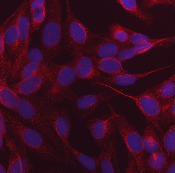
A05876-1 is listed for human IHC and IF/ICC, with IHC images from paraffin sections of human lung and mammary cancer tissue and an IF image from HeLa cells (catalog applications, reactivity, and image captions). M05876-2 is listed for IF/ICC in human, mouse, and rat, with an IF image from U20S cells (catalog applications, reactivity, and IF caption).

Which to pick: Choose rabbit A05876-1 for tissue IHC: its images show human paraffin sections processed with EDTA retrieval at pH 8.0 and 1 μg/ml primary antibody; the fixative is unreported (A05876-1 IHC captions). For IF/ICC, both A05876-1 and mouse monoclonal M05876-2 have cell images and listed IF concentrations of 4 μg/ml and 5 μg/ml, respectively (catalog applications, dilutions, and IF captions). Choose M05876-2 when mouse or rat reactivity is needed for IF/ICC; its catalog lists those species, while its supplied IF image shows U20S cells (catalog reactivity and M05876-2 IF caption).

Each figure is that product's own IHC / IF validation image from its datasheet.

References

  1. UniProt Consortium. UniProt entry Q13137 (CACO2_HUMAN, Calcium-binding and coiled-coil domain-containing protein 2).
  2. Human Protein Atlas. CALCOCO2 tissue IHC expression (reliability: Enhanced).
  3. Human Protein Atlas. CALCOCO2 subcellular location (ICC-IF): Mainly localized to the cytosol. In addition localized to vesicles..
  4. Human Protein Atlas. CALCOCO2 antibody validation summary (3 antibodies).
  5. Multi-omics data mining combined with experimental validation reveals ferroptosis- and autophagy-associated hub genes as diagnostic candidates and immune modulators in atherosclerosis. Animal models and experimental medicine 2026 — PMC13242735.
  6. Mechanistic insights into COVID-19 by global analysis of the SARS-CoV-2 3CL(pro) substrate degradome. Cell reports 2021 — PMC8501228.
  7. PubMed PMID:7540613 — UniProt-cited evidence.
  8. PubMed PMID:14702039 — UniProt-cited evidence.
  9. PubMed PMID:16625196 — UniProt-cited evidence.