CALD1 / Caldesmon · IHC design guide

Design Immunohistochemistry for CALD1

Plan chromogenic CALD1 staining in paraffin sections using cytoplasmic smooth muscle staining as the expected pattern (HPA tissue IHC). Interpret staining across cell types in light of CALD1’s distinct smooth muscle and nonmuscle isoform distributions (UniProt).

Evidence assembled Oct 2026 · For research use; verify linked source records and product datasheet before use
Immunohistochemistry protocol sheet for CALD1 (IHC for CALD1): expected localisation Cytoplasmic in smooth muscle cells (HPA tissue IHC), antibody PB9922, validated IHC image, and IHC protocol steps
Printable CALD1 IHC protocol sheet — expected localisation Cytoplasmic in smooth muscle cells (HPA tissue IHC), antibody PB9922, controls and protocol steps. Open the full CALD1 IHC guide →

CALD1 Immunohistochemistry Experimental Design Guide

Expected localisation, validated protocols, controls and antibodies — the at-a-glance facts below, then the full design guide.

Must know before staining
Expected localisation Cytoplasmic in smooth muscle cells (HPA tissue IHC)
Staining pattern Strong cytoplasmic staining in smooth muscle cells (HPA tissue IHC)
Antigen retrieval EDTA pH 8.0 HIER, heat-mediated (datasheet PB9922)
Positive control ⓘ Endometrium+4 more · see all
Negative control ⓘ Adipose tissue+4 more · see all
Important caveats
Reasons your staining may differ from the expected pattern.
Fixation Keep fixation conditions consistent across sections. (standard IHC practice; not target-specific)
Caveat Nonmuscle isoforms may broaden cellular staining (UniProt)
Regulation Isoform 1 enriched in smooth muscle (UniProt)
Isoform / epitope 6 isoforms; antibody epitope coverage unreported (UniProt; datasheet PB9922)
Section 1

Recommended CALD1 IHC & IF Protocols

The catalog antibody protocol is paired with three published CALD1 chromogenic IHC protocols from aortic valve, breast carcinoma, and bladder tumor specimens (PMC13003078; PMC4823093; PMC8164301).

Recommended immunohistochemistry (IHC-P) protocol parameters
SampleParaffin-embedded mouse intestine tissue; fixative not specified (datasheet PB9922)
FixationImage fixative and duration unreported (datasheet PB9922); verify before use.
Sectioning4–5 µm sections on charged slides (standard)
DeparaffinisationXylene, graded ethanol series to water (standard)
Antigen retrievalHeat retrieval: EDTA pH 8.0 (datasheet PB9922); 20 min, 95–100 °C (standard)
Peroxidase block3% H2O2, 10 min, room temperature (standard)
Blocking10% goat serum (datasheet PB9922)
Primary antibodyRabbit anti-CALD1, 0.5-1μg/ml (datasheet PB9922)
Primary incubationOvernight at 4 °C (datasheet PB9922)
DetectionHRP-conjugated secondary, DAB chromogen (datasheet PB9922)
CounterstainHematoxylin, blue, dehydrate and mount (standard)
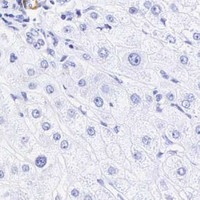
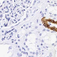
Expected resultCALD1-positive staining in ciliated epithelial cells of endometrium (HPA tissue IHC: High). HPA tissue profile: Cytoplasmic expression most abundant in smooth muscle. No signal in the no-primary control.
💡Decision noteStart with heat-mediated EDTA at pH 8.0 for the catalog antibody (PB9922 datasheet); the published citrate retrieval used a different antibody (PMC13003078).
Section 2

What Is the Expected CALD1 Staining Pattern?

CALD1 is a cytoplasmic cytoskeletal protein with no transmembrane segment (UniProt Q05682 topology and subcellular location). In paraffin IHC, expect the strongest staining in smooth muscle cells, with additional staining in selected epithelial and stromal cells (HPA: tissue IHC, Enhanced reliability). Interpret signal in its cellular context: HPA reports low tissue specificity overall, while UniProt describes smooth muscle enrichment of isoform 1 (HPA: RNA specificity; UniProt Q05682 isoforms).

What am I looking at on my slide?
Strong cytoplasmic staining in smooth muscle cells, following the contractile cell body.This fits HPA's High smooth muscle cell staining and cytoplasmic profile. CALD1 associates with thin filaments in smooth muscle (HPA: tissue IHC; UniProt Q05682 subcellular location). Compare cells within the same section before judging overall intensity.
Prominent nuclear staining, especially where cytoplasm is weak or unstained.Nuclear dominance is outside the reported CALD1 locations (UniProt Q05682 subcellular location; HPA: tissue IHC). Treat it as suspicious; review the counterstain and repeat with a no-primary control before scoring it as target signal (general IHC practice).
Strong staining in a cell population reported as negative, such as adipocytes.HPA reports CALD1 as Not detected in adipocytes (HPA: adipose tissue IHC). Check cell identity and nearby smooth muscle first. If the signal remains in adipocytes, assess possible cross-reactivity or endogenous chromogen activity with an independent antibody and a no-primary control (general IHC practice).
Diffuse color across many compartments, including empty spaces or section edges.That distribution does not match the cellular cytoplasmic pattern reported by HPA (HPA: tissue IHC). It suggests background from the detection workflow; inspect the no-primary control and optimize blocking, washing, and chromogen development (general IHC practice).
No staining in a section containing identifiable smooth muscle cells.Smooth muscle cells are a High positive reference (HPA: smooth muscle IHC). First verify tissue preservation and the positive control, then review antibody use, retrieval, and detection steps (general IHC practice). A negative run alone cannot establish CALD1 absence in the sample.
💡Expected CALD1 appearanceCall a positive result when smooth muscle cells show strong cytoplasmic staining, with appropriate staining in other HPA-positive cell types (HPA: tissue IHC); dominant nuclear color or strong signal in HPA-negative adipocytes is suspect (UniProt Q05682 subcellular location; HPA: adipose tissue IHC).
How each factor affects the staining
Cell and tissue contextHPA rates smooth muscle cells, endometrial ciliated epithelial cells, ovarian stroma cells, placental decidual cells, and testicular peritubular cells High (HPA: tissue IHC). It rates breast myoepithelial cells Medium and adipocytes Not detected (HPA: tissue IHC). Choose the cell population, not the whole organ, as the scoring unit.
Isoform coverageIsoform 1 predominates in smooth muscle; isoforms 2–5 are widely distributed in nonmuscle cells (UniProt Q05682 tissue specificity). The supplied sources do not identify which isoforms a particular IHC antibody detects, so staining outside smooth muscle cannot by itself establish isoform identity.
Antibody validationHPA lists three antibodies with Enhanced IHC validation: HPA008066, HPA017330, and CAB000006 (HPA: antibodies). Enhanced supports confidence in the reported staining pattern, but an individual run still needs suitable controls (general IHC practice).
Detection chemistryEndogenous enzyme activity can create color in chromogenic IHC if it survives the detection workflow (general IHC practice). A no-primary control helps distinguish detection background from antibody-dependent staining; HPA tissue levels alone do not identify the cause of an unexpected signal.
IF/ICC Q&A: Where is CALD1 seen?HPA reports enhanced plasma membrane and actin filament localization in ICC-IF (HPA: subcellular, Enhanced). UniProt reports cytoskeletal and stress fiber localization, including fibroblast stress fibers (UniProt Q05682 subcellular location). These observations guide IF interpretation; they do not specify an IHC retrieval or staining protocol.
Why is my staining missing, weak or wrong?
SituationLikely causeNext action
Smooth muscle positive control is blank.The run may have failed at antibody binding or detection; the supplied sources give no CALD1-specific fixation sensitivity (HPA: smooth muscle IHC; general IHC practice).Confirm the control contains smooth muscle, then check antibody identity, dilution, retrieval, secondary reagent, and chromogen against the run record (general IHC practice).
Signal is nuclear rather than cytoplasmic.Nuclear dominance conflicts with reported CALD1 localization (UniProt Q05682 subcellular location; HPA: tissue IHC). Counterstain or nonspecific signal may be misleading (general IHC practice).Review the color channels and morphology, run a no-primary control, and compare with a smooth muscle positive section (general IHC practice; HPA: smooth muscle IHC).
Adipocytes show strong color.HPA reports adipocytes as Not detected, so the result warrants review; neighboring vessels can complicate cell assignment (HPA: adipose tissue IHC; general IHC practice).Locate the stained cell boundaries, compare a no-primary control, and confirm persistent cell-specific staining with an independent IHC-validated antibody (general IHC practice; HPA: antibodies).
Color covers the section uniformly.A field-wide signal is inconsistent with HPA's cell-specific profile and may reflect detection background (HPA: tissue IHC; general IHC practice).Inspect a no-primary control, then review blocking, wash stringency, and chromogen development; judge staining only after the background is resolved (general IHC practice).
Stromal staining is called an unexpected positive.Some stromal populations are expected to stain: ovarian stroma cells and placental decidual cells are High (HPA: ovary and placenta IHC).Identify the exact cell population and compare it with the tissue-specific HPA entry before calling the pattern aberrant (HPA: tissue IHC).
IF/ICC shows filament-like signal but the IHC section looks diffuse.ICC-IF resolves actin filaments and plasma membrane localization, while HPA summarizes tissue IHC as cytoplasmic (HPA: subcellular and tissue IHC). The images use different readouts.Judge each result against its own reference pattern and controls; do not require individual filaments to be resolved in chromogenic paraffin IHC (HPA: subcellular and tissue IHC; general IHC practice).

Sample controls for CALD1 IHC & IF

🧪Run smooth muscle first; its smooth muscle cells should stain (HPA: High in smooth muscle cells). Use adipose tissue adipocytes as a negative comparator (HPA: Not detected in adipocytes); on the positive slide, score morphologically identified cells without signal as internal negatives, while allowing for CALD1 expression in nonmuscle cells (UniProt Q05682: nonmuscle isoforms are widely distributed).
Positive control tissue: Endometrium (Ciliated epithelial cells, HPA High)
Negative control tissue: Adipose tissue (HPA Not detected)
ICC-IF cell lines (HPA subcellular resource): HPA ICC-IF images show CALD1 in A-431, U-251MG, U2OS, ASC52telo, HEK293, U2OS, siRNA 1, U2OS, siRNA 2, U2OS, scrambled, with annotated localisation: Plasma membrane (enhanced), Actin filaments (enhanced) (HPA subcellular).
Technical controls: Include a no-primary, secondary-only control and an irrelevant IgG control matched to the primary antibody’s rabbit host, isotype and clonality (PB9922 tissue-IHC caption: rabbit primary); a CALD1 knockout specimen provides a biological negative. Quench endogenous peroxidase for HRP/DAB detection (PB9922 tissue-IHC caption: peroxidase secondary and DAB), and compare background with the no-primary slide.
⚠️Feasibility: A target-specific fixation window or fixation effect is unreported, and the selected PB9922 paraffin-section caption does not state a fixative. That caption reports heat retrieval in EDTA at pH 8.0, but does not establish that retrieval is universally required; the supplied evidence does not establish whether frozen sections or IF/ICC are easier (PB9922 tissue-IHC caption; HPA: ICC-IF images available). In dense smooth muscle, use morphology and counterstaining to distinguish individual stained cells when scoring (HPA: High in smooth muscle cells).

HPA tissue IHC evidence for CALD1

Comprehensive Human Protein Atlas IHC scoring per tissue (reliability: Enhanced — High consistency between antibody staining and RNA expression data. External characterization data supports antibody staining.). Rows are taken directly from the HPA tissue chart — click any row's HPA link to view the source.

Positive expression · recommended positive controls

TissueCell typeLevelEvidenceSource
Endometrium Ciliated epithelial cells High Protein (IHC) HPA →
Ovary Ovarian stroma cells High Protein (IHC) HPA →
Placenta Decidual cells High Protein (IHC) HPA →
Smooth muscle Smooth muscle cells High Protein (IHC) HPA →
Testis Peritubular cells High Protein (IHC) HPA →

Undetected expression · recommended negative controls

TissueCell typeLevelEvidenceSource
Adipose tissue Adipocytes Not detected Protein (IHC) HPA →
Adrenal gland Glandular cells Not detected Protein (IHC) HPA →
Appendix Endocrine cells Not detected Protein (IHC) HPA →
Bronchus Respiratory epithelial cells Not detected Protein (IHC) HPA →
Caudate Glial cells Not detected Protein (IHC) HPA →
Section 3

Advanced CALD1 IHC Tips

Troubleshoot CALD1 staining in paraffin sections by checking retrieval, cellular pattern, detection controls, and the isoforms your antibody can recognize.

What retrieval should I try first if CALD1 staining is weak?
Start with heat-mediated retrieval in EDTA at pH 8.0 (datasheet PB9922). The catalog antibody detected caldesmon in a paraffin-embedded mouse intestine section after that retrieval, followed by 1 μg/ml primary antibody overnight at 4°C (datasheet PB9922). If staining remains weak, adjust heating and cooling while keeping section adhesion and tissue morphology under review; compare runs against a smooth-muscle-rich region (HPA: cytoplasmic expression most abundant in smooth muscle). If needed, compare another retrieval buffer as a fallback, using matched sections and identical detection conditions (standard IHC practice). Record both signal and background before selecting a condition (standard IHC practice).
Could fixation explain weak or uneven CALD1 staining?
The selected tissue image specifies a paraffin-embedded section but does not state its fixative, so target-specific fixation sensitivity is unknown (datasheet PB9922). Record the fixative, fixation interval, tissue thickness, and processing history when comparing weak and strong sections (standard IHC practice). On adjacent sections from the same block, check whether the expected smooth-muscle cytoplasm stains consistently before changing antibody concentration (HPA: cytoplasmic expression most abundant in smooth muscle). If specimens differ in fixation history, process and stain controls alongside them and interpret intensity comparisons cautiously (standard IHC practice). Do not assign a CALD1-specific fixation effect without a controlled comparison (datasheet PB9922: fixative unreported).
Where should convincing CALD1 staining appear in tissue sections?
Expect predominantly cytoplasmic staining in smooth muscle, where high-molecular-weight caldesmon is enriched (HPA: cytoplasmic expression most abundant in smooth muscle; UniProt Q05682 isoform 1). CALD1 also associates with thin filaments in smooth muscle and stress fibers in nonmuscle cells, so inspect the relevant cell structures at high magnification (UniProt Q05682 subcellular location). A sharp nuclear-only pattern warrants review of counterstain, detection background, and antibody specificity against a matched control (UniProt Q05682 subcellular location; standard IHC practice). HPA reports high staining in ovarian stroma and decidual cells as well as smooth muscle cells (HPA: tissue IHC). Identify the stained cell population before calling a section negative (standard IHC practice).
How can CALD1 isoforms or epitope masking change the staining pattern?
CALD1 has 6 annotated isoforms; isoform 1 is predominantly expressed in smooth muscle, while isoforms 2–5 are widely distributed in nonmuscle tissues and cells (UniProt Q05682 isoforms and tissue specificity). Check the antibody’s stated immunogen or epitope and isoform coverage before interpreting a smooth-muscle-negative or broadly positive pattern (standard IHC practice). Phosphorylated residues include Ser129, Ser643, Ser656, Ser724, and Thr730, but the supplied evidence does not establish whether they alter this antibody’s staining (UniProt Q05682 modified residues; datasheet PB9922). Compare cell-type patterns under the same EDTA pH 8.0 retrieval and detection conditions (datasheet PB9922; standard IHC practice). Use a second validated epitope if specificity remains uncertain (standard IHC practice).
How should I follow up an IHC pattern with CALD1 immunofluorescence?
Use the IHC result to choose a region for IF and multiplex CALD1 with an independently validated smooth-muscle cell marker, since smooth muscle is an expected enriched cell population (HPA: cytoplasmic expression most abundant in smooth muscle; standard IF practice). Choose a fluorophore channel after checking the section’s autofluorescence and include single-label controls when separating signals (standard IF practice). CALD1 lacks a transmembrane segment and is associated with cytoskeletal structures, so use a permeabilisation condition appropriate for an intracellular epitope (UniProt Q05682 topology and subcellular location; standard IF practice). HPA reports actin-filament and plasma-membrane localisation in ICC/IF images; compare those patterns with tissue morphology cautiously (HPA: subcellular). Interpret IF through its own controls and validation (standard IF practice).
How can I reduce diffuse brown signal without losing CALD1 staining?
First distinguish diffuse deposit from the expected cytoplasmic smooth-muscle pattern on a counterstained section (HPA: tissue IHC; standard IHC practice). Run a no-primary control, review the peroxidase block, and inspect tissue edges and folds for excess DAB deposition (standard chromogenic IHC practice). The selected example used 10% goat serum blocking, 1 μg/ml primary antibody overnight at 4°C, and peroxidase-based DAB detection (datasheet PB9922). If background rises across cell types, titrate primary concentration and detection time on matched sections while retaining a known positive region (standard IHC practice). Compare changes under identical retrieval conditions (datasheet PB9922: EDTA pH 8.0; standard IHC practice).
How should I score CALD1 when a section contains mixed cell types? ⚠ ANSWER MARKED FOR VERIFICATION
Define the cell population and compartment before scoring, because CALD1 expression is most abundant in smooth-muscle cytoplasm but also occurs in other cell types (HPA: tissue IHC). For a selected population, report the percentage of positive cells and staining intensity; an H-score combines percentages at intensity grades 0–3 into a 0–300 score (standard IHC practice). Alternatively, report positive area per tissue area in mm² when boundaries are reproducible (standard IHC practice). Normalise to the relevant cell population or evaluable tissue area, and exclude folds, necrosis, and section edges by a prespecified rule (standard IHC practice). Apply identical retrieval, imaging, and thresholds across cases (standard IHC practice).
What separates a true CALD1-positive result from staining artefact?
A convincing result follows cytoplasmic structures in an anatomically plausible cell population, especially smooth muscle, rather than isolated nuclear staining (HPA: tissue IHC; UniProt Q05682 subcellular location). Caldesmon can also appear in nonmuscle cells, so cell identity and morphology matter more than brown color alone (UniProt Q05682 tissue specificity; standard IHC practice). Treat staining confined to section edges, folds, or necrotic areas as suspect and check a no-primary control for nonspecific detection (standard IHC practice). Residual endogenous enzyme activity can mimic DAB positivity; assess the peroxidase block when the control is brown (standard chromogenic IHC practice). Compare an internal smooth-muscle region before interpreting a negative result (HPA: tissue IHC; standard IHC practice).
Boster reagents

Best CALD1 / Caldesmon IHC Antibodies

Anti-CALD1 antibodies have IHC figures for human, mouse, and rat tissue and an IF figure for SiHa cells (M04578-2 and PB9922 IHC captions; PB9922 IF caption).

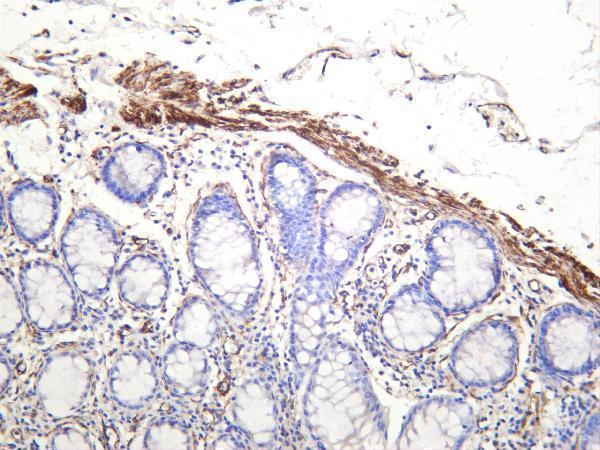
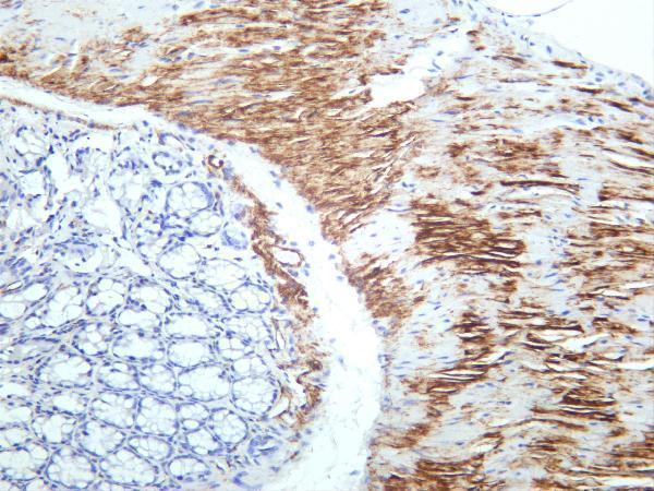
Real IHC data IHC analysis of Caldesmon using anti-Caldesmon antibody (PB9922). Caldesmon was detected in a paraffin-embedded section of mouse intestine tissue. Heat mediated antigen retrieval was performed in EDTA buffer (pH 8.0, epitope retrieval solution). The tissue section was blocked with 10% goat serum. The tissue section was then incubated with 1 μg/ml rabbit anti-Caldesmon Antibody (PB9922) overnight at 4°C. Peroxidase Conjugated Goat Anti-rabbit IgG was used as secondary antibody and incubated for 30 minutes at 37°C. The tissue section was developed using HRP Conjugated Rabbit IgG Super Vision Assay Kit (Catalog # SV0002) with DAB as the chromogen.
Anti-Caldesmon/CALD1 Antibody ®
Cat # PB9922
Real IHC data Human colon was stained with anti-Caldesmon rabbit antibody
Anti-Caldesmon Rabbit Monoclonal Antibody
Cat # M04578-2

PB9922 will render with a paraffin-section IHC figure from mouse intestine; its captions also document rat intestine and human intestinal cancer IHC, plus SiHa-cell IF (PB9922 IHC/IF captions). M04578-2 will render with a human-colon IHC figure; its captions also document mouse and rat colon IHC (M04578-2 IHC captions).

Which to pick: For paraffin-section tissue IHC, choose PB9922: its mouse-intestine caption documents EDTA retrieval at pH 8.0 and 1 μg/ml primary antibody; the fixative is unreported (PB9922 IHC caption). For IF/ICC, PB9922 has a SiHa-cell IF figure at 5 μg/ml and lists both applications; M04578-2 lists IF but has no IF figure in the payload (PB9922 IF caption; catalog applications and image alts). For IHC across human, mouse, and rat colon, choose M04578-2, a rabbit monoclonal with captions for all three species; PB9922 also has IHC captions for all three species, using different tissue examples (M04578-2 catalog and IHC captions; PB9922 IHC captions).

Each figure is that product's own IHC / IF validation image from its datasheet.

References

  1. UniProt Consortium. UniProt entry Q05682 (CALD1_HUMAN, Caldesmon).
  2. Human Protein Atlas. CALD1 tissue IHC expression (reliability: Enhanced).
  3. Human Protein Atlas. CALD1 subcellular location (ICC-IF): Localized to the plasma membrane and actin filaments..
  4. Human Protein Atlas. CALD1 antibody validation summary (3 antibodies).
  5. Caldesmon-1-mediated actin dynamics is essential for osteogenic differentiation of aortic valve interstitial cells. Scientific reports 2026 — PMC13003078.
  6. Upregulation of CALD1 predicted a poor prognosis for platinum-treated ovarian cancer and revealed it as a potential therapeutic resistance target. BMC genomics 2024 — PMC10870461.
  7. Annexin-A1 and caldesmon are associated with resistance to tamoxifen in estrogen receptor positive recurrent breast cancer. Oncotarget 2016 — PMC4823093.
  8. The cancer-associated fibroblasts related gene CALD1 is a prognostic biomarker and correlated with immune infiltration in bladder cancer. Cancer cell international 2021 — PMC8164301.
  9. PubMed PMID:1885618 — UniProt-cited evidence.
  10. PubMed PMID:1555769 — UniProt-cited evidence.
  11. PubMed PMID:1465449 — UniProt-cited evidence.