This website uses cookies to ensure you get the best experience on our website.


- Table of Contents
Use cytoplasmic staining in a subset of neurons as the CALHM1 tissue IHC benchmark (HPA tissue IHC). Start the catalog antibody at 5 μg/mL (datasheet), and score positive cells against a no-primary control (standard IHC practice).

Expected localisation, validated protocols, controls and antibodies — the at-a-glance facts below, then the full design guide.
| Expected localisation | Cytoplasmic in tissue; membrane and ER localisation annotated (HPA tissue IHC; UniProt) | |
| Staining pattern | Cytoplasmic staining in subsets of neurons and adrenal cells (HPA tissue IHC) | |
| Antigen retrieval | Citrate pH 6.0 HIER, 95–98 °C, 20 min (rule: cytoplasmic / membrane antigen) | |
| Positive control | Cerebral cortex | |
| Negative control | Adipose tissue+4 more · see all |
| Fixation | Keep fixation consistent across sections. (standard IHC practice; not target-specific) | |
| Caveat | Subset staining can make whole-field scoring misleading (HPA tissue IHC) | |
| Regulation | Predominantly expressed in adult brain (UniProt) | |
| Isoform / epitope | No isoforms or cleavage annotated; epitope side depends on topology (UniProt) |
The catalog antibody’s IHC-P protocol is paired with one published CALHM1 chromogenic protocol using taste bud sections (PMC5966022).
| Sample | Tissue sections; selected-image fixative not specified (standard IHC workflow) |
| Fixation | Image fixative and duration unreported (datasheet A03599-1); verify before use. |
| Sectioning | 4–5 µm sections on charged slides (standard) |
| Deparaffinisation | Xylene, graded ethanol series to water (standard) |
| Antigen retrieval | Heat-induced epitope retrieval in citrate buffer, pH 6.0, 20 min at 95–98 °C (standard rule: cytoplasmic / membrane antigen) |
| Peroxidase block | 3% H2O2, 10 min, room temperature (standard) |
| Blocking | 10% normal serum of the secondary host, 30 min, room temperature (standard) |
| Primary antibody | Rabbit anti-CALHM1, 5 μg/mL (datasheet A03599-1) |
| Primary incubation | Overnight at 4 °C (standard) |
| Detection | HRP-polymer secondary, DAB chromogen 5–10 min (standard) |
| Counterstain | Hematoxylin, blue, dehydrate and mount (standard) |
| Expected result | CALHM1-positive staining in neuronal cells of cerebral cortex (HPA tissue IHC: Medium). HPA tissue profile: Cytoplasmic expression in a subset of neurons and a subset of adrenal gland cells. No signal in the no-primary control. |
CALHM1 is a four-pass protein assigned to the cell, endoplasmic reticulum and basolateral membranes (UniProt Q8IU99 topology and subcellular location). In tissue IHC, expect cytoplasmic staining in a subset of neurons, especially in cerebral cortex, with weaker staining in a subset of adrenal gland cells (HPA: tissue IHC). HPA rates its tissue profile Enhanced but reports only medium consistency between antibody staining and RNA expression (HPA: reliability description).
| Subset of cortical neuronal cells shows moderate cytoplasmic chromogen, while neighboring cells differ in intensity. | This matches the observed positive tissue pattern: neuronal staining is Medium in cerebral cortex, and expression is limited to a subset of neurons (HPA: cerebral cortex; HPA: tissue profile). Cytoplasmic appearance in a section does not by itself resolve the cell membrane from intracellular membranes (UniProt Q8IU99 subcellular location; general IHC practice). |
| Staining follows cell outlines or appears within neuronal cytoplasm, with no dominant nuclear signal. | Membrane and endoplasmic reticulum localization make those distributions biologically plausible (UniProt Q8IU99 subcellular location). A dominant nuclear pattern falls outside the supplied localization evidence; assess it as a possible staining artefact before calling it CALHM1 (UniProt Q8IU99 subcellular location; general IHC practice). |
| Strong staining appears in adipocytes, appendix glandular cells or marrow hematopoietic cells. | These specified cell populations are Not detected in HPA tissue IHC (HPA: adipose tissue; HPA: appendix; HPA: bone marrow). Treat unexpected staining as possible cross-reactivity or endogenous detection activity, then check controls; an entire tissue is not necessarily a negative control for every cell type (general IHC practice). |
| Chromogen covers many cell types and extracellular areas with little distinction between structures. | A diffuse field is difficult to reconcile with the reported subset-of-neurons pattern (HPA: tissue profile). Check staining without primary antibody, blocking and chromogen development to distinguish nonspecific or detection background from a cell-associated signal (general IHC practice). |
| No neuronal staining is visible in a cerebral cortex section included as a positive control. | HPA reports Medium staining in cortical neuronal cells, so an absent signal calls for a run-level check (HPA: cerebral cortex). Confirm tissue identity and preservation, then review retrieval, primary-antibody conditions and detection controls before interpreting a study sample as negative (general IHC practice). |
| Membrane topology and epitope access (UniProt Q8IU99 topology) | CALHM1 has four transmembrane spans, cytoplasmic segments, and two extracellular loops (UniProt Q8IU99 topology). Which domain an antibody recognizes can affect access in an assay, but the supplied sources give no antibody epitope; do not prescribe permeabilisation or predict retrieval response from topology alone (general IHC practice). |
| Cell and tissue distribution (HPA: tissue IHC; UniProt Q8IU99 tissue specificity) | HPA shows Medium cortical neuronal staining and Low adrenal glandular-cell staining, with only subsets positive (HPA: cerebral cortex; HPA: adrenal gland; HPA: tissue profile). UniProt reports predominant adult-brain expression and expression in circumvallate taste bud cells; no taste-bud IHC result is supplied here (UniProt Q8IU99 tissue specificity). |
| Evidence strength (HPA: reliability description; HPA: HPA061816 IHC Enhanced) | The listed antibody, HPA061816, has an Enhanced IHC validation status, while HPA describes medium consistency with RNA expression for the tissue profile (HPA: HPA061816; HPA: reliability description). Use the reported cell pattern as a reference, and retain controls when judging staining outside it (general IHC practice). |
| Processing and glycosylation (UniProt Q8IU99 processing and glycosylation) | UniProt lists a single 1–346 chain, no signal peptide or propeptide, and one glycosylation site at residue 140 (UniProt Q8IU99 processing and glycosylation). These annotations do not identify the antibody epitope or establish a target-specific fixation or antigen-retrieval effect (general IHC practice). |
| IF/ICC localization evidence (HPA: subcellular summary) | HPA summarizes CALHM1 as Membrane for ICC-IF, but lists no main location or cell-line images in the supplied record (HPA: subcellular summary). That summary supports a localization expectation, not a demonstrated IF staining example or an IF protocol (HPA: subcellular summary). |
| Situation | Likely cause | Next action |
|---|---|---|
| Cortical neurons are unstained while the section otherwise develops. | The run may have insufficient target detection, or the selected field may miss the positive neuronal subset (HPA: cerebral cortex and tissue profile; general IHC practice). | Inspect several appropriate cortical fields and tissue quality. Review the IHC antibody instructions, retrieval conditions and detection reagents with a positive-control section before scoring the sample negative (general IHC practice). |
| Adrenal staining looks faint relative to cortical staining. | Low glandular-cell staining in the adrenal gland versus Medium cortical neuronal staining is consistent with the supplied observations (HPA: adrenal gland; HPA: cerebral cortex). | Compare like cell populations and use a cortical positive control to judge whether the run worked; avoid forcing both tissues to the same intensity by extending chromogen development (HPA: tissue IHC; general IHC practice). |
| Signal is predominantly nuclear. | The supplied locations are cell, endoplasmic reticulum and basolateral membranes; a nuclear pattern is unsupported (UniProt Q8IU99 subcellular location). | Review section morphology, primary-antibody specificity and a no-primary control. Record nuclear signal separately rather than counting it as the expected CALHM1 pattern (general IHC practice). |
| Adipocytes or appendix glandular cells stain strongly. | Those cell populations are reported as Not detected, making cross-reactivity or endogenous detection activity possibilities (HPA: adipose tissue; HPA: appendix; general IHC practice). | Check a no-primary control and the detection-system controls, then compare the distribution with a cortical positive control before interpreting the unexpected staining (general IHC practice). |
| Background obscures differences between cells. | Nonspecific antibody binding, endogenous detection activity or excessive chromogen development can obscure a subset pattern (HPA: tissue profile; general IHC practice). | Check a no-primary section; review blocking, primary-antibody concentration, washing and development time under the assay's validated conditions (general IHC practice). |
| Q: Can this IHC result be used as an IF/ICC pattern reference? | HPA gives a Membrane ICC-IF summary but supplies no main location or cell-line images here (HPA: subcellular summary). | A: Use membrane localization as a cautious expectation and assess IF/ICC with its own controls and guide; this tissue IHC pattern alone does not establish an IF/ICC result (HPA: subcellular summary; general IF practice). |


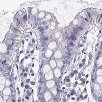
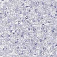
Comprehensive Human Protein Atlas IHC scoring per tissue (reliability: Enhanced — Medium consistency between antibody staining and RNA expression data.). Rows are taken directly from the HPA tissue chart — click any row's HPA link to view the source.
| Tissue | Cell type | Level | Evidence | Source |
|---|---|---|---|---|
| Cerebral cortex | Neuronal cells | Medium | Protein (IHC) | HPA → |
| Tissue | Cell type | Level | Evidence | Source |
|---|---|---|---|---|
| Adipose tissue | Adipocytes | Not detected | Protein (IHC) | HPA → |
| Appendix | Glandular cells | Not detected | Protein (IHC) | HPA → |
| Bone marrow | Hematopoietic cells | Not detected | Protein (IHC) | HPA → |
| Breast | Adipocytes | Not detected | Protein (IHC) | HPA → |
| Bronchus | Respiratory epithelial cells | Not detected | Protein (IHC) | HPA → |
Troubleshoot CALHM1 chromogenic IHC by checking retrieval, cellular distribution and controls before interpreting staining intensity.
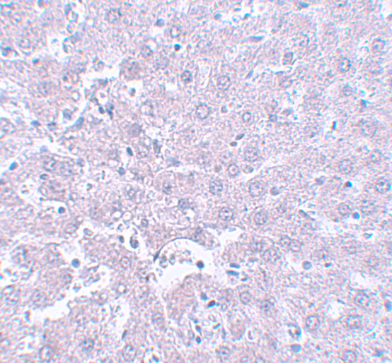
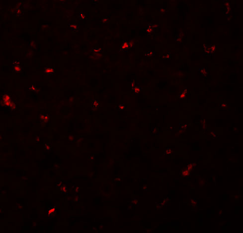
A03599-1 has IHC data from rat liver tissue and IF data from rat liver cells (catalog: image captions); listed reactivity covers human, mouse, and rat (catalog: reactivity).
A03599-1 is the rendering SKU; its IHC image shows rat liver tissue at 5 μg/mL (IHC image caption). Its IF image shows rat liver cells at 20 μg/mL (IF image caption), and its listed applications include IHC-P and IF (catalog: applications).
Which to pick: For tissue IHC, choose A03599-1: IHC-P is listed (catalog: applications), and its own image shows rat liver tissue (IHC image caption); the fixative is unreported (IHC image caption). For IF/ICC, A03599-1 has IF data from rat liver cells (IF image caption), while ICC validation is unreported (catalog: applications). For cross-species work, A03599-1 lists human, mouse, and rat reactivity (catalog: reactivity); it has a rabbit host, but clonality is unspecified (catalog: host and clone).