CAMK2A / Calcium/calmodulin-dependent protein kinase type II subunit alpha · IHC design guide

Design Immunohistochemistry for CAMK2A

Plan CAMK2A paraffin IHC around neuronal and neuropil staining in CNS (HPA tissue IHC). Use matched controls when interpreting signal because the tissue evidence cautions that staining may target protein from more than one gene (HPA tissue IHC).

Evidence assembled Oct 2026 · For research use; verify linked source records and product datasheet before use
Immunohistochemistry protocol sheet for CAMK2A (IHC for CAMK2A): expected localisation CNS neuropil; synaptic enrichment expected (HPA tissue IHC; UniProt), antibody M03241, validated IHC image, and IHC protocol steps
Printable CAMK2A IHC protocol sheet — expected localisation CNS neuropil; synaptic enrichment expected (HPA tissue IHC; UniProt), antibody M03241, controls and protocol steps. Open the full CAMK2A IHC guide →

CAMK2A Immunohistochemistry Experimental Design Guide

Expected localisation, validated protocols, controls and antibodies — the at-a-glance facts below, then the full design guide.

Must know before staining
Expected localisation CNS neuropil; synaptic enrichment expected (HPA tissue IHC; UniProt)
Staining pattern Neurons and neuropil in CNS; texture unreported (HPA tissue IHC)
Antigen retrieval EDTA pH 8.0 HIER, heat-mediated (datasheet M03241)
Positive control ⓘ Adrenal gland+4 more · see all
Negative control ⓘ Adipose tissue+4 more · see all
Important caveats
Reasons your staining may differ from the expected pattern.
Fixation Keep formalin fixation consistent across sections (standard IHC practice; not target-specific)
Caveat Staining may include protein from another gene (HPA tissue IHC)
Regulation Expression regulation unreported (UniProt)
Isoform / epitope 2 isoforms; epitope differences unreported (UniProt)
Section 1

Recommended CAMK2A IHC & IF Protocols

The catalog antibody’s IHC protocol (datasheet: M03241) sits alongside published CAMK2A protocols for rabies brain sections (PMC3660221) and human lung cancer microarrays (PMC7264342).

Recommended immunohistochemistry (IHC-P) protocol parameters
SampleParaffin-embedded human colon cancer tissue; fixative not specified (datasheet M03241)
FixationImage fixative and duration unreported (datasheet M03241); verify before use.
Sectioning4–5 µm sections on charged slides (standard)
DeparaffinisationXylene, graded ethanol series to water (standard)
Antigen retrievalHeat retrieval: EDTA pH 8.0 (datasheet M03241); 20 min, 95–100 °C (standard)
Peroxidase block3% H2O2, 10 min, room temperature (standard)
Blocking10% goat serum (datasheet M03241)
Primary antibodyRabbit monoclonal (clone AAFF-3) anti-CAMK2A, 1:50 (datasheet M03241)
Primary incubationOvernight at 4 °C (datasheet M03241)
DetectionHRP-conjugated secondary, DAB chromogen (datasheet M03241)
CounterstainHematoxylin, blue, dehydrate and mount (standard)
Expected resultCAMK2A-positive staining in glandular cells of adrenal gland (HPA tissue IHC: High). HPA tissue profile: Selective expression in neuropil and neurons in CNS. No signal in the no-primary control.
💡Decision noteStart with heat-mediated EDTA pH 8.0 (datasheet: M03241). Citrate pH 6 is a published alternative for rabies brain sections (PMC3660221).
Section 2

What Is the Expected CAMK2A Staining Pattern?

CAMK2A is a protein associated with synapses, postsynaptic densities, dendrites, and dendritic spines (UniProt Q9UQM7: subcellular location). In paraffin sections, expect selective neuronal and neuropil staining in CNS tissue (HPA: tissue IHC profile). CAMK2A has no transmembrane segment (UniProt Q9UQM7: topology). Treat the pattern as supported rather than definitive: HPA reports medium agreement with RNA data and cautions that its antibodies may target proteins from more than one gene (HPA: IHC reliability).

What am I looking at on my slide?
Neuronal cells and surrounding neuropil stain in cerebral cortex or caudate.This fits the reported CNS pattern; both sites show high neuronal-cell staining (HPA: tissue IHC). Assess distribution alongside intensity, since an intense deposit alone does not establish specificity (general IHC practice).
Neuropil or dendritic regions stain, while nuclei are not the dominant signal.This is consistent with synaptic and dendritic localization (UniProt Q9UQM7: subcellular location). Predominantly nuclear staining is outside that expected pattern; review controls and antibody specificity before interpreting it as CAMK2A (UniProt Q9UQM7; general IHC practice).
Adipocytes, squamous epithelium, or fibroblasts stain strongly.These selected cell populations were not detected in HPA tissue IHC: adipose adipocytes, esophageal or oral squamous cells, and skin or soft-tissue fibroblasts (HPA: tissue IHC). Consider cross-reactivity or endogenous detection activity; neither is established from the slide alone (general IHC practice).
A uniform chromogenic haze covers cells and extracellular spaces.Diffuse background does not resolve the selective neuronal and neuropil pattern (HPA: tissue IHC profile). Check background in a primary-antibody omission control and review blocking, washes, and detection conditions (general IHC practice).
A cerebral cortex positive-control section has no convincing neuronal signal.HPA reports high staining in cortical neuronal cells (HPA: tissue IHC). First verify section and detection controls, then review the IHC-validated antibody's supplied protocol; one blank section cannot establish absent CAMK2A expression (general IHC practice).
💡Expected CAMK2A appearanceCall a positive result when neuronal cells and neuropil show convincing, selectively distributed staining in CNS sections, with high neuronal staining possible in cerebral cortex or caudate (HPA: tissue IHC); diffuse haze or strong staining in HPA-negative cell populations is suspect (HPA: tissue IHC; general IHC practice).
How each factor affects the staining
Cellular distributionUniProt places CAMK2A at synapses, postsynaptic densities, dendrites, and dendritic spines (UniProt Q9UQM7: subcellular location). Interpret IHC at the resolution of neuronal cells and neuropil; do not require individual synapses to be resolved in chromogenic sections (general IHC practice).
Tissue choiceCerebral cortex and caudate neuronal cells, and cerebellar granular-layer cells, are reported high (HPA: tissue IHC). Adipocytes and esophageal squamous cells are reported not detected and can provide contrasting cell populations (HPA: tissue IHC).
Antibody specificityHPA assigns Supported IHC status to HPA051783, HPA051785, HPA053973, and CAB004330, while warning that the staining may reflect proteins from more than one gene (HPA: antibody validation; tissue IHC reliability). Match the antibody and controls used before attributing every positive cell to CAMK2A.
Isoforms and epitopeUniProt lists two CAMK2A isoforms, A and B, and a protein kinase domain at residues 13–271 (UniProt Q9UQM7: isoforms; domains). The supplied evidence does not locate an antibody epitope, so isoform recognition and any retrieval-specific effect cannot be predicted from these records.
IF/ICC Q: Should its pattern set the IHC expectation?A: Use it only as context. HPA lists plasma membrane and several ciliary or sperm locations as uncertain and cautions that the ICC antibodies may target proteins from multiple genes (HPA: subcellular ICC-IF). The IHC decision should rest on the tissue pattern and its controls (HPA: tissue IHC; general IHC practice).
Why is my staining missing, weak or wrong?
SituationLikely causeNext action
No staining in cerebral cortex neuronsPossible section, antibody, or detection failure; cortical neurons are reported high (HPA: tissue IHC).Confirm tissue identity and run the IHC-validated antibody with its supplied IHC-P conditions and an appropriate detection control (general IHC practice).
Strong staining appears only in nucleiA nucleus-dominant pattern does not match the reported synaptic and dendritic locations (UniProt Q9UQM7: subcellular location).Compare with neuronal and neuropil staining in a positive-control section; review primary-antibody specificity before scoring the nuclear signal (HPA: tissue IHC; general IHC practice).
Adipocytes or squamous cells stain stronglyThese selected populations are reported not detected (HPA: tissue IHC); cross-reactivity or endogenous detection activity is possible (general IHC practice).Inspect a primary-antibody omission control, check chromogen and detection background, and compare staining with a CNS positive control (general IHC practice; HPA: tissue IHC).
Diffuse brown haze obscures neuropilNonspecific background or residual endogenous enzyme activity may obscure a selective pattern (general IHC practice; HPA: tissue IHC profile).Check the omission control, endogenous-enzyme blocking where applicable, wash quality, and detection exposure before interpreting intensity (general IHC practice).
Peripheral glandular staining seems unexpectedHigh glandular-cell staining is also listed in adrenal gland, endometrium, fallopian tube, and gallbladder (HPA: tissue IHC).Do not reject the result solely because it is outside CNS; compare its cell distribution with HPA and use antibody-specific controls given the multigene-target caution (HPA: tissue IHC reliability; general IHC practice).
IF/ICC signal differs from the paraffin-section resultHPA's listed ICC-IF locations are uncertain, and its ICC summary carries a multigene-target caution (HPA: subcellular ICC-IF).Interpret the paraffin section against HPA tissue IHC and its own controls; assess IF/ICC separately on its guide page (HPA: tissue IHC; general IHC practice).

Sample controls for CAMK2A IHC & IF

🧪Run cerebral cortex first: neuronal cells should stain strongly (HPA: High in cerebral cortex neuronal cells). Use esophagus squamous epithelium as the negative tissue (HPA: Not detected in esophagus squamous epithelial cells); on the cortex slide, score non-neuronal cells as internal background comparators without assuming they are CAMK2A-negative (HPA: High is specified for neuronal cells only).
Positive control tissue: Adrenal gland (Glandular cells, HPA High)
Negative control tissue: Adipose tissue (HPA Not detected)
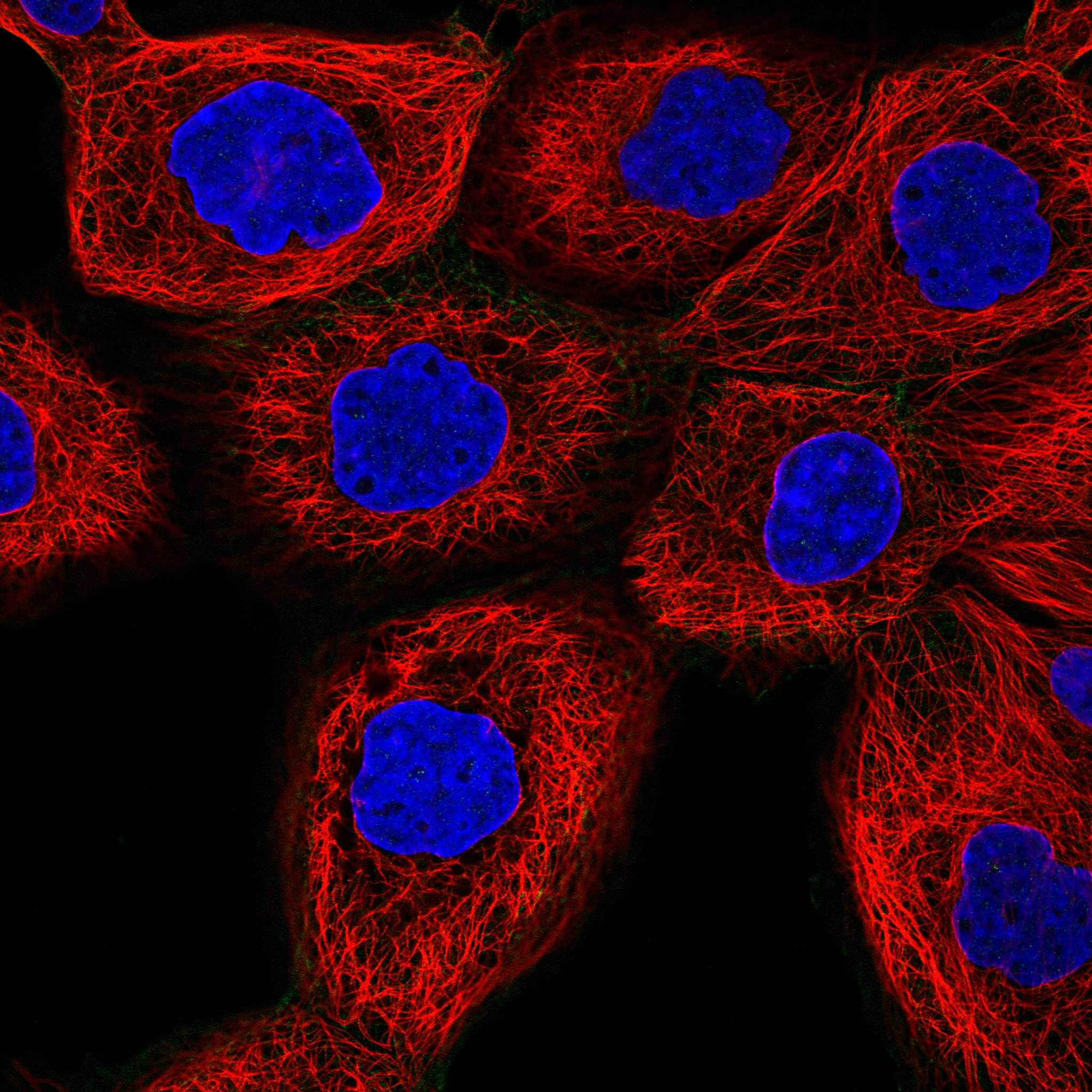
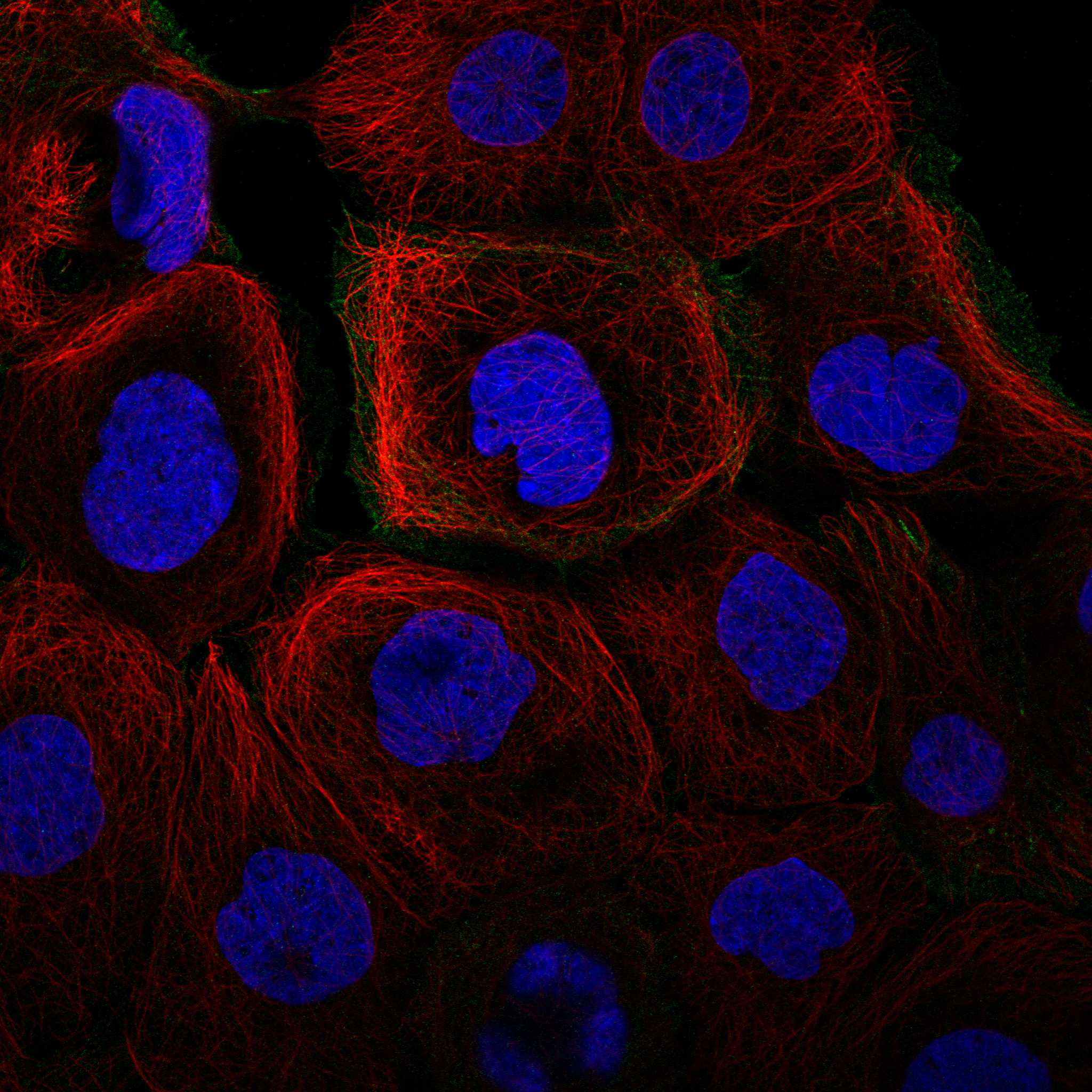
ICC-IF cell lines (HPA subcellular resource): HPA ICC-IF images show CAMK2A in A-431, U-251MG, U2OS, ASC52telo, RPTEC/TERT1, hTERT-RPE1 (serum starved), Sperm, with annotated localisation: Plasma membrane (uncertain), Primary cilium tip (uncertain), Primary cilium transition zone (uncertain), Principal piece (uncertain) (HPA subcellular).
Technical controls: Include a no-primary, secondary-only section; a concentration-matched rabbit IgG isotype control for the rabbit catalog antibody; and CAMK2A knockout tissue or cells as a biological negative (selected-SKU caption: rabbit primary; standard IHC practice). Quench endogenous peroxidase for DAB detection, and assess neuronal lipofuscin autofluorescence if using IF on cortex (selected-SKU caption: HRP/DAB; standard IHC/IF practice).
⚠️Feasibility: A target-specific fixation window or fixation effect is unreported, and the selected-SKU paraffin-section caption does not state the fixative (selected-SKU caption: fixative not stated). Heat retrieval in EDTA at pH 8.0 was used at a 1:50 primary dilution, but the caption does not establish that retrieval is required (selected-SKU caption). Frozen sections or IF are not established as easier by the supplied evidence; neuronal lipofuscin can complicate cortex IF interpretation (standard IF practice).

HPA tissue IHC evidence for CAMK2A

Comprehensive Human Protein Atlas IHC scoring per tissue (reliability: Supported — Medium consistency between antibody staining and RNA expression data. Caution, targets protein from more than one gene.). Rows are taken directly from the HPA tissue chart — click any row's HPA link to view the source.

Positive expression · recommended positive controls

TissueCell typeLevelEvidenceSource
Adrenal gland Glandular cells High Protein (IHC) HPA →
Bronchus Respiratory epithelial cells High Protein (IHC) HPA →
Caudate Neuronal cells High Protein (IHC) HPA →
Cerebellum Cells in granular layer High Protein (IHC) HPA →
Cerebral cortex Neuronal cells High Protein (IHC) HPA →

Undetected expression · recommended negative controls

TissueCell typeLevelEvidenceSource
Adipose tissue Adipocytes Not detected Protein (IHC) HPA →
Esophagus Squamous epithelial cells Not detected Protein (IHC) HPA →
Oral mucosa Squamous epithelial cells Not detected Protein (IHC) HPA →
Ovary Ovarian stroma cells Not detected Protein (IHC) HPA →
Seminal vesicle Glandular cells Not detected Protein (IHC) HPA →
Section 3

Advanced CAMK2A IHC Tips

Troubleshoot CAMK2A staining in paraffin sections by checking retrieval, compartment, antibody specificity and matched tissue controls before comparing chromogenic signal.

How should I adjust retrieval when CAMK2A staining is weak or uneven?
Start with heat-mediated retrieval in EDTA at pH 8.0 for the catalog antibody (datasheet M03241). The reported paraffin-section example used this retrieval before overnight primary incubation at 4°C (datasheet M03241); keep section thickness, heating time and cooling time consistent across the comparison. If staining remains weak, test a modest change in heating time on adjacent sections while checking for tissue lifting and loss of morphology (standard IHC practice). Compare each condition with a neuronal-rich positive control, such as cerebral cortex, and a no-primary control; neuronal staining there is reported as high (HPA: cerebral cortex neuronal cells, High).
Could fixation explain weak or patchy CAMK2A staining?
Target-specific fixation sensitivity is unknown: the selected paraffin-section caption does not state a fixative (datasheet M03241). Record the actual fixative, duration and processing history for each specimen, then compare sections processed together under the same EDTA pH 8.0 retrieval conditions (datasheet M03241; standard IHC practice). If a batch differs, examine morphology and control-tissue staining before attributing reduced signal to CAMK2A loss (standard IHC practice). Avoid treating a change in chromogenic intensity alone as evidence of altered protein abundance, especially when fixation or processing differs between groups (standard IHC interpretation practice).
Where should convincing CAMK2A staining appear in a paraffin section?
Assess signal in neuronal cell bodies and neuropil, with particular attention to dendritic and synaptic regions (HPA: selective expression in CNS neurons and neuropil; UniProt Q9UQM7: dendrite, dendritic spine and postsynaptic density). Because CAMK2A has no transmembrane segment, a sharp membrane-only rim should prompt review rather than automatic acceptance as its expected pattern (UniProt Q9UQM7 topology). Compare the suspected signal with adjacent morphology and a neuronal-rich control on the same run (standard IHC practice; HPA: cerebral cortex neuronal cells, High). Document cytoplasmic, process-associated and unexpected nuclear staining separately so that differences in compartment are not hidden in a single positive score (standard IHC scoring practice).
How can I judge whether staining represents CAMK2A rather than another CaMKII chain?
CAMK2A has 2 recorded isoforms, A and B, and CaMKII holoenzymes can contain chains encoded by CAMK2A, CAMK2B, CAMK2G and CAMK2D (UniProt Q9UQM7). Request the catalog antibody's immunogen or mapped epitope and check whether its sequence distinguishes the intended isoforms and related chains (standard antibody validation practice). CAMK2A also carries multiple phosphorylation sites, including Thr-286; epitope accessibility may therefore need empirical assessment if the mapped epitope overlaps a modified region (UniProt Q9UQM7; standard antibody validation practice). Treat staining as antibody-defined until specificity is supported by an orthogonal control, because the available HPA tissue assessment cautions that its antibodies target proteins from more than one gene (HPA: tissue reliability description).
How should I check a CAMK2A pattern by multiplex IF?
On a matched section, pair CAMK2A with an independently validated neuronal marker and examine whether signal follows neuronal bodies and neuropil (HPA: selective expression in CNS neurons and neuropil; standard IF practice). Choose fluorophores after inspecting unstained tissue autofluorescence, and place the weaker target signal in a channel with good separation from that background (standard IF practice). CAMK2A lacks a transmembrane segment and is associated with dendrites and postsynaptic densities, so permeabilisation should permit access to intracellular epitopes without erasing fine processes (UniProt Q9UQM7 topology and subcellular location; standard IF practice). Titrate permeabilisation on matched sections and include single-label controls to assess bleed-through (standard IF practice).
What should I change when DAB obscures CAMK2A-positive processes?
First inspect a no-primary section for secondary-reagent, endogenous peroxidase or pigment signal, and check whether DAB development has saturated fine structures (standard chromogenic IHC practice). The selected paraffin-section example used 10% goat-serum block, 1:50 primary antibody overnight at 4°C, and an HRP/DAB detection workflow (datasheet M03241). Use these as documented starting conditions, then titrate the primary and shorten chromogen development on matched sections if background remains high (datasheet M03241; standard IHC practice). Compare suspected neuronal processes with a no-primary control and tissue morphology before counting diffuse deposits as CAMK2A signal (HPA: CNS neuronal and neuropil profile; standard IHC practice).
How should I quantify CAMK2A IHC across specimens? ⚠ ANSWER MARKED FOR VERIFICATION
Define the anatomical region and cell population before scoring, and keep section thickness, retrieval and DAB development consistent across specimens (standard IHC quantification practice). For discrete cells, report percent positive and an H-score; for neuropil-rich regions, report positive area or signal density per mm² of the same tissue compartment (standard IHC quantification practice). Normalise counts to the number of evaluable cells or measured tissue area, and exclude folds, necrosis and damaged edges using prespecified rules (standard IHC quantification practice). Report neuronal and non-neuronal compartments separately because the tissue profile emphasises CNS neurons and neuropil while also listing staining in some glandular cells (HPA: tissue profile and positive-cell listings).
How do I distinguish true CAMK2A staining from tissue artefact?
Give greatest weight to reproducible staining that follows neuronal cells and neuropil in a cerebral cortex or caudate control (HPA: CNS profile; cerebral cortex and caudate neuronal cells, High). Review an isolated membrane-only rim or diffuse nuclear signal against morphology and controls, since CAMK2A is recorded at dendrites, spines and postsynaptic densities and has no transmembrane segment (UniProt Q9UQM7 subcellular location and topology). Discount staining confined to cut edges, necrotic areas or folds, and investigate deposits that persist without primary antibody as possible endogenous enzyme or detection background (standard IHC interpretation practice). In non-neural tissue, avoid declaring every stained cell erroneous: reported positive glandular populations coexist with an HPA specificity caution about antibodies targeting more than one gene (HPA: positive-cell listings and tissue reliability description).
Boster reagents

Best CAMK2A / Calcium/calmodulin-dependent protein kinase type II subunit alpha IHC Antibodies

The catalog includes CAMK2A antibodies with IHC images from human tissue and rat brain, and IF images from human MCF-7 cells and rat brain (catalog image captions).

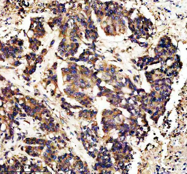
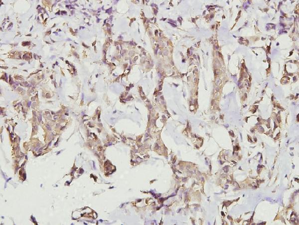
Real IHC data IHC analysis of CAMK2A using anti-CAMK2A antibody (M03241). CAMK2A was detected in a paraffin-embedded section of human colon cancer tissue. Heat mediated antigen retrieval was performed in EDTA buffer (pH 8.0, epitope retrieval solution). The tissue section was blocked with 10% goat serum. The tissue section was then incubated with 1:50 rabbit anti-CAMK2A Antibody (M03241) overnight at 4°C. Peroxidase Conjugated Goat Anti-rabbit IgG was used as secondary antibody and incubated for 30 minutes at 37°C. The tissue section was developed using HRP Conjugated Rabbit IgG Super Vision Assay Kit (Catalog # SV0002) with DAB as the chromogen.
Anti-CaMKII alpha CAMK2A Rabbit Monoclonal Antibody
Cat # M03241
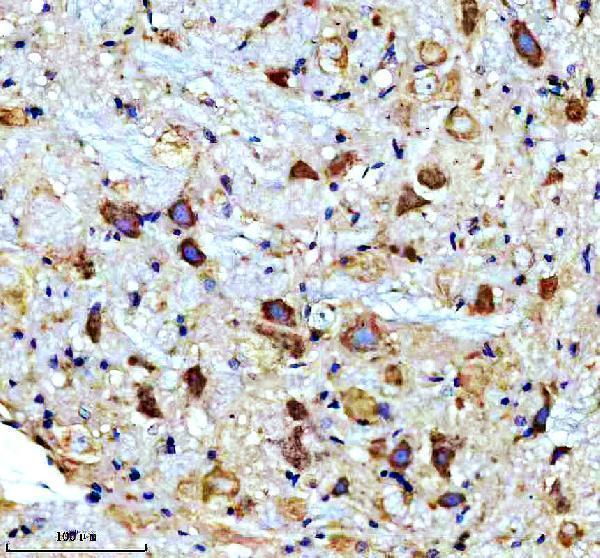
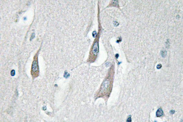
Real IHC data Human brain was stained with anti-CaMKII α rabbit antibody
Anti-CaMKII α Rabbit Monoclonal Antibody
Cat # M03241-3
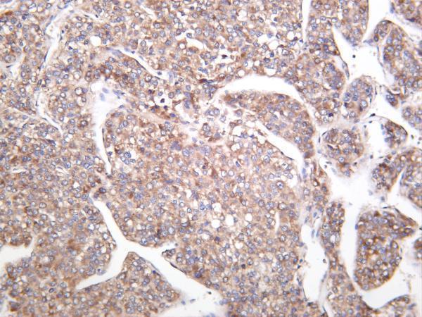
Real IHC data Immunohistochemistry (IHC) analyzes of CaMKIIα/δ (M281) pAb in paraffin-embedded human breast carcinoma tissue at 1:100.
Anti-CaMKII Alpha / Delta (M281) CAMK2A Antibody
Cat # A03241-3
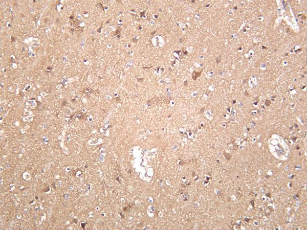
Real IHC data Immunohistochemistry (IHC) analyzes of p-CaMKIIα/δ (T286) pAb in paraffin-embedded human brain tissue.
Anti-CaMKII Alpha / Delta (phospho-T286) CAMK2A Antibody
Cat # A03241T286
Real IF data IF analysis of CaMKII alpha/CAMK2A using anti-CaMKII alpha/CAMK2A antibody (A03241-2). CaMKII alpha/CAMK2A was detected in immunocytochemical section of MCF-7 cells. Enzyme antigen retrieval was performed using IHC enzyme antigen retrieval reagent (AR0022) for 15 mins. The cells were blocked with 10% goat serum. And then incubated with 5μg/mL rabbit anti-CaMKII alpha/CAMK2A Antibody (A03241-2) overnight at 4°C. DyLight®488 Conjugated Goat Anti-Rabbit IgG (BA1127) was used as secondary antibody at 1:100 dilution and incubated for 30 minutes at 37°C. The section was counterstained with DAPI. Visualize using a fluorescence microscope and filter sets appropriate for the label used.
Anti-CaMKII alpha/CAMK2A Antibody ®
Cat # A03241-2

M03241 has IHC images from paraffin-embedded human colon cancer and rat brain; M03241-3 has IHC images from human brain, hepatocellular carcinoma, and mouse brain (catalog image captions). A03241-3 has a paraffin-embedded human breast carcinoma IHC image, A03241T286 has a paraffin-embedded human brain IHC image, and A03241-2 has IF images from MCF-7 cells and paraffin-embedded rat brain (catalog image captions).

Which to pick: For paraffin-section IHC, M03241 offers a rabbit monoclonal with a documented 1:50 IHC example using heat retrieval in EDTA at pH 8.0; the caption does not report the fixative (M03241 catalog entry and IHC caption). For IF/ICC, choose A03241-2 at the listed 5 μg/mL, supported by an MCF-7 cell IF image; it has no listed IHC application (A03241-2 catalog entry and IF caption). For work spanning species, M03241 has human and rat IHC images and listed human, mouse, and rat reactivity; its mouse IHC performance still needs validation in your sample (M03241 catalog entry and IHC captions).

Each figure is that product's own IHC / IF validation image from its datasheet.