CARD9 / Caspase recruitment domain-containing protein 9 · IHC design guide

Design Immunohistochemistry for CARD9

CARD9 shows cytoplasmic tissue staining, including in lung macrophages and spleen red pulp cells (HPA tissue IHC). Use this guide to plan paraffin-section staining and assess cell-specific signal alongside the reported off-target staining caveat (HPA tissue IHC).

Evidence assembled Oct 2026 · For research use; verify linked source records and product datasheet before use
Immunohistochemistry protocol sheet for CARD9 (IHC for CARD9): expected localisation Cytoplasmic staining in several tissues (HPA tissue IHC), antibody A01410-1, validated IHC image, and IHC protocol steps
Printable CARD9 IHC protocol sheet — expected localisation Cytoplasmic staining in several tissues (HPA tissue IHC), antibody A01410-1, controls and protocol steps. Open the full CARD9 IHC guide →

CARD9 Immunohistochemistry Experimental Design Guide

Expected localisation, validated protocols, controls and antibodies — the at-a-glance facts below, then the full design guide.

Must know before staining
Expected localisation Cytoplasmic staining in several tissues (HPA tissue IHC)
Staining pattern Cytoplasmic staining in immune cells (HPA tissue IHC)
Antigen retrieval EDTA pH 8.0 HIER, heat-mediated (datasheet A01410-1)
Positive control ⓘ Testis+3 more · see all
Negative control ⓘ Adipose tissue+4 more · see all
Important caveats
Reasons your staining may differ from the expected pattern.
Fixation Keep paraffin-section fixation consistent (standard IHC practice; not target-specific)
Caveat Presumed off-target staining can confound scoring (HPA tissue IHC)
Regulation Expression regulation is unreported (UniProt)
Isoform / epitope 3 isoforms; map the epitope across variants (UniProt)
Section 1

Recommended CARD9 IHC & IF Protocols

The catalog antibody uses heat-mediated EDTA retrieval (datasheet A01410-1). The published CARD9 IHC protocols below cover esophageal carcinoma, paraffin sections, mouse heart, and lung adenocarcinoma (PMC7105841; PMC4085533; PMC3139445; PMC7498929).

Recommended immunohistochemistry (IHC-P) protocol parameters
SampleParaffin-embedded human rectal cancer tissue; fixative not specified (datasheet A01410-1)
FixationImage fixative and duration unreported (datasheet A01410-1); verify before use.
Sectioning4–5 µm sections on charged slides (standard)
DeparaffinisationXylene, graded ethanol series to water (standard)
Antigen retrievalHeat retrieval: EDTA pH 8.0 (datasheet A01410-1); 20 min, 95–100 °C (standard)
Peroxidase block3% H2O2, 10 min, room temperature (standard)
Blocking10% goat serum (datasheet A01410-1)
Primary antibodyRabbit anti-CARD9, 0.5-1μg/ml (datasheet A01410-1)
Primary incubationOvernight at 4 °C (datasheet A01410-1)
DetectionStreptavidin-biotin complex (SABC), DAB chromogen (datasheet A01410-1)
CounterstainHematoxylin, blue, dehydrate and mount (standard)
Expected resultCARD9-positive staining in elongated or late spermatids of testis (HPA tissue IHC: High). HPA tissue profile: Cytoplasmic expression in several tissues, including immune cells. No signal in the no-primary control.
💡Decision noteStart with EDTA pH 8.0 heat retrieval for the catalog antibody (datasheet A01410-1). A lung adenocarcinoma protocol used pH 7.8 Tris-EDTA-citrate retrieval (PMC7498929 methods).
Section 2

What Is the Expected CARD9 Staining Pattern?

CARD9 is a cytoplasmic protein with no transmembrane segment (UniProt Q9H257: subcellular location and topology). Expect staining mainly in phagocytes, including macrophages, monocytes and dendritic cells (UniProt Q9H257: tissue specificity). HPA reports cytoplasmic staining in several tissues, including immune cells; its tissue IHC reliability is Enhanced, with medium agreement between staining and RNA data and presumed off-target staining disregarded (HPA: tissue IHC reliability and profile).

What am I looking at on my slide?
Discrete cytoplasmic staining in lung macrophages, with nearby cell types less conspicuous (HPA: lung macrophages, Medium).This fits the reported IHC pattern and UniProt localisation (HPA: lung macrophages, Medium; UniProt Q9H257: cytoplasm). Score identifiable macrophages separately from whole-section colour: a mixed tissue can look weak even when its relevant cells stain (standard IHC practice).
Cytoplasmic signal in spleen red-pulp cells or tonsil non-germinal-center cells (HPA: both Medium).These are useful additional positive-pattern checks (HPA: spleen and tonsil, Medium). UniProt reports high expression in spleen and restriction to several phagocyte populations; do not assign every stained red-pulp or tonsillar cell a lineage from colour alone (UniProt Q9H257: tissue specificity; standard IHC practice).
Dominant nuclear, crisp membrane, or extracellular staining in the cells being scored (UniProt Q9H257: cytoplasm; no transmembrane segment).That distribution conflicts with the expected compartment and warrants review as possible artefact or nonspecific staining (UniProt Q9H257: localisation and topology; standard IHC practice). Check morphology, the negative reagent control and whether any convincing cytoplasmic signal remains.
Broad staining of cell types reported as negative, such as adipocytes or bronchial basal cells (HPA: both Not detected).Treat this as a specificity warning, particularly if it overwhelms staining in expected immune cells (HPA: tissue IHC profile). Possible explanations include cross-reactivity or endogenous chromogen-producing activity (standard IHC practice). HPA notes presumed off-target binding was observed and disregarded (HPA: reliability description).
No staining in lung macrophages or spleen red-pulp cells (HPA: both Medium).An absent expected signal can reflect assay failure, section quality or a missed positive cell population (standard IHC practice). Check the control section, morphology and staining run before interpreting a study specimen as negative; HPA's Medium levels do not guarantee every cell or section will stain (HPA: lung and spleen).
💡Expected CARD9 appearanceCall a result consistent when identifiable phagocytes show cytoplasmic staining, typically around the reported Medium level in lung macrophages; dominant nuclear, membrane or broad negative-cell staining is suspect (UniProt Q9H257: localisation and tissue specificity; HPA: lung macrophages, Medium; HPA: negative-cell examples; standard IHC practice).
How each factor affects the staining
Compartment and cell identityCARD9 is cytoplasmic and lacks a transmembrane segment (UniProt Q9H257: localisation and topology). Interpret signal within morphologically identified cells; a diffuse tissue-wide score can conceal the phagocyte-associated pattern (UniProt Q9H257: tissue specificity; standard IHC practice).
Choice of positive and negative tissue areasLung macrophages, spleen red-pulp cells and tonsil non-germinal-center cells have Medium HPA staining; elongated or late spermatids have High staining (HPA: tissue IHC). The spermatid result is an observed pattern, while UniProt describes phagocyte-restricted expression; use cellular context and orthogonal evidence before treating it as a general lineage expectation (UniProt Q9H257: tissue specificity; HPA: reliability description).
Strength of antibody evidenceThe listed antibody HPA059502 has Enhanced IHC validation, while HPA reports only medium staining-to-RNA consistency and presumed off-target binding (HPA: antibody validation and reliability description). An Enhanced label supports use of the reported pattern but does not make unexpected staining self-validating (HPA: reliability description; standard IHC practice).
Molecular variants and assay limitsUniProt lists 3 isoforms, a CARD domain at residues 6–98, and modified residues (UniProt Q9H257: isoforms, domains and modified residues). These facts alone do not identify the catalog antibody's epitope, predict isoform detection, or establish a CARD9-specific antigen-retrieval or fixation effect; those outcomes require assay evidence.
Why is my staining missing, weak or wrong?
SituationLikely causeNext action
Expected macrophage signal is absent in a staining run (HPA: lung macrophages, Medium).The positive-cell population may be missed, or a general IHC step may have failed (standard IHC practice).Review macrophage morphology and a concurrently stained known-positive section; then check retrieval, antibody incubation and chromogen development against the assay instructions (standard IHC practice). No CARD9-specific retrieval condition is supplied.
Background obscures cytoplasmic boundaries (UniProt Q9H257: cytoplasm).Excess primary or detection reagent, inadequate blocking or incomplete washing can raise general IHC background (standard IHC practice).Compare a negative reagent control, then adjust blocking, dilution, washing and detection exposure one variable at a time (standard IHC practice). No dilution is supplied here.
Brown signal persists where the primary antibody is omitted (standard IHC practice).Endogenous detection activity or nonspecific secondary-system signal may contribute (standard chromogenic IHC practice).Check the omission control and apply the detection system's appropriate endogenous-activity block; interpret remaining tissue signal only after that control is clean (standard chromogenic IHC practice).
Signal is mainly nuclear or membrane-associated (UniProt Q9H257: cytoplasm; no transmembrane segment).The compartment conflicts with the UniProt assignment; artefact or nonspecific staining is possible (UniProt Q9H257: localisation and topology; standard IHC practice).Recheck focus, counterstain and cell borders; compare positive and negative controls, and seek an independent specificity check before calling that pattern CARD9 (standard IHC practice).
Adipocytes or bronchial basal cells stain broadly (HPA: both Not detected).Off-target binding or detection background is plausible; HPA reports presumed off-target binding in its IHC assessment (HPA: reliability description; standard IHC practice).Score those cell types separately from nearby immune cells and compare omission and known-positive controls; do not convert their staining into a positive CARD9 call without independent support (HPA: tissue IHC; standard IHC practice).
IF/ICC Q&A: where should CARD9 fluorescence appear?UniProt assigns CARD9 to cytoplasm, but HPA supplies no ICC-IF images or assigned main location (UniProt Q9H257: subcellular location; HPA: subcellular record).Use cytoplasmic localisation as the provisional interpretation and validate it with IF-specific controls; this IHC section supplies no IF protocol or image-based HPA localisation claim (UniProt Q9H257: subcellular location; HPA: subcellular record; standard IF practice).

Sample controls for CARD9 IHC & IF

🧪Run lung first: macrophages should show cytoplasmic CARD9 staining (HPA: Medium in lung macrophages; UniProt Q9H257: cytoplasm). Use adrenal gland glandular cells as a negative tissue (HPA: Not detected in adrenal gland glandular cells); on the lung slide, nonmacrophage cells should remain at background staining, judged against the no-primary control (standard IHC practice).
Positive control tissue: Testis (Elongated or late spermatids, HPA High)
Negative control tissue: Adipose tissue (HPA Not detected)
ICC-IF cell lines (HPA subcellular resource): HPA carries no ICC-IF cell line for CARD9; derive a cell-line control from the positive tissue's cell type (Elongated or late spermatids) and confirm it by RNA or western blot first.
Technical controls: Include a no-primary, secondary-only control, concentration-matched nonimmune rabbit IgG as an isotype control, and CARD9-knockout material as a biological negative if available (caption: rabbit primary; standard IHC controls). Block endogenous peroxidase and biotin in the lung section before detection (caption: streptavidin–biotin complex with DAB; standard IHC practice).
⚠️Feasibility: A paraffin-section example uses heat retrieval in EDTA at pH 8.0, but retrieval dependence has not been established (selected-SKU IHC caption). The caption does not report a fixative, and the supplied evidence reports no CARD9-specific fixation window or fixation effect (selected-SKU IHC caption). Paraffin IHC has a documented example; the supplied evidence does not establish that frozen sections or IF are easier, and lung macrophage pigment should be distinguished from DAB staining using the no-primary slide (selected-SKU IHC caption; HPA: Medium in lung macrophages; standard IHC practice).

HPA tissue IHC evidence for CARD9

Comprehensive Human Protein Atlas IHC scoring per tissue (reliability: Enhanced — Medium consistency between antibody staining and RNA expression data. Presumed off target binding observed and disregarded.). Rows are taken directly from the HPA tissue chart — click any row's HPA link to view the source.

Positive expression · recommended positive controls

TissueCell typeLevelEvidenceSource
Testis Elongated or late spermatids High Protein (IHC) HPA →
Lung Macrophages Medium Protein (IHC) HPA →
Spleen Cells in red pulp Medium Protein (IHC) HPA →
Tonsil Non-germinal center cells Medium Protein (IHC) HPA →

Undetected expression · recommended negative controls

TissueCell typeLevelEvidenceSource
Adipose tissue Adipocytes Not detected Protein (IHC) HPA →
Adrenal gland Glandular cells Not detected Protein (IHC) HPA →
Appendix Glandular cells Not detected Protein (IHC) HPA →
Bronchus Basal cells Not detected Protein (IHC) HPA →
Caudate Glial cells Not detected Protein (IHC) HPA →
Section 3

Advanced CARD9 IHC Tips

Troubleshoot CARD9 staining in paraffin sections by checking retrieval, cell identity and cytoplasmic localisation alongside appropriate controls.

What retrieval should I use if CARD9 staining is weak in paraffin sections?
Start with heat-mediated antigen retrieval in EDTA at pH 8.0 (datasheet A01410-1). The catalog antibody detected CARD9 in a paraffin section after this retrieval, followed by 1 μg/ml primary antibody overnight at 4°C (datasheet A01410-1). If staining is weak, check heating and cooling consistency, section adhesion and reagent performance before comparing retrieval conditions on adjacent sections (standard IHC practice). Judge any apparent gain against cytoplasmic staining in the expected cell population and any increase in background; CARD9 is cytoplasmic, with expression reported in phagocyte populations (UniProt Q9H257).
Could fixation explain weak or uneven CARD9 staining?
Target-specific fixation sensitivity is unknown: the selected tissue caption describes a paraffin section but does not state its fixative (datasheet A01410-1). Record the fixative, fixation duration, tissue thickness and processing history for each block, then compare sections processed together using EDTA retrieval at pH 8.0 (datasheet A01410-1; standard IHC practice). Uneven staining warrants a check for drying, folds and inconsistent reagent coverage before changing the primary antibody concentration (standard IHC practice). Assess any processing change using morphology, background and cytoplasmic signal in plausible cells, rather than treating a brighter section alone as proof of improved CARD9 detection (UniProt Q9H257; standard IHC practice).
Where should convincing CARD9 staining appear in a tissue section?
Look primarily for cytoplasmic staining: CARD9 is annotated in the cytoplasm and has no transmembrane segment (UniProt Q9H257). Macrophages, monocytes and dendritic cells are plausible expressing populations, while the tissue atlas reports medium staining in lung macrophages and spleen red-pulp cells (UniProt Q9H257; HPA tissue IHC). Identify cells by morphology and, when needed, a separate lineage marker rather than assigning identity from brown signal alone (standard IHC practice). Predominantly nuclear or crisp membrane outlining should prompt review of background, counterstain and cell identification; record the compartment and proportion of stained cells before calling such a pattern CARD9 positive (UniProt Q9H257; standard IHC practice).
How could isoforms or epitope accessibility change the IHC result?
CARD9 has 3 annotated isoforms and a CARD domain at residues 6–98, so epitope coverage matters when comparing antibodies (UniProt Q9H257). Check the catalog antibody's stated immunogen or mapped epitope before assuming that all isoforms are detected; no epitope position is supplied here (standard IHC practice; UniProt Q9H257). Phosphorylated residues are annotated, including positions 231 and 277, but their effect on this antibody's staining is unreported (UniProt Q9H257). If results disagree between antibodies, compare serial sections with matched retrieval and controls, then interpret agreement in cell type and cytoplasmic pattern alongside epitope information (standard IHC practice; UniProt Q9H257).
How should I investigate a CARD9 pattern with multiplex immunofluorescence?
Use this as a complementary localisation check, because the supplied catalog evidence is for chromogenic staining in a paraffin section, and no HPA cell line IF images are supplied (datasheet A01410-1; HPA subcellular). Pair CARD9 with a validated marker for an expected phagocyte population, and assess overlap at the single-cell level (UniProt Q9H257; standard IF practice). Choose fluorophores after checking tissue autofluorescence and include single-stain and no-primary controls to set channel exposure and assess bleed-through (standard IF practice). Since CARD9 is cytoplasmic and lacks a transmembrane segment, choose permeabilisation that permits intracellular antibody access, then verify that cell boundaries remain interpretable (UniProt Q9H257; standard IF practice).
How can I reduce diffuse brown staining without losing CARD9 signal?
First compare the stained section with a no-primary control and inspect whether brown signal follows tissue edges, damaged areas or endogenous pigment (standard IHC practice). Use an appropriate peroxidase block and serum or protein block, then optimise washing and detection conditions on matched sections (standard IHC practice). The selected caption used 10% goat serum, 1 μg/ml primary antibody overnight at 4°C, and a biotin-based detection system with DAB (datasheet A01410-1). If diffuse signal persists, assess possible endogenous biotin with detection controls; retain conditions that show interpretable cytoplasmic staining in plausible cells while keeping control staining low (standard IHC practice; UniProt Q9H257).
What should I score when comparing CARD9 across sections? ⚠ ANSWER MARKED FOR VERIFICATION
Define the cell compartment and scoring area before reviewing groups, because CARD9 is cytoplasmic and expected expression varies with phagocyte content (UniProt Q9H257; standard IHC practice). For a defined cell population, report the percentage of positive cells and, if intensity is reproducible, an H-score from 0–300 (standard IHC practice). For tissue comparisons, also report positive-cell density per mm² of viable tissue or normalise positive cells to the counted lineage population (standard IHC practice). Apply one threshold and sampling rule across sections, exclude folds and necrosis, and report cell abundance separately from staining intensity (standard IHC practice).
How do I distinguish true CARD9 staining from artefact?
A credible result combines cytoplasmic signal with an identified, plausible cell population; CARD9 is cytoplasmic and expressed in several phagocyte populations (UniProt Q9H257). The atlas reports medium staining in lung macrophages and spleen red-pulp cells, while also noting presumed off-target binding and only medium staining-to-RNA consistency (HPA tissue IHC). Treat nuclear-only staining, strong signal in an unexpected cell population, edge accentuation and necrotic deposits as reasons to investigate rather than as evidence of CARD9 expression (UniProt Q9H257; standard IHC practice). Compare morphology, a no-primary control, peroxidase blocking and, where feasible, an independent marker or antibody before assigning a biological interpretation (standard IHC practice).
Boster reagents

Best CARD9 / Caspase recruitment domain-containing protein 9 IHC Antibodies

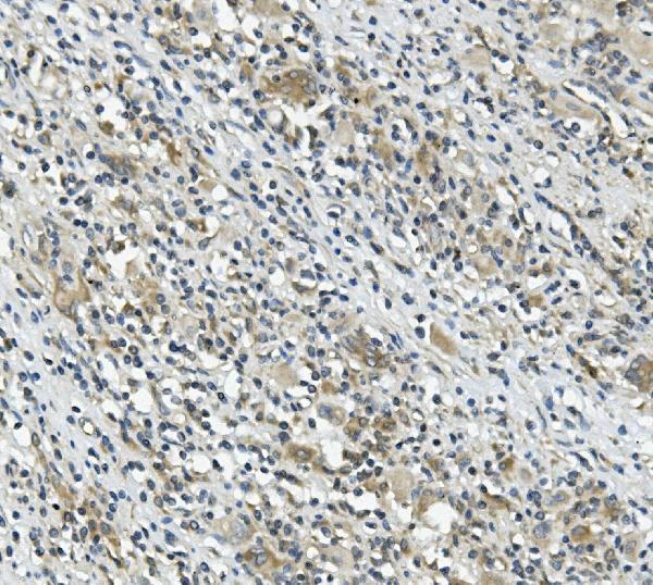
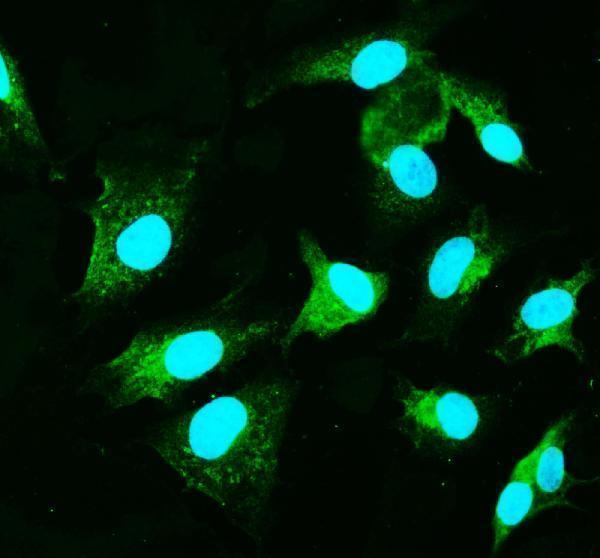
The IHC-validated antibody A01410-1 has IHC data from a human paraffin-embedded tissue section and IF/ICC data from A549 cells (catalog image captions).

Real IHC data IHC analysis of CARD9 using anti-CARD9 antibody (A01410-1). CARD9 was detected in paraffin-embedded section of human rectal cancer tissue. Heat mediated antigen retrieval was performed in EDTA buffer (pH8.0, epitope retrieval solution). The tissue section was blocked with 10% goat serum. The tissue section was then incubated with 1μg/ml rabbit anti-CARD9 Antibody (A01410-1) overnight at 4°C. Biotinylated goat anti-rabbit IgG was used as secondary antibody and incubated for 30 minutes at 37°C. The tissue section was developed using Strepavidin-Biotin-Complex (SABC) (Catalog # SA1022) with DAB as the chromogen.
Anti-CARD9 ® Antibody
Cat # A01410-1

A01410-1 will render with IHC data from a paraffin-embedded human rectal cancer tissue section (IHC image caption). Its IF/ICC data show CARD9 detection in A549 cells (IF image caption).

Which to pick: Choose A01410-1 for human tissue IHC: its image caption documents a paraffin-embedded section, while the fixative is unreported (IHC image caption; catalog: Human reactivity). The same SKU is listed for IF/ICC and has an A549 cell IF image (catalog: IF/ICC applications; IF image caption). Cross-species use is unsupported by the listed Human reactivity, and clonality is unreported.

Each figure is that product's own IHC / IF validation image from its datasheet.

References

  1. UniProt Consortium. UniProt entry Q9H257 (CARD9_HUMAN, Caspase recruitment domain-containing protein 9).
  2. Human Protein Atlas. CARD9 tissue IHC expression (reliability: Enhanced).
  3. Human Protein Atlas. CARD9 subcellular location (ICC-IF): Highest expression in THP-1: 63.3 nTPM.
  4. Human Protein Atlas. CARD9 antibody validation summary (1 antibodies).
  5. Caspase recruitment domain family member 9 expression is a promising biomarker in esophageal squamous cell carcinoma. Annals of gastroenterological surgery 2020 — PMC7105841.
  6. Tumor cell-activated CARD9 signaling contributes to metastasis-associated macrophage polarization. Cell death and differentiation 2014 — PMC4085533.
  7. Proinflammatory protein CARD9 is essential for infiltration of monocytic fibroblast precursors and cardiac fibrosis caused by Angiotensin II infusion. American journal of hypertension 2011 — PMC3139445.
  8. Caspase Recruitment Domain-Containing Protein 9 Expression is a Novel Prognostic Factor for Lung Adenocarcinoma. OncoTargets and therapy 2020 — PMC7498929.
  9. PubMed PMID:11053425 — UniProt-cited evidence.
  10. PubMed PMID:14702039 — UniProt-cited evidence.
  11. PubMed PMID:15164053 — UniProt-cited evidence.