This website uses cookies to ensure you get the best experience on our website.


- Table of Contents
Plan CARF staining in paraffin sections using its observed nuclear and cytoplasmic tissue pattern (HPA tissue IHC). This guide covers fixation, controls and scoring, with high-staining reference cells including cerebellar Purkinje cells and pancreatic exocrine glandular cells (HPA tissue IHC).

Expected localisation, validated protocols, controls and antibodies — the at-a-glance facts below, then the full design guide.
| Expected localisation | Nuclear and cytoplasmic tissue staining (HPA tissue IHC); nuclear location (UniProt) | |
| Staining pattern | Positive tissue cells show nuclear and cytoplasmic staining (HPA tissue IHC) | |
| Antigen retrieval | Tris-EDTA pH 9.0 HIER, 95–98 °C, 20 min (rule: nuclear antigen) | |
| Positive control | Appendix+4 more · see all | |
| Negative control | None in HPA (detected in all 45 tissues); use no-primary + isotype controls |
| Fixation | Keep fixation consistent across paraffin sections. (standard IHC practice; not target-specific) | |
| Caveat | Purkinje cells stain high; hippocampal neurons stain low (HPA tissue IHC) | |
| Regulation | CARF expression regulation is not annotated (UniProt) | |
| Isoform / epitope | 3 isoforms; no extracellular/cytoplasmic split; epitope unknown (UniProt; datasheet) |
Compare the catalog antibody’s IHC-P protocol with 2 published CARF IHC workflows: mouse paraffin sections (PMC12040762) and HCC tissues (PMC5348329).
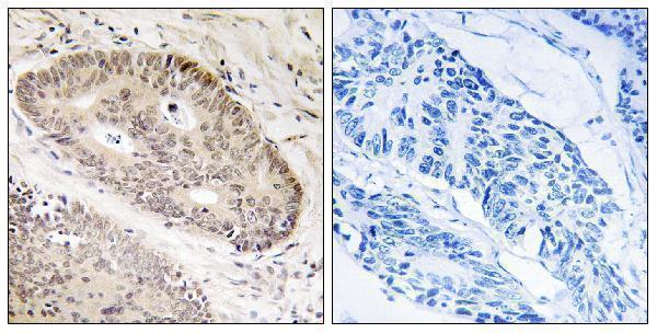
| Sample | Paraffin-embedded human lung carcinoma tissue; fixative not specified (datasheet A30570) |
| Fixation | Image fixative and duration unreported (datasheet A30570); verify before use. |
| Sectioning | 4–5 µm sections on charged slides (standard) |
| Deparaffinisation | Xylene, graded ethanol series to water (standard) |
| Antigen retrieval | Heat-induced epitope retrieval in Tris-EDTA buffer, pH 9.0, 20 min at 95–98 °C (standard rule: nuclear antigen) |
| Peroxidase block | 3% H2O2, 10 min, room temperature (standard) |
| Blocking | 10% normal serum of the secondary host, 30 min, room temperature (standard) |
| Primary antibody | Rabbit anti-CARF, 1:100 - 1:300 (datasheet A30570) |
| Primary incubation | Overnight at 4 °C (standard) |
| Detection | HRP-polymer secondary, DAB chromogen 5–10 min (standard) |
| Counterstain | Hematoxylin, blue, dehydrate and mount (standard) |
| Expected result | CARF-positive staining in glandular cells of appendix (HPA tissue IHC: High). HPA tissue profile: Nuclear and cytoplasmic expression. No signal in the no-primary control. |
In paraffin-section IHC, expect nuclear and cytoplasmic CARF staining in selected epithelial, glandular, neuronal and decidual cells (HPA tissue IHC). UniProt places CARF in the nucleus and annotates no transmembrane segment (UniProt Q8N187). HPA rates its tissue staining Approved, with medium consistency against RNA data and external verification pending (HPA tissue IHC). Interpret the stained cell type and compartment together.
| Nuclear and cytoplasmic signal in pancreatic exocrine glandular cells or cerebellar Purkinje cells. | These are high-staining cell populations (HPA tissue IHC). Score signal in the identified cells, using tissue morphology and the counterstain to distinguish nuclei from adjacent cytoplasm (general IHC practice). |
| Only sharp cell-surface staining, with no convincing nuclear or cytoplasmic component. | That distribution does not match the reported IHC profile (HPA tissue IHC) or nuclear annotation and lack of a transmembrane segment (UniProt Q8N187). Check morphology, detection background and antibody specificity before calling it CARF. |
| Strong staining is concentrated in an unexpected cell population. | Compare with the named HPA cell populations; unexpected staining can reflect cross-reactivity or endogenous chromogenic activity (general IHC practice). It is not automatically false: HPA reports low tissue specificity at the RNA level and supplies no validated negative tissue (HPA tissue IHC). |
| Diffuse chromogen covers many cells or tissue spaces without clear cellular boundaries. | Treat the pattern as background until a no-primary control and the tissue morphology support specific staining (general IHC practice). HPA's nuclear and cytoplasmic description refers to cellular localisation, not a diffuse field-wide deposit (HPA tissue IHC). |
| No signal appears in a tissue with a reported high-staining cell population. | First confirm that the relevant cells are present; for example, pancreatic exocrine glandular cells are reported High (HPA tissue IHC). Then check the positive control, retrieval, primary-antibody conditions and detection reagents (general IHC practice). |
| Cell population and tissue choice | HPA reports High staining in eight listed cell populations, including respiratory epithelium and decidual cells, but Low staining in six listed populations, including hippocampal neurons (HPA tissue IHC). Choose controls by cell type, not tissue name alone (general IHC practice). |
| Antibody validation | HPA034557 is Approved for IHC; HPA043191 has no IHC status in this record (HPA antibodies). The tissue profile is Approved with medium RNA agreement and pending external verification (HPA tissue IHC), so review discordant staining cautiously. |
| Sequence and topology | CARF has three annotated isoforms, one 1–725 chain, no annotated glycosylation sites and no transmembrane segment (UniProt Q8N187). The payload gives no antibody epitope, so it cannot establish which isoforms an IHC antibody detects. |
| Target-specific fixation sensitivity | No fixation-effect evidence is supplied for CARF (UniProt Q8N187; HPA tissue IHC). If staining fails, assess retrieval and processing against controls as general IHC troubleshooting, without attributing the result to a proven CARF-specific masking effect. |
| How should IF/ICC be interpreted? | HPA reports mainly vesicular localisation, with additional nucleoli fibrillar-center and cytosolic signal in ICC-IF (HPA subcellular). That observation comes from a different application; use the IHC tissue profile to judge paraffin-section chromogenic staining. |
| Chromogenic detection | Endogenous enzyme activity or insufficient blocking can imitate antibody signal (general IHC practice). A no-primary control helps identify detection-derived deposit; it does not establish CARF expression in a particular cell. |
| Situation | Likely cause | Next action |
|---|---|---|
| A reported High population stains weakly or not at all. | The relevant cells may be absent from the section, or the IHC workflow may have lost sensitivity (general IHC practice). | Confirm cell identity and run a positive tissue control alongside the section; then review retrieval, primary-antibody conditions and detection (general IHC practice). HPA's High designation is observational, not a guaranteed result in every specimen (HPA tissue IHC). |
| Chromogen appears throughout the section, including areas without identifiable target cells. | Nonspecific reagent binding or endogenous detection activity is possible (general IHC practice). | Inspect a no-primary control, blocking and washing; score only cell-associated staining with discernible morphology (general IHC practice). |
| Staining appears exclusively along cell borders. | The pattern conflicts with nuclear and cytoplasmic tissue staining (HPA tissue IHC) and the lack of a transmembrane segment (UniProt Q8N187). | Check for edge artifact and detection background, then reassess with an IHC-validated antibody and appropriate controls (general IHC practice; HPA antibodies: HPA034557 Approved). |
| Unexpected cell types stain strongly while the expected population does not. | Cross-reactivity, endogenous activity or mistaken cell identification are possibilities (general IHC practice); HPA provides no validated negative tissue in this payload (HPA tissue IHC). | Recheck morphology and the no-primary control; compare the same named cell population in a reported High tissue before interpreting the unexpected signal (HPA tissue IHC; general IHC practice). |
| An IHC section lacks the vesicular pattern described for ICC-IF. | The vesicular observation comes from HPA's ICC-IF record, whereas its tissue IHC profile is nuclear and cytoplasmic (HPA subcellular; HPA tissue IHC). | Judge the chromogenic section by the IHC cell and compartment profile; do not require an ICC-IF pattern in paraffin tissue. |
| A Low population stains faintly beside a strongly stained control population. | HPA reports different staining levels by cell population, including Low in hippocampal neurons and High in Purkinje cells (HPA tissue IHC). | Score each identified population separately and check that the stronger signal remains cell-associated; avoid treating faint staining alone as assay failure (HPA tissue IHC; general IHC practice). |


Comprehensive Human Protein Atlas IHC scoring per tissue (reliability: Approved — Medium consistency between antibody staining and RNA expression data. Pending external verification.). Rows are taken directly from the HPA tissue chart — click any row's HPA link to view the source.
| Tissue | Cell type | Level | Evidence | Source |
|---|---|---|---|---|
| Appendix | Glandular cells | High | Protein (IHC) | HPA → |
| Bronchus | Respiratory epithelial cells | High | Protein (IHC) | HPA → |
| Cerebellum | Purkinje cells | High | Protein (IHC) | HPA → |
| Gallbladder | Glandular cells | High | Protein (IHC) | HPA → |
| Nasopharynx | Respiratory epithelial cells | High | Protein (IHC) | HPA → |
| Tissue | Cell type | Level | Evidence | Source |
|---|---|---|---|---|
| None in HPA: CARF is detected in all 45 scored tissues. Use a no-primary (secondary-only) and an isotype control instead. | ||||
Troubleshoot CARF staining in paraffin sections by checking retrieval, compartment, controls and scoring before interpreting chromogenic signal.
The catalog lists A30570 for IHC, IF and ICC (catalog applications), with human lung carcinoma paraffin-section IHC data (catalog image caption) and Human, Mouse and Rat reactivity (catalog reactivity).
A30570 has an IHC image from a human lung carcinoma paraffin section, including a peptide-blocked comparison (catalog image caption). It is listed for IHC, IF and ICC in Human, Mouse and Rat (catalog applications; catalog reactivity); no IF image is supplied (catalog IF image alts).
Which to pick: Choose A30570 for paraffin-section tissue IHC because its own image caption documents that preparation (catalog image caption); the fixative is unreported (catalog image caption). Choose A30570 for IF/ICC because those applications are listed, while no IF image is supplied (catalog applications; catalog IF image alts). It is also the listed cross-species option for Human, Mouse and Rat, and is polyclonal (catalog reactivity; catalog dilution record).