This website uses cookies to ensure you get the best experience on our website.


- Table of Contents
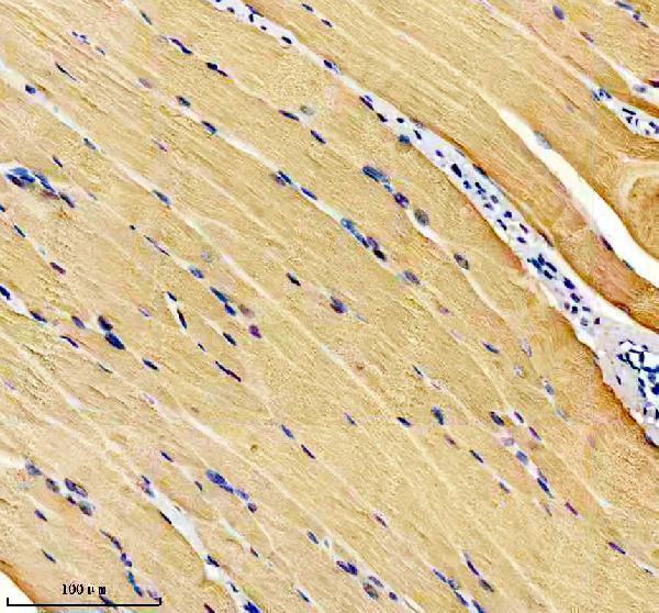
Plan CASQ1 paraffin IHC around selective cytoplasmic staining in skeletal myocytes (HPA tissue IHC). The catalog antibody was used at 1:50 with chromogenic detection in a mouse skeletal muscle section (datasheet M05235).

Expected localisation, validated protocols, controls and antibodies — the at-a-glance facts below, then the full design guide.
| Expected localisation | Myocyte cytoplasm (HPA tissue IHC); SR lumen expected (UniProt) | |
| Staining pattern | Selective skeletal myocyte cytoplasmic staining (HPA tissue IHC) | |
| Antigen retrieval | EDTA pH 8.0 HIER, heat-mediated (datasheet M05235) | |
| Positive control | Skeletal muscle | |
| Negative control | Adipose tissue+4 more · see all |
| Fixation | Keep fixation consistent across sections (standard IHC practice; not target-specific) | |
| Caveat | Low neuronal staining may complicate interpretation (HPA tissue IHC) | |
| Regulation | Expression regulation unreported (UniProt) | |
| Isoform / epitope | No annotated isoforms; mature chain starts at residue 35 (UniProt) |
The catalog antibody protocol is accompanied by one published paraffin-section IHC protocol for CASQ1/2 staining in tissue biopsies (PMC12955825 methods).
| Sample | Paraffin-embedded mouse skeletal muscle tissue; fixative not specified (datasheet M05235) |
| Fixation | Image fixative and duration unreported (datasheet M05235); verify before use. |
| Sectioning | 4–5 µm sections on charged slides (standard) |
| Deparaffinisation | Xylene, graded ethanol series to water (standard) |
| Antigen retrieval | Heat retrieval: EDTA pH 8.0 (datasheet M05235); 20 min, 95–100 °C (standard) |
| Peroxidase block | 3% H2O2, 10 min, room temperature (standard) |
| Blocking | 10% goat serum (datasheet M05235) |
| Primary antibody | Rabbit monoclonal (clone AEGE-3) anti-CASQ1, 1:50 (datasheet M05235) |
| Primary incubation | Overnight at 4 °C (datasheet M05235) |
| Detection | HRP-conjugated secondary, DAB chromogen (datasheet M05235) |
| Counterstain | Hematoxylin, blue, dehydrate and mount (standard) |
| Expected result | CASQ1-positive staining in myocytes of skeletal muscle (HPA tissue IHC: High). HPA tissue profile: Selective cytoplasmic expression in skeletal muscle. No signal in the no-primary control. |
CASQ1 should give strong cytoplasmic staining in skeletal-muscle myocytes on IHC sections (HPA: High in skeletal-muscle myocytes; selective cytoplasmic expression). Its principal muscle location is the sarcoplasmic-reticulum lumen, especially terminal cisternae of fast skeletal-muscle cells (UniProt P31415: subcellular location). It has no transmembrane segment (UniProt P31415: topology). HPA rates the tissue IHC profile Enhanced, citing high consistency between antibody staining and RNA expression data (HPA: tissue IHC reliability).
| Strong cytoplasmic staining in skeletal-muscle myocytes, with little staining in neighboring nonmyocytes. | This matches the reported positive tissue and cell type (HPA: High in skeletal-muscle myocytes). At light-microscope resolution, a cytoplasmic result is compatible with CASQ1 in the sarcoplasmic-reticulum lumen; the IHC pattern alone does not resolve terminal cisternae (UniProt P31415: subcellular location). |
| Predominantly nuclear staining, or a membrane-outline pattern, in skeletal-muscle myocytes. | Treat this as a compartment mismatch requiring validation: tissue IHC is reported as cytoplasmic (HPA: tissue IHC profile), while UniProt places muscle CASQ1 chiefly in the sarcoplasmic-reticulum lumen and reports no transmembrane segment (UniProt P31415: location and topology). |
| Strong staining in adipocytes, hematopoietic cells, or glandular cells while myocytes are weak. | Review specificity and detection controls. Those cell populations were reported as Not detected in the listed HPA tissues (HPA: adipose tissue, bone marrow, cervix). Cross-reactivity or endogenous detection activity are possible explanations under general IHC practice; this pattern does not establish CASQ1 expression. |
| Diffuse color across the section, including cells expected to be negative. | The loss of cell-type contrast makes CASQ1 interpretation unreliable. HPA reports selective cytoplasmic expression in skeletal muscle and Not detected staining in several other cell types (HPA: tissue IHC profile). Under general IHC practice, assess nonspecific antibody binding and detection-system background. |
| No staining in skeletal-muscle myocytes on an otherwise interpretable section. | This conflicts with the HPA High result for those cells (HPA: skeletal-muscle myocytes). Check the positive control, antibody application, retrieval, and chromogenic detection as general IHC troubleshooting steps. The supplied sources do not establish a CASQ1-specific retrieval requirement or fixation sensitivity. |
| Cell-type distribution | Skeletal-muscle myocytes are a positive reference (HPA: High); adipocytes and bone-marrow hematopoietic cells are reported Not detected (HPA: tissue IHC). HPA also lists low staining in some neuronal, glandular, and respiratory epithelial cells, so every nonmuscle signal should not automatically be called false. |
| Subcellular interpretation | UniProt places CASQ1 in the sarcoplasmic-reticulum lumen of fast skeletal-muscle cells and also lists ER and mitochondrial-matrix locations (UniProt P31415: subcellular location). HPA's tissue IHC summary supports a cytoplasmic reading, without resolving those organelles (HPA: tissue IHC profile). |
| IHC antibody evidence | The listed antibodies HPA007845, HPA026823, and CAB015170 each have Enhanced IHC status (HPA: antibody validation). This supports the reported tissue pattern; it does not make an unexpected compartment or cell type positive without controls. |
| Protein processing and epitope | UniProt annotates a signal peptide at residues 1–34 and the mature chain at 35–396 (UniProt P31415: processing). No antibody epitope is supplied, so these annotations cannot predict recognition after retrieval, or explain a weak section by themselves. |
| IF/ICC Q&A: should nuclear or mitochondrial IF be expected in this IHC guide? | HPA approves nucleoplasm and mitochondria as ICC-IF locations in RT-4 images (HPA: subcellular ICC-IF), but tissue IHC reports selective cytoplasmic skeletal-muscle staining (HPA: tissue IHC). HPA007845 has Uncertain ICC status (HPA: antibody validation). Interpret IF on its own guide page; no IF protocol is established here. |
| Situation | Likely cause | Next action |
|---|---|---|
| Skeletal-muscle myocytes show no chromogenic signal. | The result disagrees with HPA's High myocyte staining (HPA: tissue IHC); a failed staining or detection step is possible under general IHC practice. | Run a known-positive skeletal-muscle section alongside the sample. Verify primary-antibody application and chromogen development, then review retrieval against the antibody's IHC-P instructions. Do not assume CASQ1-specific fixation sensitivity: none is supplied. |
| Myocytes are weak, but the entire section has a faint wash of color. | Low contrast may reflect general IHC background or insufficient usable signal; the supplied sources do not identify a CASQ1-specific chemical cause. | Compare with a no-primary control, inspect blocking and wash steps, and adjust antibody or detection conditions only within validated IHC-P guidance. Score the myocyte pattern against the HPA High reference after background is controlled (HPA: skeletal-muscle myocytes). |
| Nuclei dominate the stain in a skeletal-muscle section. | The compartment conflicts with HPA's cytoplasmic tissue pattern (HPA: tissue IHC profile). HPA's approved nucleoplasmic observation comes from ICC-IF, a different application (HPA: subcellular ICC-IF). | Check the tissue positive control and a no-primary control. Reassess specificity before scoring nuclear IHC as CASQ1; the ICC-IF observation alone cannot validate that tissue IHC pattern. |
| Adipocytes or bone-marrow hematopoietic cells stain as strongly as myocytes. | Those cells are reported Not detected by HPA, whereas skeletal-muscle myocytes are High (HPA: tissue IHC). Cross-reactivity or endogenous detection activity remain possibilities under general IHC practice. | Compare matched control sections, including a no-primary control, and check whether color appears without primary antibody. Recheck the antibody's IHC validation before attributing the unexpected cell staining to CASQ1. |
| An apparent membrane outline replaces the myocyte cytoplasmic pattern. | A membrane-dominant result does not fit the reported tissue IHC profile (HPA: selective cytoplasmic expression) or the luminal muscle location and absent transmembrane segment (UniProt P31415: location and topology). | Inspect section morphology and control staining, then repeat with the IHC-validated antibody under its documented conditions. Record the compartment as discordant until a reproducible, specifically controlled result supports interpretation. |
| A nonmuscle section shows a few weakly stained cells. | HPA lists Low staining in several nonmuscle cell populations, including neuronal and respiratory epithelial cells (HPA: tissue IHC). Weak signal therefore needs cell-specific review rather than an automatic false-positive label. | Identify the cell type and compare its HPA level with the observed intensity. Check background controls and require the expected strong skeletal-muscle myocyte signal on the positive control before interpreting weak nonmuscle staining. |
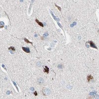
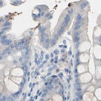
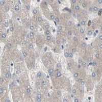
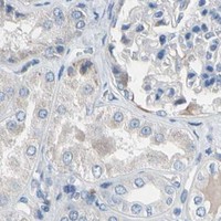


Comprehensive Human Protein Atlas IHC scoring per tissue (reliability: Enhanced — High consistency between antibody staining and RNA expression data.). Rows are taken directly from the HPA tissue chart — click any row's HPA link to view the source.
| Tissue | Cell type | Level | Evidence | Source |
|---|---|---|---|---|
| Skeletal muscle | Myocytes | High | Protein (IHC) | HPA → |
| Tissue | Cell type | Level | Evidence | Source |
|---|---|---|---|---|
| Adipose tissue | Adipocytes | Not detected | Protein (IHC) | HPA → |
| Bone marrow | Hematopoietic cells | Not detected | Protein (IHC) | HPA → |
| Breast | Adipocytes | Not detected | Protein (IHC) | HPA → |
| Cervix | Glandular cells | Not detected | Protein (IHC) | HPA → |
| Epididymis | Glandular cells | Not detected | Protein (IHC) | HPA → |
Use the paraffin-section IHC example as the starting workflow, then judge CASQ1 staining against skeletal muscle expression and sarcoplasmic reticulum localisation (datasheet M05235; HPA tissue IHC; UniProt P31415).
M05235 has real IHC data from mouse skeletal muscle paraffin sections (M05235 IHC caption). Listed reactivity covers human, mouse and rat (catalog: M05235 reactivity).
M05235 is a rabbit monoclonal antibody listed for IHC, with an image from mouse skeletal muscle paraffin sections (catalog: M05235 host and clone; M05235 applications; M05235 IHC caption). Its listed reactivity is human, mouse and rat; no IF image is supplied (catalog: M05235 reactivity and IF images).
Which to pick: For tissue IHC, choose M05235: its own image caption documents paraffin sections, EDTA retrieval at pH 8.0 and a 1:50 primary dilution; the fixative is unreported (M05235 IHC caption). There is no supported IF/ICC choice among the supplied products because M05235 has no listed IF/ICC application or IF image (catalog: M05235 applications and IF images). For cross-species planning, M05235 lists human, mouse and rat reactivity, while its IHC image documents mouse tissue only (catalog: M05235 reactivity; M05235 IHC caption).