This website uses cookies to ensure you get the best experience on our website.


- Table of Contents
Plan chromogenic CBX1 IHC in paraffin sections with a nuclear readout (HPA tissue IHC). Start the IHC-validated antibody at 2–5 μg/mL (datasheet A04206-2), then score within defined cell populations (standard IHC practice).

Expected localisation, validated protocols, controls and antibodies — the at-a-glance facts below, then the full design guide.
| Expected localisation | Nuclear staining across tissues (HPA tissue IHC) | |
| Staining pattern | Nuclei stain across many cell types (HPA tissue IHC) | |
| Antigen retrieval | EDTA pH 8.0 HIER, heat-mediated (datasheet A04206-2) | |
| Positive control | Bone marrow+4 more · see all | |
| Negative control | None in HPA (detected in all 45 tissues); use no-primary + isotype controls |
| Fixation | Keep fixation conditions consistent across sections (standard IHC practice; not target-specific) | |
| Caveat | Widespread nuclear staining complicates cell-specific scoring (HPA tissue IHC) | |
| Regulation | No specific staining regulator reported (UniProt) | |
| Isoform / epitope | No annotated isoforms; one 1–185 chain (UniProt) |
The catalog antibody protocol uses EDTA pH 8.0 retrieval (datasheet A04206-2). Two published CBX1 IHC protocols provide tissue microarray and lymphoma section examples (PMC11461582; PMC10331996).
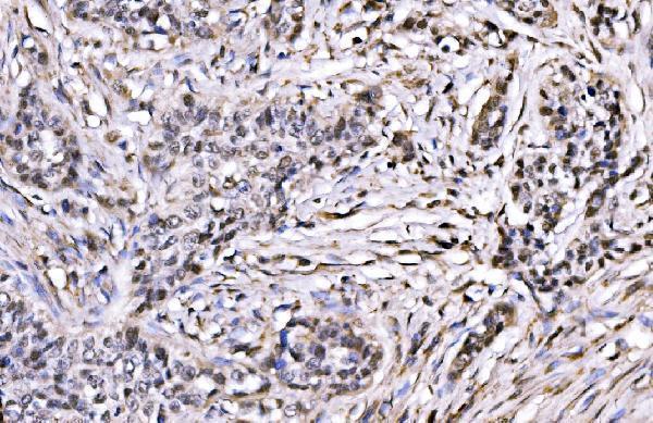
| Sample | Paraffin-embedded human lung cancer tissue; fixative not specified (datasheet A04206-2) |
| Fixation | Image fixative and duration unreported (datasheet A04206-2); verify before use. |
| Sectioning | 4–5 µm sections on charged slides (standard) |
| Deparaffinisation | Xylene, graded ethanol series to water (standard) |
| Antigen retrieval | Heat retrieval: EDTA pH 8.0 (datasheet A04206-2); 20 min, 95–100 °C (standard) |
| Peroxidase block | 3% H2O2, 10 min, room temperature (standard) |
| Blocking | 10% goat serum (datasheet A04206-2) |
| Primary antibody | Rabbit anti-CBX1, 2-5 μg/ml (datasheet A04206-2) |
| Primary incubation | Overnight at 4 °C (datasheet A04206-2) |
| Detection | Streptavidin-biotin complex (SABC), DAB chromogen (datasheet A04206-2) |
| Counterstain | Hematoxylin, blue, dehydrate and mount (standard) |
| Expected result | CBX1-positive staining in hematopoietic cells of bone marrow (HPA tissue IHC: High). HPA tissue profile: Ubiquitous nuclear expression. No signal in the no-primary control. |
CBX1 should appear predominantly in nuclei across many cell types: UniProt places it in the nucleus and reports expression in all adult and embryonic tissues (UniProt P83916). Tissue IHC shows ubiquitous nuclear expression with staining supported by agreement with RNA data (HPA: Supported; ubiquitous nuclear expression). CBX1 has no transmembrane segment, so a membrane pattern is unexpected (UniProt P83916 topology).
| Nuclear staining across several cell types, with strong signal in bone marrow hematopoietic cells. | This fits the reported broad nuclear pattern and a documented high-staining cell population; compare nuclei within the same section when judging intensity (HPA: ubiquitous nuclear expression; High in bone marrow hematopoietic cells). |
| Membrane or predominantly cytoplasmic staining, with little nuclear signal. | The compartment conflicts with the reported nuclear location and absence of a transmembrane segment (UniProt P83916 localization and topology; HPA: ubiquitous nuclear expression). Review detection background and antibody specificity before scoring it as CBX1 (standard IHC practice). |
| Staining appears confined to one cell type while nearby intact nuclei are unstained. | A restricted pattern needs review because CBX1 expression is broad; it may reflect cross-reactivity, endogenous detection activity, or technical variation (UniProt P83916 tissue specificity; HPA: ubiquitous nuclear expression; standard IHC practice). Do not designate the unstained cells biologically negative from this pattern alone. |
| Color covers tissue and spaces between cells, obscuring nuclear boundaries. | Diffuse deposition cannot establish CBX1 localization (HPA: ubiquitous nuclear expression). Excess detection signal or incomplete blocking or washing can produce background; assess a control section processed without primary antibody (standard IHC practice). |
| No nuclear signal in an otherwise intact, known-positive section. | An absent result conflicts with documented high staining in the selected positive cell population, such as bronchial respiratory epithelium (HPA: High in bronchus respiratory epithelial cells). Check tissue identity and assay controls before interpreting the specimen as CBX1-negative (standard IHC practice). |
| Tissue and cell context | CBX1 is broadly expressed, while HPA specifically reports high staining in hematopoietic, respiratory epithelial, glial and several other cell populations (UniProt P83916 tissue specificity; HPA: tissue IHC). Use a documented high-staining population to assess an absent result. |
| Cell-cycle context | UniProt reports CBX1 as unassociated with chromosomes during mitosis (UniProt P83916 localization). Judge its expected nuclear pattern in interphase cells; staining on condensed mitotic chromosomes need not match that pattern (UniProt P83916 localization). |
| Antibody validation | CAB012265 has Supported IHC status, whereas HPA071387 has no listed IHC validation in this payload (HPA: antibody validation). Treat an IHC result obtained with HPA071387 as lacking the cited IHC support; validation status alone does not prove every observed deposit is specific. |
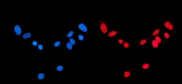
| IF/ICC expectation? | In IF/ICC, expect mainly nucleoplasmic and nuclear-body signal (HPA: subcellular ICC-IF, both supported). This describes the separate IF/ICC application; it does not establish how finely nuclear bodies will resolve in chromogenic paraffin IHC. |
| Epitope and retrieval evidence | The supplied record gives CBX1 domains and phosphosites but no antibody epitope or target-specific retrieval response (UniProt P83916 domains and modified residues). Choose retrieval conditions from the antibody's IHC-P instructions, then assess tissue controls; do not infer a CBX1 fixation effect from these records (standard IHC practice). |
| Situation | Likely cause | Next action |
|---|---|---|
| Known-positive tissue has no nuclear staining. | A failed staining or detection step is possible when a documented high-staining population is blank (HPA: High in bone marrow hematopoietic cells; standard IHC practice). | Confirm the positive tissue and primary-antibody application, then check retrieval and detection against the antibody's IHC-P instructions and run a suitable positive control (standard IHC practice). |
| Color is widespread and nuclei cannot be distinguished. | Background from blocking, washing or chromogen development may obscure the expected nuclear pattern (HPA: ubiquitous nuclear expression; standard IHC practice). | Inspect a section processed without primary antibody; optimize blocking, washing and detection development using that control (standard IHC practice). |
| Signal appears mainly at cell membranes or in cytoplasm. | This conflicts with CBX1's nuclear location and lack of a transmembrane segment; nonspecific staining is a possibility (UniProt P83916 localization and topology). | Compare nuclear signal in a documented positive tissue and examine a no-primary control before assigning the non-nuclear color to CBX1 (HPA: tissue IHC; standard IHC practice). |
| Only one cell population stains in an otherwise intact section. | The restriction differs from the reported broad expression and may reflect uneven staining or an off-target or endogenous signal (UniProt P83916 tissue specificity; HPA: ubiquitous nuclear expression; standard IHC practice). | Check whether staining is nuclear, compare a documented high-staining cell population, and inspect the no-primary control; avoid declaring adjacent cells negative solely by contrast (HPA: tissue IHC; standard IHC practice). |
| A strongly colored nucleus is seen on a mitotic figure. | CBX1 is reported to be unassociated with chromosomes during mitosis, so interphase nuclear localization does not establish a matching chromosome pattern (UniProt P83916 localization). | Base localization assessment on identifiable interphase nuclei and examine whether color on mitotic figures persists in controls (UniProt P83916 localization; standard IHC practice). |
| An IF/ICC image shows nuclear puncta that seem different from IHC. | HPA reports supported localization in both nucleoplasm and nuclear bodies by ICC-IF; the appearance of these structures can differ between fluorescence images and chromogenic sections (HPA: subcellular ICC-IF; standard microscopy practice). | Interpret the IF/ICC image using nucleoplasmic and nuclear-body localization, and score the paraffin IHC section for nuclear staining using its own controls (HPA: subcellular ICC-IF; HPA: tissue IHC; standard IHC practice). |


Comprehensive Human Protein Atlas IHC scoring per tissue (reliability: Supported — High consistency between antibody staining and RNA expression data.). Rows are taken directly from the HPA tissue chart — click any row's HPA link to view the source.
| Tissue | Cell type | Level | Evidence | Source |
|---|---|---|---|---|
| Bone marrow | Hematopoietic cells | High | Protein (IHC) | HPA → |
| Bronchus | Respiratory epithelial cells | High | Protein (IHC) | HPA → |
| Caudate | Glial cells | High | Protein (IHC) | HPA → |
| Cerebellum | Cells in granular layer | High | Protein (IHC) | HPA → |
| Cerebral cortex | Glial cells | High | Protein (IHC) | HPA → |
| Tissue | Cell type | Level | Evidence | Source |
|---|---|---|---|---|
| None in HPA: CBX1 is detected in all 45 scored tissues. Use a no-primary (secondary-only) and an isotype control instead. | ||||
Troubleshoot CBX1 staining in paraffin sections by checking retrieval, nuclear localisation, controls, and scoring against the documented IHC conditions.
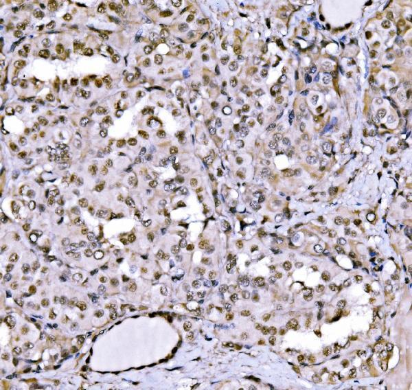
A04206-2 has paraffin-section IHC images from human lung cancer, thyroid papillary carcinoma, mouse intestine and rat brain (catalog IHC captions), plus IF/ICC imaging in CACO-2 cells (catalog IF caption).
A04206-2 is listed for IHC in human, mouse and rat, with paraffin-section images from human lung cancer, human thyroid papillary carcinoma, mouse intestine and rat brain (catalog applications, reactivity and IHC captions). The same SKU is listed for human IF and has a CACO-2 cell image (catalog applications, reactivity and IF caption).
Which to pick: For paraffin-section tissue IHC, choose A04206-2; its own captions document EDTA retrieval at pH 8.0 and primary antibody at 2 μg/ml, while the fixative is unreported (catalog IHC captions). For IF/ICC, A04206-2 has a CACO-2 cell image and a listed human IF concentration of 5 μg/ml (catalog IF caption and datasheet). For cross-species IHC, A04206-2 lists human, mouse and rat reactivity and has paraffin-section images for all three species; its host is rabbit and its clonality is unreported (catalog reactivity, IHC captions and host/clone fields).