CBX2 / Chromobox protein homolog 2 · IHC design guide

Design Immunohistochemistry for CBX2

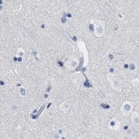
Use late spermatids in testis as a high-signal control for nuclear CBX2 staining (HPA tissue IHC). This guide covers paraffin-section controls, fixation consistency and cell-level chromogenic scoring.

Evidence assembled Oct 2026 · For research use; verify linked source records and product datasheet before use
Immunohistochemistry protocol sheet for CBX2 (IHC for CBX2): expected localisation Nuclear staining in several tissues (HPA tissue IHC), antibody A06356-2, validated IHC image, and IHC protocol steps
Printable CBX2 IHC protocol sheet — expected localisation Nuclear staining in several tissues (HPA tissue IHC), antibody A06356-2, controls and protocol steps. Open the full CBX2 IHC guide →

CBX2 Immunohistochemistry Experimental Design Guide

Expected localisation, validated protocols, controls and antibodies — the at-a-glance facts below, then the full design guide.

Must know before staining
Expected localisation Nuclear staining in several tissues (HPA tissue IHC)
Staining pattern Nuclear staining in several tissues; late spermatids high in testis (HPA tissue IHC)
Antigen retrieval EDTA pH 8.0 HIER, heat-mediated (datasheet A06356-2)
Positive control ⓘ Testis
Negative control ⓘ Adipose tissue+4 more · see all
Important caveats
Reasons your staining may differ from the expected pattern.
Fixation Matched tissue-IHC evidence does not establish the fixation claim. Validate the specimen-specific method before use. (selected-SKU IHC image A06356-2)
Caveat Testis signal varies by cell type; late spermatids are high (HPA tissue IHC)
Regulation RNA enhanced in lymphoid tissue, testis (HPA tissue RNA)
Isoform / epitope 2 isoforms; epitope differences are unreported (UniProt; datasheet A06356-2)
Section 1

Recommended CBX2 IHC & IF Protocols

The catalog antibody’s IHC-P protocol comes from its datasheet (datasheet A06356-2). The four published CBX2 IHC protocols below report the methods available in their excerpts (PMC12804991; PMC6639449; PMC11835617; PMC5403172).

Recommended immunohistochemistry (IHC-P) protocol parameters
SampleParaffin-embedded human the renal pelvis is squamous metaplasia tissue; fixative not specified (datasheet A06356-2)
FixationImage fixative and duration unreported (datasheet A06356-2); verify before use.
Sectioning4–5 µm sections on charged slides (standard)
DeparaffinisationXylene, graded ethanol series to water (standard)
Antigen retrievalHeat retrieval: EDTA pH 8.0 (datasheet A06356-2); 20 min, 95–100 °C (standard)
Peroxidase block3% H2O2, 10 min, room temperature (standard)
Blocking10% goat serum (datasheet A06356-2)
Primary antibodyRabbit anti-CBX2, 2-5 μg/ml (datasheet A06356-2)
Primary incubationOvernight at 4 °C (datasheet A06356-2)
DetectionStreptavidin-biotin complex (SABC), DAB chromogen (datasheet A06356-2)
CounterstainHematoxylin, blue, dehydrate and mount (standard)
Expected resultCBX2-positive staining in elongated or late spermatids of testis (HPA tissue IHC: High). HPA tissue profile: Nuclear expression in several tissues, including testis. No signal in the no-primary control.
💡Decision noteStart with EDTA pH 8.0 heat retrieval for the catalog antibody (datasheet A06356-2). The published citrate protocols provide alternatives when adapting their methods (PMC12804991; PMC11835617; PMC5403172).
Section 2

What Is the Expected CBX2 Staining Pattern?

CBX2 is a nuclear, chromatin-associated protein with no transmembrane segment (UniProt Q14781: location and topology). In paraffin-section IHC, expect nuclear staining in several tissues, with high staining in elongated or late spermatids of testis (HPA tissue IHC: Approved; testis, High). Interpret staining by cell type and compartment; HPA reports several cell types with no detected signal (HPA tissue IHC: negative entries).

What am I looking at on my slide?
Strong staining in elongated or late spermatids, concentrated in nuclei.This fits the reported high testicular cell staining and the broader nuclear tissue pattern (HPA tissue IHC: testis, High; profile). UniProt places CBX2 in the nucleus and on chromosomes, including chromatin foci (UniProt Q14781: subcellular location). Score the relevant cells and their nuclear signal, rather than judging the whole section by its overall darkness.
Predominantly cytoplasmic or membrane-like staining, with little nuclear signal.Treat this as a localization mismatch: CBX2 is nuclear and chromatin-associated, with no transmembrane segment (UniProt Q14781: location and topology). Review the negative control, detection background and staining distribution before assigning it to CBX2 (general IHC practice).
Strong stain in a cell type recorded as not detected, such as adipocytes.Adipocytes are listed as not detected (HPA tissue IHC: adipose tissue). Consider cross-reactivity or endogenous detection activity, especially if staining also appears in controls (general IHC practice). A negative HPA entry is a useful comparator for this cell type, but does not establish that every specimen must be blank.
Diffuse color covers nuclei, cytoplasm and spaces between cells.This distribution does not match the reported nuclear tissue pattern or nucleoplasmic IF localization (HPA tissue IHC: profile; HPA subcellular: supported). Examine primary-antibody omission and detection controls, wash quality and counterstain before interpreting faint nuclear color beneath the background (general IHC practice).
No stain in testis elongated or late spermatids.Absence of signal in the reported high cell population makes the run inconclusive for CBX2 detection (HPA tissue IHC: testis, High). Check tissue identity and preservation, then the antibody and chromogenic detection steps using appropriate run controls (general IHC practice). Do not score other tissues as negative until the positive control works.
💡Expected CBX2 appearanceCall a result consistent with CBX2 when high staining marks nuclei of elongated or late spermatids in testis (HPA tissue IHC: testis, High; nuclear profile); broad cytoplasmic or membrane-like color is a localization mismatch (UniProt Q14781: location and topology).
How each factor affects the staining
Subnuclear distributionUniProt describes distinct chromatin foci and chromocenters; HPA IF reports a supported nucleoplasmic location (UniProt Q14781: location; HPA subcellular: supported). Nuclear signal may therefore look uneven. The IF record does not specify how clearly individual foci should resolve in chromogenic paraffin-section IHC.
Cell-specific tissue patternHPA reports high staining in elongated or late spermatids, low staining in several other named cell types, and no detected staining in others (HPA tissue IHC: positive, low and negative entries). Match the observed cell population to its entry before treating a weak field or a dark neighboring cell as a discrepancy.
Strength of IHC evidenceThe tissue IHC reliability label is Approved, with external characterization supporting antibody staining (HPA tissue IHC: reliability). The listed antibody HPA023083 is Approved for IHC and Supported for ICC (HPA antibodies). These labels support interpretation of the reported patterns; they do not validate every staining condition or specimen.
Isoforms and antibody epitopeUniProt lists two CBX2 isoforms (UniProt Q14781: isoforms). The supplied evidence does not locate this antibody's epitope or show whether its IHC signal distinguishes isoforms. Report a nuclear CBX2 staining pattern without attributing it to a particular isoform.
Why is my staining missing, weak or wrong?
SituationLikely causeNext action
The positive testis control is blank.The expected high cell population is absent from the section, or the IHC run failed (HPA tissue IHC: testis, High; general IHC practice).Confirm that elongated or late spermatids are present; review the antibody, retrieval and detection steps against the run controls (general IHC practice). Interpret study negatives only after the control shows the expected staining.
Signal is mainly outside nuclei.The distribution conflicts with CBX2's nuclear and chromatin location (UniProt Q14781: subcellular location).Check a primary-antibody omission control and inspect the chromogen and counterstain separately (general IHC practice). If extranuclear color persists, record it as nonspecific-looking staining rather than a CBX2 localization result.
A listed negative cell type stains strongly.Cross-reactivity or endogenous detection activity may explain an unexpected signal (general IHC practice; HPA tissue IHC: negative entries).Compare the same cell type in control and study sections; check the detection-system controls and repeat with an independently validated antibody if available (general IHC practice). Do not transfer HPA's cell-specific negative call to an entire organ.
Background obscures nuclear detail.Diffuse detection color can hide the compartment that matters for CBX2 interpretation (general IHC practice; HPA tissue IHC: nuclear profile).Use omission and detection controls to locate the background source; review blocking, washes and detection exposure using the assay's instructions (general IHC practice). Reassess only after nuclei can be distinguished.
Nearly every cell appears equally nuclear-positive.That uniform result needs scrutiny against HPA's cell-specific high, low and not-detected entries (HPA tissue IHC: positive, low and negative entries).Compare named cell populations within the section and include a listed negative comparator where available (HPA tissue IHC: negative entries; general IHC practice). Check counterstain and nonspecific nuclear background before assigning CBX2 positivity.
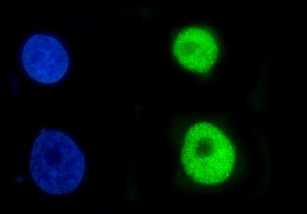
Q: What should an IF/ICC image show?HPA reports supported nucleoplasmic localization in ICC-IF, while UniProt also describes chromatin foci (HPA subcellular: supported; UniProt Q14781: location).A: Look for nuclear, chiefly nucleoplasmic signal and compare it with a nuclear counterstain (HPA subcellular: supported; general IF practice). Use the separate IF/ICC guide for assay setup; this IHC tissue pattern does not supply an IF protocol.

Sample controls for CBX2 IHC & IF

🧪Run testis first and expect staining in elongated or late spermatids (HPA: High); use adipose tissue as the negative tissue and expect adipocytes to lack detectable staining (HPA: Not detected). On the testis slide, use any unstained cells as an internal background reference, without assuming a particular cell type is CBX2-negative (HPA: positive cell type specified only as elongated or late spermatids).
Positive control tissue: Testis (Elongated or late spermatids, HPA High)
Negative control tissue: Adipose tissue (HPA Not detected)
ICC-IF cell lines (HPA subcellular resource): HPA ICC-IF images show CBX2 in A-431, U-251MG, U2OS, KOLF2.1J, with annotated localisation: Nucleoplasm (supported) (HPA subcellular).
Technical controls: Include a no-primary, secondary-only control, a host- and isotype-matched nonimmune rabbit IgG control for the catalog antibody, and CBX2-knockout material as a biological negative (hero caption: rabbit primary; standard IHC practice). Quench endogenous peroxidase and check for endogenous biotin before interpreting DAB staining in testis, because the caption uses biotinylated secondary antibody and a streptavidin–biotin detection system (hero caption; standard IHC practice).
⚠️Feasibility: A target-specific fixation window or fixation effect is unreported, and the selected A06356-2 tissue-IHC caption does not state the fixative (hero caption). The demonstrated paraffin-section procedure uses heat retrieval in EDTA at pH 8.0, but the evidence does not establish whether retrieval is required or whether frozen sections or IF are easier (hero caption). Judge testis staining by the specified spermatid cell type and nuclear localization, while controlling for testis background and biotin-related signal (HPA: High in elongated or late spermatids; UniProt Q14781: nucleus and chromatin foci; hero caption: biotin-based DAB detection).

HPA tissue IHC evidence for CBX2

Comprehensive Human Protein Atlas IHC scoring per tissue (reliability: Approved — External characterization data supports antibody staining.). Rows are taken directly from the HPA tissue chart — click any row's HPA link to view the source.

Positive expression · recommended positive controls

TissueCell typeLevelEvidenceSource
Testis Elongated or late spermatids High Protein (IHC) HPA →

Undetected expression · recommended negative controls

TissueCell typeLevelEvidenceSource
Adipose tissue Adipocytes Not detected Protein (IHC) HPA →
Adrenal gland Glandular cells Not detected Protein (IHC) HPA →
Caudate Glial cells Not detected Protein (IHC) HPA →
Cerebellum Cells in granular layer Not detected Protein (IHC) HPA →
Cerebral cortex Endothelial cells Not detected Protein (IHC) HPA →
Section 3

Advanced CBX2 IHC Tips

Troubleshoot CBX2 staining in paraffin sections by checking retrieval, nuclear localisation, controls and cell-specific scoring.

How should I retrieve CBX2 in paraffin sections with weak nuclear staining?
Start with heat-mediated EDTA retrieval at pH 8.0 for the catalog antibody (datasheet A06356-2). Its paraffin-section caption pairs that retrieval with 2 μg/ml primary antibody overnight at 4°C, so keep those conditions consistent while investigating weak signal (datasheet A06356-2). Compare a known positive control and a no-primary control on the same run, then adjust heating time in small increments if nuclear signal remains weak (standard IHC practice). Evaluate whether staining sharpens within nuclei; CBX2 is associated with chromatin foci, and diffuse nonnuclear colour should not be rescued by more retrieval alone (UniProt Q14781 localisation).
Could fixation explain inconsistent CBX2 staining between paraffin blocks?
The catalog image identifies a paraffin-embedded section but does not state its fixative, so target-specific CBX2 fixation sensitivity is unknown (datasheet A06356-2). Record each block’s actual fixative, fixation duration and processing history before comparing staining across blocks (standard IHC practice). Run sections from the same block together using EDTA at pH 8.0 and the same primary incubation, 2 μg/ml overnight at 4°C, to limit procedural variation (datasheet A06356-2). If nuclei vary between blocks, assess tissue preservation and retrieval consistency with an appropriate control before attributing the difference to CBX2 abundance (standard IHC practice).
What localisation should count as convincing CBX2 staining in IHC?
Score CBX2 primarily in nuclei: tissue IHC reports nuclear expression, while the protein record describes chromatin foci and chromocenters (HPA tissue IHC; UniProt Q14781 localisation). Nucleoplasmic localisation is also supported by cellular imaging, although individual foci may be difficult to resolve with chromogenic detection (HPA subcellular; standard IHC practice). Compare the DAB signal with a nuclear counterstain and count only cells whose nuclear boundary and staining can be assigned confidently (standard IHC practice). Because CBX2 has no transmembrane segment, a predominantly membranous pattern should prompt checks for nonspecific staining and tissue artefact (UniProt Q14781 topology; standard IHC practice).
Can isoforms or epitope access account for different CBX2 staining patterns?
CBX2 has 2 annotated isoforms and a chromodomain at residues 12–70, but the supplied antibody caption does not identify its epitope (UniProt Q14781 isoforms and domains; datasheet A06356-2). Do not assign an isoform to stained nuclei or assume that retrieval exposes both isoforms equally without epitope mapping (standard IHC practice). Compare adjacent sections under the same EDTA pH 8.0 retrieval and primary conditions before interpreting a changed pattern (datasheet A06356-2; standard IHC practice). If isoform specificity matters, seek documented immunogen or epitope information and verify the assignment with an independent method (standard IHC practice).
How can IF help assess ambiguous CBX2 IHC localisation?
Use IF on a separate, appropriately validated sample to examine whether CBX2 signal overlaps nuclei or chromatin-associated regions seen in IHC (UniProt Q14781 localisation; standard IF practice). Multiplex with a marker for the expected cell population, such as a validated spermatid marker when examining testis, where high staining is reported in elongated or late spermatids (HPA tissue IHC; standard IF practice). Choose a fluorophore whose emission avoids the sample’s measured autofluorescence, and include single-colour controls when channels overlap (standard IF practice). Because CBX2 has no transmembrane segment and is nuclear, validate permeabilisation sufficient for antibody access to the nuclear epitope (UniProt Q14781 topology and localisation; standard IF practice).
How do I reduce diffuse brown background without losing nuclear CBX2 signal?
First compare the stained section with no-primary and known positive controls to distinguish detection background from a weak nuclear signal (standard IHC practice). The catalog example used a 10% goat-serum block, 2 μg/ml rabbit primary overnight at 4°C, and biotinylated secondary for 30 minutes at 37°C (datasheet A06356-2). Check peroxidase blocking, washes and DAB development time, and assess endogenous biotin when using a biotin-based detection system (standard IHC practice; datasheet A06356-2 detection). Reduce development or primary concentration systematically if background persists, while preserving interpretable nuclear staining in the positive control (standard IHC practice; HPA tissue IHC).
How should I quantify CBX2 across samples with different cellular composition? ⚠ ANSWER MARKED FOR VERIFICATION
Define the cell population and analysis area before scoring, then record the percentage of CBX2-positive nuclei and nuclear intensity (standard IHC practice; HPA tissue IHC nuclear profile). An H-score can combine intensity categories with their percentages, while positive nuclei per mm² is useful when cell density differs (standard IHC practice). Normalise to the number of evaluable nuclei within the same annotated cell compartment, and report excluded necrotic or damaged areas (standard IHC practice). Keep retrieval, imaging and thresholds consistent across batches, and report cell-type composition because high staining is documented in elongated or late spermatids in testis (datasheet A06356-2 retrieval; HPA tissue IHC).
When is apparent CBX2 positivity more likely to be an artefact?
A credible result should place signal in nuclei and, when morphology permits, align it with the cell population being assessed; CBX2 is a nuclear chromatin-associated protein (HPA tissue IHC; UniProt Q14781 localisation). High staining in testis elongated or late spermatids offers a documented positive reference, whereas adipocytes are reported as not detected and can inform comparison in a matched assay (HPA tissue IHC). Treat dominant cytoplasmic or membranous colour, section-edge staining, and staining confined to necrotic areas as reasons to review specificity and tissue quality (UniProt Q14781 topology and localisation; standard IHC practice). If colour persists without primary antibody, investigate endogenous enzyme activity or detection-system background before calling cells positive (standard IHC practice).
Boster reagents

Best CBX2 / Chromobox protein homolog 2 IHC Antibodies

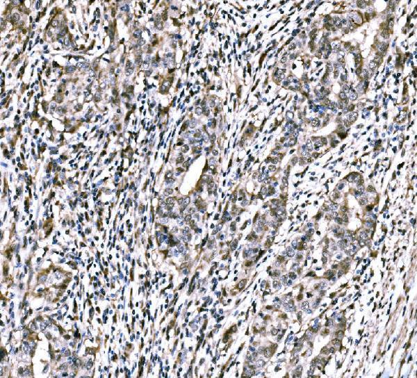
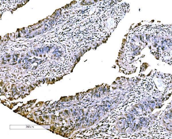
A06356-2 has IHC images from human renal pelvis, rectum and placenta, and mouse testis (catalog IHC captions); IF/ICC data are from A431 cells (catalog IF caption).

Real IHC data IHC analysis of CBX2 using anti-CBX2 antibody (A06356-2). CBX2 was detected in a paraffin-embedded section of human the renal pelvis is squamous metaplasia tissue. Heat mediated antigen retrieval was performed in EDTA buffer (pH 8.0, epitope retrieval solution). The tissue section was blocked with 10% goat serum. The tissue section was then incubated with 2 μg/ml rabbit anti-CBX2 Antibody (A06356-2) overnight at 4°C. Biotinylated goat anti-rabbit IgG was used as secondary antibody and incubated for 30 minutes at 37°C. The tissue section was developed using Strepavidin-Biotin-Complex (SABC) (Catalog # SA1022) with DAB as the chromogen.
Anti-CBX2 Antibody ®
Cat # A06356-2

A06356-2 is listed for human and mouse IHC (catalog applications/reactivity), with paraffin-section images from human renal pelvis, rectum and placenta, and mouse testis (A06356-2 IHC captions). A06356-2 is also listed for IF/ICC (catalog applications), with an IF image from A431 cells (A06356-2 IF caption).

Which to pick: For tissue IHC, choose A06356-2 at a starting concentration within 2–5 μg/ml (datasheet: IHC dilution); its IHC images use paraffin sections, and the fixative is unreported (A06356-2 IHC captions). For IF/ICC, A06356-2 has an A431-cell image at 5 μg/ml (A06356-2 IF caption). For human and mouse work, A06356-2 lists both species as reactive and has a mouse testis IHC image; its clone is unreported (catalog reactivity/clone; A06356-2 IHC caption).

Each figure is that product's own IHC / IF validation image from its datasheet.

References

  1. UniProt Consortium. UniProt entry Q14781 (CBX2_HUMAN, Chromobox protein homolog 2).
  2. Human Protein Atlas. CBX2 tissue IHC expression (reliability: Approved).
  3. Human Protein Atlas. CBX2 subcellular location (ICC-IF): Localized to the nucleoplasm..
  4. Human Protein Atlas. CBX2 antibody validation summary (1 antibodies).
  5. CBX2 promoted oral squamous cell carcinoma via increasing CEP55/NF-κB/METTL3/SHP2 signaling induced metastasis/proliferation and angiogenesis. Scientific reports 2025 — PMC12804991.
  6. CBX2 is a functional target of miRNA let-7a and acts as a tumor promoter in osteosarcoma. Cancer medicine 2019 — PMC6639449.
  7. CBX2 promotes cervical cancer cell proliferation and resistance to DNA-damaging treatment via maintaining cancer stemness. The Journal of biological chemistry 2025 — PMC11835617.
  8. Chromobox homolog 2 protein: A novel biomarker for predicting prognosis and Taxol sensitivity in patients with breast cancer. Oncology letters 2017 — PMC5403172.
  9. PubMed PMID:16625196 — UniProt-cited evidence.
  10. PubMed PMID:15489334 — UniProt-cited evidence.
  11. PubMed PMID:7782071 — UniProt-cited evidence.