This website uses cookies to ensure you get the best experience on our website.


- Table of Contents
Plan CBX8 staining in paraffin sections using nuclear localisation as the molecular expectation (UniProt) and nuclear plus cytoplasmic staining as the observed tissue pattern (HPA tissue IHC). Rectal glandular cells stain strongly, while ovarian stromal cells are reported as undetected (HPA tissue IHC).

Expected localisation, validated protocols, controls and antibodies — the at-a-glance facts below, then the full design guide.
| Expected localisation | Nuclear expected (UniProt); nuclear and cytoplasmic staining observed (HPA tissue IHC) | |
| Staining pattern | General cytoplasmic and nuclear staining (HPA tissue IHC) | |
| Antigen retrieval | EDTA pH 8.0 HIER, heat-mediated (datasheet A05234-2) | |
| Positive control | Bone marrow+4 more · see all | |
| Negative control | Hippocampus+3 more · see all |
| Fixation | Keep fixation consistent across sections. (standard IHC practice; not target-specific) | |
| Caveat | Antibody staining and RNA show medium consistency (HPA tissue IHC) | |
| Regulation | Low tissue specificity (HPA tissue RNA) | |
| Isoform / epitope | No annotated isoforms; one 1–389 chain (UniProt) |
The catalog antibody protocol is followed by published CBX8 IHC methods for hepatocellular carcinoma and colorectal cancer specimens (PMC6361915; PMC4279409).
| Sample | Paraffin-embedded human rectal cancer tissue; fixative not specified (datasheet A05234-2) |
| Fixation | Image fixative and duration unreported (datasheet A05234-2); verify before use. |
| Sectioning | 4–5 µm sections on charged slides (standard) |
| Deparaffinisation | Xylene, graded ethanol series to water (standard) |
| Antigen retrieval | Heat retrieval: EDTA pH 8.0 (datasheet A05234-2); 20 min, 95–100 °C (standard) |
| Peroxidase block | 3% H2O2, 10 min, room temperature (standard) |
| Blocking | 10% goat serum (datasheet A05234-2) |
| Primary antibody | Rabbit anti-CBX8, 0.5-1μg/ml (datasheet A05234-2) |
| Primary incubation | Overnight at 4 °C (datasheet A05234-2) |
| Detection | Streptavidin-biotin complex (SABC), DAB chromogen (datasheet A05234-2) |
| Counterstain | Hematoxylin, blue, dehydrate and mount (standard) |
| Expected result | CBX8-positive staining in hematopoietic cells of bone marrow (HPA tissue IHC: High). HPA tissue profile: General cytoplasmic and nuclear expression. No signal in the no-primary control. |
CBX8 is a nuclear protein with no transmembrane segment (UniProt Q9HC52: subcellular location and topology). In paraffin-section IHC, HPA reports general cytoplasmic and nuclear staining, with high staining in colon glandular cells, bone-marrow hematopoietic cells and several other specified cell populations (HPA: tissue IHC). Treat the pattern as a guide to interpretation: HPA rates its IHC evidence Approved, with medium consistency between staining and RNA expression (HPA: tissue IHC reliability).
| Nuclear staining in colon glandular cells, with some cytoplasmic staining. | This fits the reported high level in colon glandular cells and HPA’s general nuclear-and-cytoplasmic IHC profile (HPA: tissue IHC). Nuclear localisation also agrees with UniProt (UniProt Q9HC52: subcellular location). Compare cells within the same section before judging intensity. |
| Strong cytoplasmic staining with little or no nuclear signal. | Interpret cautiously: HPA reports a cytoplasmic component in tissue IHC, but UniProt places CBX8 in the nucleus and HPA ICC-IF localises it to the nucleoplasm (HPA: tissue IHC and subcellular; UniProt Q9HC52). Review controls before accepting a cytoplasm-only result. |
| Staining in cholangiocytes, while the expected cell population is unclear. | HPA lists liver cholangiocytes as not detected, so staining there warrants review for cross-reactivity or endogenous chromogen activity (HPA: tissue IHC; general IHC practice). That observation alone does not establish either cause or mean that every liver cell must be negative. |
| Broad, diffuse colour obscures nuclei and tissue boundaries. | The distribution cannot be scored reliably for the reported CBX8 compartments or cell populations (HPA: tissue IHC). Inspect a control without primary antibody and assess blocking, washing and detection background before assigning cellular staining (general IHC practice). |
| No signal in colon glandular cells or bone-marrow hematopoietic cells. | Both are listed as High by HPA, making them useful reference populations for a failed or weak run (HPA: tissue IHC). Check that the reference cells are present, then review detection and the catalog antibody’s IHC-P instructions (general IHC practice). |
| Compartment evidence | UniProt assigns CBX8 to the nucleus, and HPA ICC-IF refines this to the nucleoplasm; HPA tissue IHC reports nuclear plus cytoplasmic staining (UniProt Q9HC52; HPA: subcellular and tissue IHC). Use this distinction when evaluating a cytoplasm-only IHC result. |
| Cell-population reference | HPA reports High staining in rectal glandular cells, skin melanocytes and round or early testicular spermatids, among other listed populations (HPA: tissue IHC). Its not-detected calls apply to named populations, including hippocampal glial cells and liver cholangiocytes, rather than entire organs (HPA: tissue IHC). |
| Evidence strength | The tissue IHC profile is Approved with medium staining-to-RNA consistency; the listed antibody HPA031462 has IHC Approved and ICC Enhanced status (HPA: tissue IHC and antibodies). ICC enhancement does not upgrade the IHC validation status, so assess each application against its own evidence. |
| Protein structure and epitope limits | UniProt records one chain spanning residues 1–389, a chromo domain at 11–69, no annotated isoforms and no signal peptide or propeptide (UniProt Q9HC52). These annotations do not identify this antibody’s epitope or establish a CBX8-specific antigen-retrieval condition. |
| Chromogenic background | Endogenous enzyme activity or nonspecific detection can contribute colour independently of primary-antibody binding (general IHC practice). A control without primary antibody helps distinguish this background from the cell-specific HPA pattern; it cannot by itself prove that all remaining staining is CBX8-specific (HPA: tissue IHC; general IHC practice). |
| Situation | Likely cause | Next action |
|---|---|---|
| High reference tissue stains weakly or not at all. | The expected cells may be absent from the section, or the IHC detection run may have failed (HPA: High in colon glandular cells and bone-marrow hematopoietic cells; general IHC practice). | Confirm the named cells are present; check the catalog antibody’s IHC-P instructions, reagent activity and detection controls before interpreting the sample as CBX8-negative (general IHC practice). |
| Signal appears only in cytoplasm. | HPA tissue IHC includes cytoplasmic staining, while UniProt and HPA ICC-IF support a nuclear location (HPA: tissue IHC and subcellular; UniProt Q9HC52). The discrepancy alone does not identify its cause. | Compare nuclear and cytoplasmic signal in a reported High cell population and inspect the control without primary antibody; qualify a cytoplasm-only result if nuclear signal remains absent (HPA: tissue IHC; general IHC practice). |
| A not-detected reference population stains strongly. | For example, HPA reports ovarian stroma cells as not detected; cross-reactivity or endogenous detection activity are possible explanations, not established diagnoses (HPA: tissue IHC; general IHC practice). | Verify the stained cell identity, compare a reported High population in the same run and inspect the control without primary antibody before assigning the signal to CBX8 (HPA: tissue IHC; general IHC practice). |
| Brown colour spreads across tissue without clear cell boundaries. | Diffuse deposition or nonspecific detection may prevent compartment-level scoring (general IHC practice). It cannot be matched confidently to HPA’s reported nuclear-and-cytoplasmic cell pattern (HPA: tissue IHC). | Inspect the control without primary antibody and review blocking, washing and chromogen development under the IHC workflow; score only interpretable cellular staining (general IHC practice). |
| A low-staining population is treated as a negative control. | HPA labels lung macrophages, small-intestinal glandular cells and adipocytes Low, rather than not detected (HPA: tissue IHC). Residual staining in these populations is therefore compatible with the supplied profile. | Use HPA’s named not-detected populations for a closer comparison, while keeping the call specific to those cells; retain a separate control without primary antibody for detection background (HPA: tissue IHC; general IHC practice). |
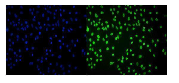
| IF/ICC question: What location should a separate fluorescence image show? | HPA’s ICC-IF summary localises CBX8 to the nucleoplasm, and HPA031462 has ICC Enhanced status (HPA: subcellular and antibodies). These are fluorescence observations, separate from the Approved tissue IHC profile. | Look for nucleoplasmic signal when interpreting IF/ICC images (HPA: subcellular); use the dedicated IF/ICC guide for that application, while interpreting paraffin IHC against the tissue IHC evidence (HPA: tissue IHC). |


Comprehensive Human Protein Atlas IHC scoring per tissue (reliability: Approved — Medium consistency between antibody staining and RNA expression data.). Rows are taken directly from the HPA tissue chart — click any row's HPA link to view the source.
| Tissue | Cell type | Level | Evidence | Source |
|---|---|---|---|---|
| Bone marrow | Hematopoietic cells | High | Protein (IHC) | HPA → |
| Bronchus | Ciliated cells (cell body) | High | Protein (IHC) | HPA → |
| Colon | Glandular cells | High | Protein (IHC) | HPA → |
| Fallopian tube | Ciliated cells (cell body) | High | Protein (IHC) | HPA → |
| Nasopharynx | Ciliated cells (cell body) | High | Protein (IHC) | HPA → |
| Tissue | Cell type | Level | Evidence | Source |
|---|---|---|---|---|
| Hippocampus | Glial cells | Not detected | Protein (IHC) | HPA → |
| Liver | Cholangiocytes | Not detected | Protein (IHC) | HPA → |
| Ovary | Ovarian stroma cells | Not detected | Protein (IHC) | HPA → |
| Parathyroid gland | Glandular cells | Not detected | Protein (IHC) | HPA → |
Troubleshoot CBX8 staining in paraffin sections by checking retrieval, nuclear localisation, detection controls and cell-specific scoring.
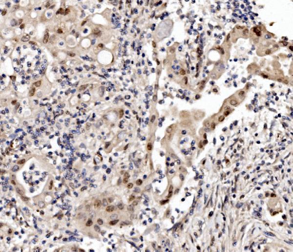
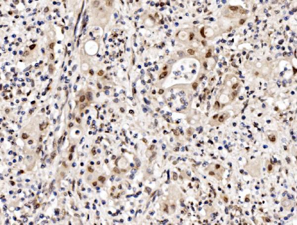
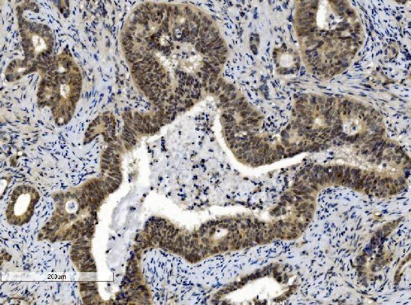
The catalog documents CBX8 staining in human paraffin sections and cells, plus mouse and rat paraffin sections (A05234-2 and M05234 IHC captions; A05234-2 IF captions).
A05234-2 has IHC data from human rectal cancer and mouse and rat small intestine paraffin sections, plus IF data from A549 cells and human rectal cancer paraffin sections (A05234-2 image captions). M05234 has IHC data from human gallbladder adenocarcinoma paraffin sections (M05234 IHC caption).
Which to pick: For tissue IHC, choose A05234-2 for human, mouse or rat paraffin sections (A05234-2 IHC captions), or monoclonal M05234 for human paraffin sections (M05234 catalog entry and IHC caption). For IF/ICC, choose A05234-2, which lists both applications and has human cell and paraffin-section IF images (A05234-2 catalog entry and IF captions). For cross-species IHC, choose A05234-2 because its IHC captions show all three species (A05234-2 IHC captions); the fixative is unreported in both products’ paraffin-section captions (A05234-2 and M05234 IHC captions).