CCK / Cholecystokinin · IHC design guide

Design Immunohistochemistry for CCK

Plan CCK chromogenic IHC in paraffin sections using intestinal endocrine cells and hypothalamic projections as reference staining patterns (HPA tissue IHC). This guide covers fixation consistency, staining interpretation and the implications of a secreted, processed peptide (UniProt).

Evidence assembled Oct 2026 · For research use; verify linked source records and product datasheet before use
Immunohistochemistry protocol sheet for CCK (IHC for CCK): expected localisation Secreted (UniProt); endocrine cells and neuronal projections (HPA tissue IHC), antibody A01098, validated IHC image, and IHC protocol steps
Printable CCK IHC protocol sheet — expected localisation Secreted (UniProt); endocrine cells and neuronal projections (HPA tissue IHC), antibody A01098, controls and protocol steps. Open the full CCK IHC guide →

CCK Immunohistochemistry Experimental Design Guide

Expected localisation, validated protocols, controls and antibodies — the at-a-glance facts below, then the full design guide.

Must know before staining
Expected localisation Secreted (UniProt); endocrine cells and neuronal projections (HPA tissue IHC)
Staining pattern Intestinal endocrine cells and hypothalamic projections (HPA tissue IHC)
Antigen retrieval Citrate pH 6.0 HIER, 95–98 °C, 20 min (rule: cytoplasmic / membrane antigen)
Positive control ⓘ Duodenum+2 more · see all
Negative control ⓘ Adipose tissue+4 more · see all
Important caveats
Reasons your staining may differ from the expected pattern.
Fixation Keep fixation consistent across paraffin sections. (standard IHC practice; not target-specific)
Caveat Secretion can separate CCK protein from its RNA source (HPA tissue IHC)
Regulation No expression regulator specified (UniProt)
Isoform / epitope No isoforms annotated; processing may affect epitopes (UniProt)
Section 1

Recommended CCK IHC & IF Protocols

The catalog antibody’s IHC-P protocol is paired with published CCK IHC procedures for rat brain, myocardial tissue, and white sea bream gut (PMC3638803; PMC8405328; PMC3527491).

Recommended immunohistochemistry (IHC-P) protocol parameters
SampleParaffin-embedded human colorectal carcinoma tissue; fixative not specified (datasheet A01098)
FixationImage fixative and duration unreported (datasheet A01098); verify before use.
Sectioning4–5 µm sections on charged slides (standard)
DeparaffinisationXylene, graded ethanol series to water (standard)
Antigen retrievalHeat-induced epitope retrieval in citrate buffer, pH 6.0, 20 min at 95–98 °C (standard rule: cytoplasmic / membrane antigen)
Peroxidase block3% H2O2, 10 min, room temperature (standard)
Blocking10% normal serum of the secondary host, 30 min, room temperature (standard)
Primary antibodyRabbit anti-CCK, 1:50-1:200 (datasheet A01098)
Primary incubationOvernight at 4 °C (standard)
DetectionHRP-polymer secondary, DAB chromogen 5–10 min (standard)
CounterstainHematoxylin, blue, dehydrate and mount (standard)
Expected resultCCK-positive staining in endocrine cells of duodenum (HPA tissue IHC: High). HPA tissue profile: Expressed in hypothalamus and neuroendocrine cells in small intestine and duodenum. No signal in the no-primary control.
💡Decision noteFor paraffin sections, start with citrate HIER at pH 6.0, 95–98 °C for 20 min (page antigen-retrieval rule).
Section 2

What Is the Expected CCK Staining Pattern?

CCK is a secreted peptide with no transmembrane segment (UniProt P06307 topology). In paraffin-section IHC, expect staining in duodenal and small-intestinal endocrine cells and hypothalamic neuronal projections, all reported as High (HPA tissue IHC). HPA rates the tissue profile Enhanced, while noting that secretion can make protein staining differ from tissue RNA distribution (HPA tissue IHC).

What am I looking at on my slide?
Strong staining in scattered duodenal or small-intestinal endocrine cells, or in hypothalamic neuronal projections (HPA tissue IHC).This matches the reported High cell-specific patterns (HPA tissue IHC). Assess the labeled cells or projections, rather than treating uniform staining across the section as the expected result.
Predominantly nuclear staining in the expected positive cells.A nuclear pattern is unexpected for a secreted peptide without a transmembrane segment (UniProt P06307 topology). Check antibody specificity and the detection controls before interpreting it as CCK.
Strong staining in unrelated cells, including adipocytes in adipose tissue or glandular cells in adrenal gland (HPA tissue IHC).Those cell populations are reported as Not detected (HPA tissue IHC). Cross-reactivity or endogenous chromogenic detection activity is possible; compare with a no-primary control.
Diffuse color across cells, stroma, and section edges, with little distinction between labeled and unlabeled structures.This is background rather than the restricted endocrine-cell and projection pattern (HPA tissue IHC). Review blocking, washes, chromogen development, and section handling as general IHC checks.
No staining in duodenal or small-intestinal endocrine cells, or hypothalamic neuronal projections (HPA tissue IHC).A negative result in a reported High-positive population needs a run check (HPA tissue IHC). Review the catalog antibody's IHC-P instructions and positive control before concluding that the sample lacks CCK.
💡Expected CCK appearanceCall the IHC result positive when distinct, strong staining marks duodenal or small-intestinal endocrine cells or hypothalamic neuronal projections (HPA tissue IHC); uniform nuclear or unrelated-cell staining is suspect (UniProt P06307 topology; HPA tissue IHC).
How each factor affects the staining
Secretion and tissue contextCCK is secreted (UniProt P06307), and HPA cautions that tissue RNA and protein locations may differ (HPA tissue IHC); score the observed cell pattern rather than requiring RNA and protein maps to coincide.
Precursor processing and epitopeCCK has a signal peptide, annotated propeptide regions, and modified residues (UniProt P06307). The supplied record gives no antibody epitope, so it cannot establish which processed forms the IHC stain detects.
Strength of IHC evidenceThe tissue profile is Enhanced; HPA lists HPA069515 as IHC Enhanced and HPA045039 as IHC Supported (HPA tissue IHC; HPA antibodies). Validation status supports interpretation but does not make every sample positive.
IF/ICC: is the same pattern established?HPA calls CCK Secreted but provides no ICC-IF main location or cell-line images (HPA subcellular). Use the IHC tissue pattern as context; a specific IF compartment pattern is unverified in this payload.
Why is my staining missing, weak or wrong?
SituationLikely causeNext action
A reported High-positive tissue has no visible signal (HPA tissue IHC).The run may have missed a detectable endocrine-cell or projection signal; the supplied sources do not identify a CCK-specific fixation effect.Check the catalog antibody's IHC-P instructions, positive-control section, detection reagents, and counterstain; adjust general IHC steps only after confirming the run.
Color is broad and weak instead of restricted to expected cells or projections (HPA tissue IHC).Nonspecific background or excess chromogen development may obscure the tissue pattern; this is a general IHC interpretation, not an HPA-reported CCK mechanism.Compare no-primary and positive controls, then review blocking, washes, antibody concentration, and chromogen development as general IHC checks.
Adipocytes in adipose tissue or adrenal glandular cells stain strongly (HPA tissue IHC).Both populations are reported as Not detected (HPA tissue IHC); cross-reactivity or endogenous detection activity is possible.Inspect a no-primary control for endogenous signal and compare the same run with a reported High-positive tissue (HPA tissue IHC).
A hypothalamus section lacks clearly labeled neuronal projections (HPA tissue IHC).The expected High pattern is in projections, so a whole-field score can miss the relevant structures (HPA tissue IHC).Review projection-rich areas and morphology, then compare a positive control before assigning a negative score; avoid calling unrelated nuclear color positive.
A low-signal brain or epithelial sample appears negative.HPA reports Low staining in cerebral cortical and hippocampal neuronal cells, bronchial respiratory epithelium, and bladder urothelium (HPA tissue IHC).Use a reported High-positive tissue to assess run performance; do not use a Low population alone to judge antibody failure (HPA tissue IHC).
IF/ICC gives a compartment pattern that differs from the IHC slide.HPA supplies no ICC-IF main location or cell-line images for CCK, so that IF pattern lacks confirmation here (HPA subcellular).Treat the IF result as provisional and assess its controls separately; retain the reported endocrine-cell and hypothalamic projection patterns as the IHC reference (HPA tissue IHC).

Sample controls for CCK IHC & IF

🧪Run duodenum first: its endocrine cells should stain for CCK (HPA: High in duodenal endocrine cells). Use appendix endocrine cells as a negative tissue comparator (HPA: Not detected in appendix endocrine cells); adjacent non-endocrine cells on the duodenal slide should show only background staining relative to the endocrine cells (HPA: High in duodenal endocrine cells).
Positive control tissue: Duodenum (Endocrine cells, HPA High)
Negative control tissue: Adipose tissue (HPA Not detected)
ICC-IF cell lines (HPA subcellular resource): HPA carries no ICC-IF cell line for CCK; derive a cell-line control from the positive tissue's cell type (Endocrine cells) and confirm it by RNA or western blot first.
Technical controls: Include no-primary (secondary-only) and host-species-matched immunoglobulin controls appropriate to the primary antibody’s clonality, plus CCK-knockout tissue if available or an immunizing-peptide block when the peptide is known (standard IHC control practice). For chromogenic duodenal IHC, quench endogenous peroxidase and assess endogenous biotin if using avidin–biotin detection (standard IHC control practice).
⚠️Feasibility: The selected A01098 paraffin-section caption reports 1:50 but no fixative; a CCK-specific fixation window or fixation effect is unreported, and paraffin embedding alone does not establish fixation (selected-SKU tissue-IHC caption). Antigen-retrieval dependence is unreported in the supplied evidence (selected-SKU tissue-IHC caption). The supplied evidence does not establish that frozen sections or IF are easier than paraffin IHC; duodenal autofluorescence can complicate IF interpretation, so assess an unstained section (standard IF practice). The selected A01098 tissue-IHC caption documents paraffin sections, but does not specify the fixative (selected-SKU IHC image A01098).

HPA tissue IHC evidence for CCK

Comprehensive Human Protein Atlas IHC scoring per tissue (reliability: Enhanced — Secreted protein, tissue location of RNA and protein is expected to differ. External characterization data supports antibody staining.). Rows are taken directly from the HPA tissue chart — click any row's HPA link to view the source.

Positive expression · recommended positive controls

TissueCell typeLevelEvidenceSource
Duodenum Endocrine cells High Protein (IHC) HPA →
Hypothalamus Neuronal projections High Protein (IHC) HPA →
Small intestine Endocrine cells High Protein (IHC) HPA →

Undetected expression · recommended negative controls

TissueCell typeLevelEvidenceSource
Adipose tissue Adipocytes Not detected Protein (IHC) HPA →
Adrenal gland Glandular cells Not detected Protein (IHC) HPA →
Appendix Endocrine cells Not detected Protein (IHC) HPA →
Bone marrow Hematopoietic cells Not detected Protein (IHC) HPA →
Breast Adipocytes Not detected Protein (IHC) HPA →
Section 3

Advanced CCK IHC Tips

Troubleshoot CCK staining by comparing signal with expected endocrine and neuronal patterns, while keeping antibody validation and tissue processing in view.

What retrieval should I try when CCK staining is weak in paraffin sections?
Use citrate pH 6.0 heat-induced epitope retrieval at 95–98 °C for 20 min (page retrieval setting). Let sections cool in the retrieval buffer before washing, and keep heating and cooling conditions consistent across the run (standard IHC practice). Compare the weak section with duodenal endocrine cells or hypothalamic neuronal projections, where staining is reported as high (HPA tissue IHC). The A01098 image shows staining in paraffin-embedded colorectal carcinoma at 1:50, but its caption does not report retrieval conditions (A01098 caption). If signal remains weak, test a different retrieval condition on paired sections and assess morphology alongside staining (standard IHC practice).
Could fixation explain inconsistent CCK staining across paraffin blocks?
The A01098 caption identifies paraffin-embedded tissue but does not state its fixative, so target-specific fixation sensitivity is unknown (A01098 caption). Record fixative, fixation duration and processing history for each block before comparing staining intensity (standard IHC practice). For a controlled comparison, process matched specimens with the same 10% neutral buffered formalin workflow and hold retrieval at citrate pH 6.0, 95–98 °C for 20 min (standard IHC practice; page retrieval setting). Include a consistently processed positive control, such as duodenal endocrine cells (HPA tissue IHC). Do not infer a CCK-specific fixation effect from its tissue distribution or peptide processing annotations (HPA tissue IHC; UniProt P06307 processing).
Where should I expect CCK signal, and when is its location suspicious?
Assess staining in duodenal and small-intestinal endocrine cells and in hypothalamic neuronal projections, which show high signal in the tissue atlas (HPA tissue IHC). CCK is a secreted peptide with a signal sequence and no transmembrane segment, so intracellular secretory-pathway staining may be plausible, while a crisp membrane-only pattern needs scrutiny (UniProt P06307 localisation and topology). Extracellular signal can also complicate assignment to a particular producing cell because secreted protein and tissue RNA locations may differ (UniProt P06307 localisation; HPA reliability description). Compare the pattern with an adjacent section stained at the catalog image’s 1:50 dilution only as a starting point for that exact antibody (A01098 caption; standard IHC practice).
How could CCK processing change what my antibody detects?
CCK has 0 annotated isoforms, but its 115-amino-acid precursor undergoes signal-peptide removal and propeptide processing (UniProt P06307 processing and isoforms). Its annotations also include sulfotyrosines and an amidated phenylalanine, so epitope accessibility may depend on which molecular form the antibody recognizes (UniProt P06307 modified residues; standard IHC practice). Check the antibody’s stated immunogen or epitope before interpreting staining as mature peptide rather than precursor; that distinction is not established by the A01098 tissue caption (A01098 caption). Compare adjacent positive-control sections under the same citrate pH 6.0 retrieval conditions, changing only one staining variable at a time (page retrieval setting; standard IHC practice).
How should I extend this CCK tissue result to multiplex immunofluorescence?
Treat IF performance as unestablished for this antibody: the A01098 caption documents paraffin-section IHC at 1:50, and the supplied subcellular record lists no ICC/IF images (A01098 caption; HPA subcellular). Pair CCK with a separately validated endocrine-cell marker in intestinal tissue, then check whether the signals identify the same cells (HPA tissue IHC; standard IF practice). Choose a far-red fluorophore when tissue autofluorescence affects shorter wavelengths, and inspect unstained sections in every imaging channel (standard IF practice). If the antibody recognizes an intracellular precursor epitope, test mild permeabilisation; a secreted-peptide epitope does not justify assuming the same access requirement (UniProt P06307 processing and localisation; standard IF practice).
How can I reduce diffuse or misleading CCK chromogenic background?
Run a no-primary control and inspect staining at tissue edges, necrotic areas and endogenous-pigment deposits before attributing diffuse color to CCK (standard IHC practice). For peroxidase detection, include a peroxide block and assess whether background persists without primary antibody; these are general chromogenic controls (standard IHC practice). Titrate the catalog antibody around the image’s 1:50 dilution on comparable paraffin sections, keeping citrate pH 6.0 retrieval constant (A01098 caption; page retrieval setting; standard IHC practice). Judge improvements against endocrine cells in duodenum or small intestine rather than overall section darkness (HPA tissue IHC; standard IHC practice). Because CCK is secreted, diffuse extracellular color alone does not identify its producing cells (UniProt P06307 localisation).
What should I score when comparing CCK staining across tissue sections? ⚠ ANSWER MARKED FOR VERIFICATION
Define the region and cell population before scoring; duodenal or small-intestinal endocrine cells and hypothalamic neuronal projections require different sampling units (HPA tissue IHC; standard IHC practice). For endocrine cells, report the percentage positive and an H-score based on staining intensity; normalize counts to the total eligible endocrine-cell population examined (standard IHC practice). For neuronal projections, measure positive area or signal density per mm² within a prespecified region and normalize to sampled tissue area (standard IHC practice). Keep antibody dilution, retrieval and chromogen development consistent, including citrate pH 6.0 retrieval for 20 min when using this page’s setting (page retrieval setting; standard IHC practice).
How do I distinguish genuine CCK staining from artefact?
Look for a reproducible pattern in duodenal or small-intestinal endocrine cells or hypothalamic neuronal projections, the reported high-staining populations (HPA tissue IHC). CCK is secreted and lacks a transmembrane segment, so a uniform membrane rim without the expected cell context needs independent validation (UniProt P06307 localisation and topology; HPA tissue IHC). Compare matched sections with a no-primary control, and reject edge-only color, necrotic deposits or residual endogenous-peroxidase staining as evidence of target expression (standard IHC practice). The A01098 colorectal carcinoma image at 1:50 establishes an example staining condition, but its caption alone does not establish the identity of every stained cell (A01098 caption).
Boster reagents

Best CCK / Cholecystokinin IHC Antibodies

A01098 has IHC data from paraffin-embedded human colorectal carcinoma tissue (IHC image caption). Its listed reactivity covers human, mouse and rat (catalog reactivity).

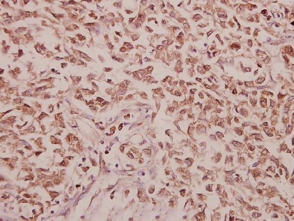
Real IHC data Immunohistochemistry (IHC) analyzes of CCK (V46) pAb in paraffin-embedded human colorectal carcinoma tissue at 1:50.
Anti-CCK (V46) Antibody
Cat # A01098

A01098 is listed for IHC in human, mouse and rat (catalog applications and reactivity). Its own image shows CCK staining in paraffin-embedded human colorectal carcinoma tissue at 1:50 (IHC image caption).

Which to pick: Choose A01098 for tissue IHC on paraffin sections: it is a rabbit pAb with an IHC image from human colorectal carcinoma tissue (catalog host; IHC image caption). No IF/ICC option is supported here (catalog applications: IHC; IF images: none). A01098 lists mouse and rat reactivity alongside human, although its shown IHC evidence is human tissue only; the image caption does not report the fixative (catalog reactivity; IHC image caption).

Each figure is that product's own IHC / IF validation image from its datasheet.

References

  1. UniProt Consortium. UniProt entry P06307 (CCKN_HUMAN, Cholecystokinin).
  2. Human Protein Atlas. CCK tissue IHC expression (reliability: Enhanced).
  3. Human Protein Atlas. CCK subcellular location (ICC-IF): Secreted.
  4. Human Protein Atlas. CCK antibody validation summary (2 antibodies).
  5. Localization of Cholecystokinin/Sulfakinin Neuropeptides in Biomphalaria glabrata, an Intermediate Host for Schistosomiasis. The Journal of comparative neurology 2025 — PMC11841659.
  6. The dorsomedial hypothalamus mediates stress-induced hyperalgesia and is the source of the pronociceptive peptide cholecystokinin in the rostral ventromedial medulla. Neuroscience 2013 — PMC3638803.
  7. Cholecystokinin Expression in the Development of Myocardial Hypertrophy. Scanning 2021 — PMC8405328.
  8. Cholecystokinin in white sea bream: molecular cloning, regional expression, and immunohistochemical localization in the gut after feeding and fasting. PloS one 2012 — PMC3527491.
  9. PubMed PMID:3856870 — UniProt-cited evidence.
  10. PubMed PMID:15489334 — UniProt-cited evidence.
  11. PubMed PMID:9371719 — UniProt-cited evidence.