This website uses cookies to ensure you get the best experience on our website.


- Table of Contents
Plan chromogenic CCL19 IHC in paraffin sections using the IHC-validated antibody (datasheet A01605-2). Compare staining with HPA tissue controls, and interpret cytoplasmic immune-cell signal in light of CCL19 secretion (HPA tissue IHC; UniProt).

Expected localisation, validated protocols, controls and antibodies — the at-a-glance facts below, then the full design guide.
| Expected localisation | Immune-cell cytoplasm (HPA tissue IHC) | |
| Staining pattern | Cytoplasmic staining in immune cells (HPA tissue IHC) | |
| Antigen retrieval | EDTA pH 8.0 HIER, heat-mediated (datasheet A01605-2) | |
| Positive control | Appendix+2 more · see all | |
| Negative control | Adipose tissue+4 more · see all |
| Fixation | Keep fixation consistent across sections. (standard IHC practice; not target-specific) | |
| Caveat | Secreted CCL19 may stain away from its RNA source (HPA tissue IHC) | |
| Regulation | High in lymph nodes, thymus, appendix (UniProt) | |
| Isoform / epitope | 0 isoforms annotated; map epitopes to mature chain 22–98 (UniProt) |
The catalog antibody uses EDTA pH 8.0 heat retrieval (datasheet: A01605-2). The options summarize published CCL19 IHC protocols (PMC3923395; PMC11102069; PMC10683612; PMC4572056).
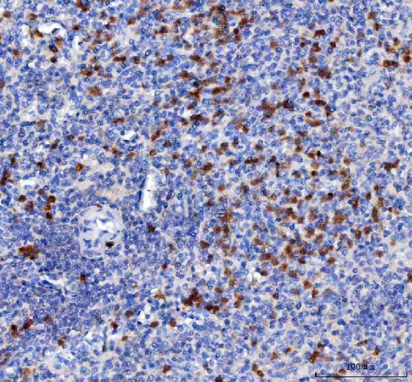
| Sample | Paraffin-embedded human spleen tissue; fixative not specified (datasheet A01605-2) |
| Fixation | Image fixative and duration unreported (datasheet A01605-2); verify before use. |
| Sectioning | 4–5 µm sections on charged slides (standard) |
| Deparaffinisation | Xylene, graded ethanol series to water (standard) |
| Antigen retrieval | Heat retrieval: EDTA pH 8.0 (datasheet A01605-2); 20 min, 95–100 °C (standard) |
| Peroxidase block | 3% H2O2, 10 min, room temperature (standard) |
| Blocking | 10% goat serum (datasheet A01605-2) |
| Primary antibody | Rabbit anti-CCL19, 2-5 μg/ml (datasheet A01605-2) |
| Primary incubation | Overnight at 4 °C (datasheet A01605-2) |
| Detection | HRP-conjugated secondary, DAB chromogen (datasheet A01605-2) |
| Counterstain | Hematoxylin, blue, dehydrate and mount (standard) |
| Expected result | CCL19-positive staining in germinal center cells of appendix (HPA tissue IHC: Medium). HPA tissue profile: Cytoplasmic expression in immune cells. No signal in the no-primary control. |
CCL19 is a secreted protein with no transmembrane segment (UniProt Q99731). In tissue IHC, expect chiefly cytoplasmic staining in immune cells, including medium staining in appendix and tonsil germinal center cells; small intestine endocrine cells also show medium staining (HPA tissue IHC). HPA rates the tissue pattern Enhanced, while noting medium agreement between antibody staining and RNA expression because secretion can separate protein from its site of synthesis (HPA tissue IHC).
| Cytoplasmic chromogen in appendix or tonsil germinal center cells. | This fits the reported medium staining and immune cell profile (HPA tissue IHC). Judge the cells and compartment together; staining elsewhere does not establish its source in a secreted protein (UniProt Q99731; HPA tissue IHC). |
| Predominantly nuclear staining or a crisp, continuous cell membrane outline. | Neither is the reported tissue pattern (HPA tissue IHC). CCL19 is secreted and lacks a transmembrane segment (UniProt Q99731); review morphology and controls before calling such a pattern specific. |
| Strong staining in cells reported as undetected, such as adipose adipocytes or bronchial respiratory epithelium. | This conflicts with those HPA tissue observations (HPA tissue IHC). Consider cross-reactivity or endogenous chromogenic detection activity, and compare a no-primary control (general IHC practice). |
| Broad, poorly localized color across cells and extracellular spaces. | CCL19 secretion makes extracellular location plausible (UniProt Q99731), but diffuse color alone cannot establish specific staining. Check whether the signal follows tissue structures and whether a no-primary control also colors them (general IHC practice). |
| No staining in appendix or tonsil germinal center cells. | Those cells show medium staining in HPA tissue IHC (HPA tissue IHC). A blank result warrants a check of tissue preservation, retrieval, antibody and detection conditions, and the positive control; it does not alone prove CCL19 absence (general IHC practice). |
| Secreted protein and precursor processing (UniProt Q99731). | CCL19 has a signal peptide at residues 1–21 and a mature chain at 22–98, with no transmembrane segment (UniProt Q99731). Assess cytoplasmic tissue staining without assuming all extracellular signal marks the producing cell (HPA tissue IHC; UniProt Q99731). |
| Tissue and cell context (HPA tissue IHC). | Appendix and tonsil germinal center cells and small intestine endocrine cells have medium staining; spleen red pulp and lymph node non-germinal center cells have low staining (HPA tissue IHC). Select and compare controls at the cell level. |
| RNA and protein agreement (HPA tissue IHC; UniProt Q99731). | HPA calls the IHC pattern Enhanced but reports medium consistency with RNA because secreted protein and its RNA can occupy different tissue locations (HPA tissue IHC). UniProt reports high expression in lymph nodes, thymus, and appendix (UniProt Q99731); that does not require equally strong staining in every cell. |
| Antibody validation scope (HPA antibodies). | HPA067758 is listed as rabbit polyclonal with Enhanced IHC validation (HPA antibodies). That status supports the reported tissue pattern; it does not establish specificity for every new specimen, staining condition, or unexpected compartment (HPA tissue IHC; general IHC practice). |
| IF/ICC Q&A: is a cellular fluorescence pattern established? | HPA labels CCL19 secreted but gives no main subcellular location or cell line ICC-IF images, and lists no ICC status for HPA067758 (HPA subcellular; HPA antibodies). Use the tissue IHC pattern as context, not as an IF/ICC validation claim. |
| Situation | Likely cause | Next action |
|---|---|---|
| Appendix or tonsil germinal center cells are blank (HPA tissue IHC). | A failed staining run or unsuitable section can mimic absence (general IHC practice); these cells have medium HPA staining (HPA tissue IHC). | Confirm the positive control and tissue morphology, then review the chosen retrieval, primary antibody, and detection steps against their validated IHC instructions (general IHC practice). |
| Signal is faint but confined to lymph node non-germinal center cells or spleen red pulp (HPA tissue IHC). | HPA reports low staining in those cells (HPA tissue IHC); weak color there alone need not indicate a failed run. | Compare an appendix or tonsil germinal center positive control in the same run before changing detection conditions (HPA tissue IHC; general IHC practice). |
| Diffuse color obscures cellular boundaries. | Background staining may obscure interpretation (general IHC practice); secretion also prevents extracellular location alone from identifying producing cells (UniProt Q99731). | Inspect the no-primary control and tissue morphology; review blocking, washing, and detection conditions if control color is present (general IHC practice). |
| Adipose adipocytes or bronchial respiratory epithelial cells stain strongly. | Both are reported as undetected in HPA tissue IHC (HPA tissue IHC); cross-reactivity or endogenous detection activity is possible (general IHC practice). | Check a no-primary control, assess whether the color follows the reported immune cell pattern, and verify the unexpected result independently before scoring it as CCL19 (HPA tissue IHC; general IHC practice). |
| Nuclear or continuous membrane staining dominates. | It conflicts with the reported cytoplasmic immune cell profile (HPA tissue IHC) and with the absence of a transmembrane segment (UniProt Q99731). | Recheck cell identification and no-primary controls; treat the compartment as unconfirmed until it is independently supported (general IHC practice). |
| Lymph node staining appears lower than expected from its expression description. | UniProt reports high lymph node expression, while HPA reports low staining in lymph node non-germinal center cells and cautions that secreted protein may differ in location from RNA (UniProt Q99731; HPA tissue IHC). | Compare the same cell population across sections and a positive tissue control; report the observed cells and intensity instead of inferring protein distribution from tissue-wide expression (HPA tissue IHC; general IHC practice). |


Comprehensive Human Protein Atlas IHC scoring per tissue (reliability: Enhanced — Medium consistency between antibody staining and RNA expression data. Secreted protein, tissue location of RNA and protein is expected to differ.). Rows are taken directly from the HPA tissue chart — click any row's HPA link to view the source.
| Tissue | Cell type | Level | Evidence | Source |
|---|---|---|---|---|
| Appendix | Germinal center cells | Medium | Protein (IHC) | HPA → |
| Small intestine | Endocrine cells | Medium | Protein (IHC) | HPA → |
| Tonsil | Germinal center cells | Medium | Protein (IHC) | HPA → |
| Tissue | Cell type | Level | Evidence | Source |
|---|---|---|---|---|
| Adipose tissue | Adipocytes | Not detected | Protein (IHC) | HPA → |
| Adrenal gland | Glandular cells | Not detected | Protein (IHC) | HPA → |
| Bone marrow | Hematopoietic cells | Not detected | Protein (IHC) | HPA → |
| Breast | Adipocytes | Not detected | Protein (IHC) | HPA → |
| Bronchus | Respiratory epithelial cells | Not detected | Protein (IHC) | HPA → |
Troubleshoot CCL19 staining in paraffin sections by checking retrieval, controls, cellular context and detection before interpreting signal intensity.
The catalog has one human-reactive anti-CCL19 antibody with real IHC data from a paraffin-embedded human spleen section (A01605-2 image caption; catalog reactivity).
A01605-2 is listed for human IHC, flow cytometry and ELISA (catalog applications and reactivity). Its IHC figure shows a paraffin-embedded human spleen section (A01605-2 image caption).
Which to pick: For tissue IHC, choose A01605-2, a rabbit polyclonal antibody listed at 2–5 μg/ml for paraffin sections (catalog host, clonality and IHC dilution). Its spleen IHC caption reports EDTA retrieval at pH 8.0 and a 2 μg/ml primary antibody concentration; the fixative is unreported (A01605-2 image caption). There is no listed IF/ICC or cross-species option: A01605-2 lists human reactivity and IHC, flow cytometry and ELISA, with no IF dilution or figure (catalog applications, reactivity and IF fields).