This website uses cookies to ensure you get the best experience on our website.
- Table of Contents
3 Citations 10 Q&As
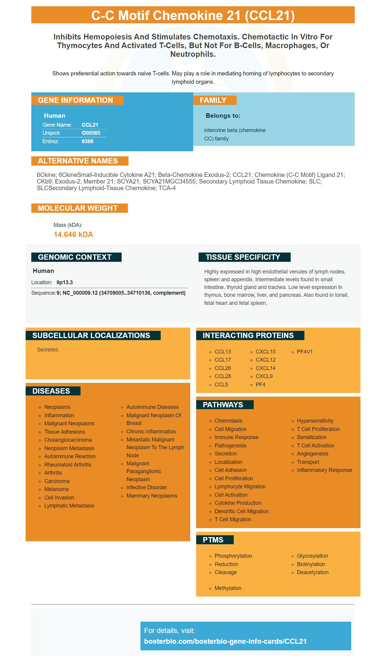
Facts about C-C motif chemokine 21.
Shows preferential action towards naive T-cells. May play a role in mediating homing of lymphocytes to secondary lymphoid organs.
Human | |
---|---|
Gene Name: | CCL21 |
Uniprot: | O00585 |
Entrez: | 6366 |
Belongs to: |
---|
intercrine beta (chemokine CC) family |
6Ckine; 6CkineSmall-inducible cytokine A21; Beta-chemokine exodus-2; CCL21; chemokine (C-C motif) ligand 21; CKb9; exodus-2; member 21; SCYA21; SCYA21MGC34555; secondary lymphoid tissue chemokine; SLC; SLCSecondary lymphoid-tissue chemokine; TCA-4
Mass (kDA):
14.646 kDA
Human | |
---|---|
Location: | 9p13.3 |
Sequence: | 9; NC_000009.12 (34709005..34710136, complement) |
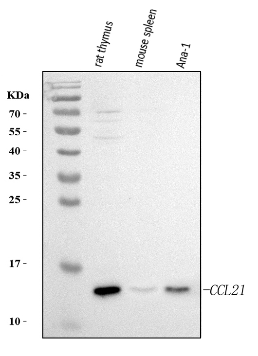
Highly expressed in high endothelial venules of lymph nodes, spleen and appendix. Intermediate levels found in small intestine, thyroid gland and trachea. Low level expression in thymus, bone marrow, liver, and pancreas. Also found in tonsil, fetal heart and fetal spleen.
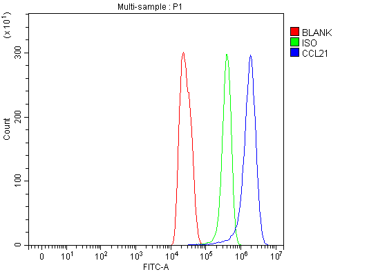
Secreted.
If you are interested in using CCL21 as dendritic cell marker, then you have come to the right spot. In this article, we will examine what this marker is and how it operates and what uses it could be used for. We will also explore how it interacts with dendritic cells. We will also talk about how CCL21 can be utilized in research.
CCL21-producing cells are found in synovial tissue of the rheumatoid joint. CCL21-expressing cells co-localize to the CD31 and CD34 mRNAs. CD31 and CD34. The immunostained sections of tissues that have been stained for CCL21, CD31, and CD34 show the presence of synovial lymphoid aggregates. The insets illustrate the same vascular structures as those seen in parallel sections.
The present study revealed that CCL21 co-localizes in lymphoid nodes that are SMA-positive and dendritiform cells. These cells dispersed throughout the lymph node, connecting HEVs. 92.7+/- 3.4% SMA+ cells co-localized with MLNs in CCL21+ cells. The proportion of SMA+ cells that co-localized with CCL21+ cells was 84.2+/1% in the CLNs and the ALNs. In the tonsils, however CCL21+ cells lacked SMA-positive cells, but they co-localized with podoplanin.
While the precise mechanism behind the CCL21-producing dendritic cells is not understood completely but it is probable that CCL21 is a crucial homeostatic chemokine that is involved in the recruitment and organization of T cells in lymphoid T zones. CCL21 is released by the cells that circulate throughout the T zone of human SLOs. These tissues lack high endothelial veins. Therefore, cells that are not lymphoid-specific replicate the characteristics of SLOs.
In the present study, the presence of CCL21 on dendritic cells was connected to the synthesis of IFN-g and GM-CSF. Further evidence of the impact CCL21 had on dendritic cell was due to its binding to Gi receptors with a protein. This discovery opens the door to future clinical research in this field. CCL21 was identified as a marker for dendritic cell proliferation in this study.
Inflammatory tissues, CCL21 induces rapid mobilization of antigen-charged dendritic cell (DC). These DCs move through the afferent Lymphatics. CCL21 is vital for DC migration and triggers integrins by its CCR7 receptor. However, recent studies have suggested that CCL21 is a non-integrin-dependent factor.
Induction of CCL21 by TNFa triggers the production of CCL21 by HDLECs that are primary. Ex vivo, HDLECs stimulated by TNFa release 50 pg ml-1 CCL21. CCL21-producing cells in later passages didn't secrete CCL21 when TNFa was added. These results suggest that TNFa could be a more potent stimulator of CCL21 than was previously thought.
Although the mechanism that explains this phenomenon isn't fully understood however, recent research has shown that HDLEC secrete higher levels of CCL21 in a culture. However, when they are removed from their native tissue environment they lose the control over chemokine action. The chemokine produced by HDLEC may play a part in DC migration in lymphatic tissues. The inflammatory response may also be affected by peripheral lymphatics which release the chemical.
To determine the presence of CCL21, the cells were grown in duplicate wells with TNFa and actinomycin-D. The cells were lysed in the buffer consisting of 50 mM Tris pH 7.4 100 mM NaCl and 1 percent NP-40, and protease inhibitor cocktail. Semi-quantitative westernblots were utilized to determine CCL21 levels.
CCL21 is a chemokine the antigen-presenting DCs secrete into the lymphatic system. CCL21 is produced by endothelial cells in the lymphatic capillary and forms a gradient that disintegrates into the interstitium. The exact mechanism for CCL21's gradient formation is not clear however, it is believed to work through chemotactic interactions with DCs.
Expression of CCL21 was examined in peripheral tissues. Patients suffering from RA were able to have their synovial tissues analyzed. Also minor salivary glands that were normal were extracted from patients suspected to have amyloidosis. A sample of the skin from a mastectomy was also collected. All tissue samples were fixed with formalin and embedded in paraffin for analysis. Four additional RA synovial tissues were selected for further study for the presence of lymphoid aggregates. In addition two human tonsils and a MLN were embedded in OCT and analyzed for the presence of lymphoma.
In addition to examining chemokine response The company also looked into the phenotype of dendritic cells. In particular, they assessed the expression levels of E2 integrin (OX62) and MHC-II. After five days in CCL21-positive media The cells were washed in PBS and then stained with 5-(and-6) carboxytetramethyl Rhodamine succimidyl ester. The cells were then suspended in R10 + GM-CSF hybridoma supernatant.
Additionally, these results suggest that the CCL21 marker connects to the CCR7 receptor on dendritic cells . It's associated with the maturation of dendritic cells. Researchers believe that CCL21 could also regulate immune interactions through binding to dendritic cells. CCL21 has also been found to protect mice from EAE, suggesting that it interacts with dendritic cells to regulate the immune system's activities.
CCL21 production has been found to be preserved in human lymphoid tissues. This suggests that chronic inflammation disorders are frequently associated with the production of this chemokine. Chronic inflammation is also associated with ECs that produce CCL21. These findings also suggest that CCL21 may act with dendritic cells in the lymphatic system.
HEVs also were associated with the ECs that contain synovial tissue that express CCL21. The mRNA as well as the protein levels of CCL21 were colocalized in the case of human MLNs. Both markers appeared to be in close proximity in a organized group of HEVs. Double IF stainings showed that CCL21 and PNAd are strongly located in close proximity to each other and with HEV shape.
These results also suggest that a mutation in CCL21 can reduce the interstitial CCL21 gradient. This would result in a longer decay time and reduced DC circulation to lymphatic capillaries. Researchers hoped to test this hypothesis using the genetic deletion of lymphatic endothelial cells lines to see if CCL21 could actually interact with dendritic cells.
CCL21 is an inflammatory protein produced by dendritic cells that plays a critical role in the development of the immune system. It also serves as a vital marker for thymocytes that are postnatal day-10 subsets. CCL21-/ mice lack CCL21a. However MTECs as well TECs express CCL21a. There are two kinds of mTECs. They express CCL21a or CCL21b. Thymic stromal cell are the mTEC subsets that express CCL21a or CCL21a.
HDLECs produce endogenous CCL21 and stimulate DC movement through lymphatic systems. It facilitates translymphatic migration in resting and inflammatory conditions. The inflammatory pathway is believed to involve activation of b2integrin however, the chemokine CCL21 is also essential for lymphatic passage, where DCs interact with T cells.
To color dendritic cell dendrites, PBMCs were obtained from healthy donors and then subjected to positive selection using anti-CD14 magnetic microbeads. After positive selection, monocytes were grown in RPMI-10% FCS for five days. The monocytes were then pre-fixed with 2% formaldehyde, which inhibits the activation of integrins. The cells were then treated with goat anti-mouse IgG-488 Alexa fluor(r). Images were then analysed using Image J software.
To determine CCL21 Supernatants from primary HDLEC were used for ELISA in triplicate. The cells were then lysed with lysis buffer (50mM Tris pH 7.4 100mM NaCl, 100mM NP-40 and 1 percent N-40) and a protease inhibitor combination. ELISAs were then used to identify CCL21 and Lyve-1.
When CCL21 is interacting with DCs, lamellipodium gets distributed on their surfaces by DCs that express CCL21. Inflammed DCs also retract their protrusions. CCL21 expression was also suppressed in inflamed cells, indicating that CCL21 may be inducible. These findings also confirm earlier studies using morphology.
CCL21 as well as CCL19 are weak antagonists. CCL21 when liganded induces differential signaling via CCR7, which is a vital receptor in the immune system. CCL21 can be broken down using DC-released proteases, which is similar to CCL19 however, it is far less powerful. Its effects are more defined than CCL19.
PMID: 9235955 by Nagira M., et al. Molecular cloning of a novel human CC chemokine secondary lymphoid- tissue chemokine that is a potent chemoattractant for lymphocytes and mapped to chromosome 9p13.
PMID: 9257816 by Hedrick J.A., et al. Identification and characterization of a novel beta chemokine containing six conserved cysteines.
*More publications can be found for each product on its corresponding product page