This website uses cookies to ensure you get the best experience on our website.


- Table of Contents
Plan chromogenic CCL25 IHC-P using the catalog antibody's 1:50–1:200 dilution range (datasheet). Use small-intestinal glandular cells as a positive reference for cytoplasmic staining (HPA tissue IHC), and account for secretion when interpreting the pattern (UniProt).

Expected localisation, validated protocols, controls and antibodies — the at-a-glance facts below, then the full design guide.
| Expected localisation | Cytoplasmic staining in a few tissues (HPA tissue IHC) | |
| Staining pattern | Cytoplasmic staining in small-intestinal glandular cells (HPA tissue IHC) | |
| Antigen retrieval | Citrate pH 6.0 HIER, 95–98 °C, 20 min (rule: cytoplasmic / membrane antigen) | |
| Positive control | Small intestine | |
| Negative control | Adipose tissue+4 more · see all |
| Fixation | Keep fixation consistent across sections (standard IHC practice; not target-specific) | |
| Caveat | Secretion may separate protein staining from RNA location (HPA tissue IHC) | |
| Regulation | No expression regulator specified (UniProt) | |
| Isoform / epitope | 3 isoforms; signal peptide 1–23 is removed; no cytoplasmic domain (UniProt) |
The catalog antibody’s IHC-P protocol is followed by published CCL25 IHC methods for mouse heart and human ileal sections (PMC5009347; PMC9536268).
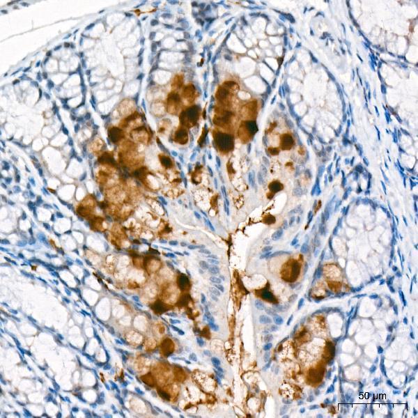
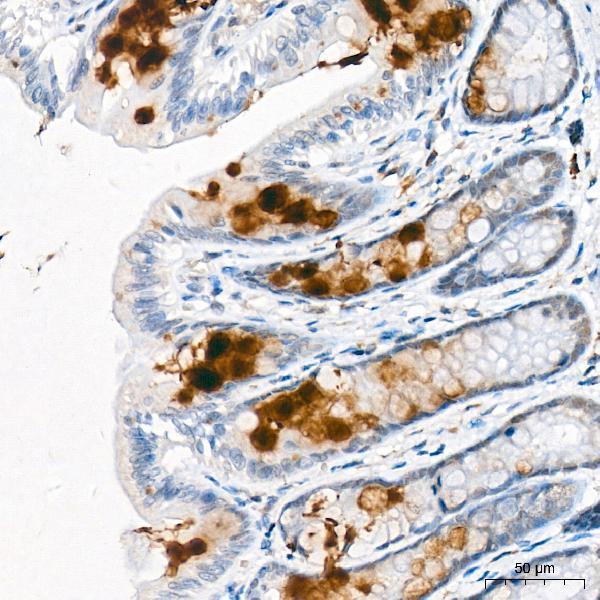
| Sample | Paraffin-embedded Mouse large intestine tissue; fixative not specified (datasheet A04077) |
| Fixation | Image fixative and duration unreported (datasheet A04077); verify before use. |
| Sectioning | 4–5 µm sections on charged slides (standard) |
| Deparaffinisation | Xylene, graded ethanol series to water (standard) |
| Antigen retrieval | Heat-induced epitope retrieval in citrate buffer, pH 6.0, 20 min at 95–98 °C (standard rule: cytoplasmic / membrane antigen) |
| Peroxidase block | 3% H2O2, 10 min, room temperature (standard) |
| Blocking | 10% normal serum of the secondary host, 30 min, room temperature (standard) |
| Primary antibody | Rabbit anti-CCL25, 1:50-1:200 (datasheet A04077) |
| Primary incubation | Overnight at 4 °C (standard) |
| Detection | HRP-polymer secondary, DAB chromogen 5–10 min (standard) |
| Counterstain | Hematoxylin, blue, dehydrate and mount (standard) |
| Expected result | CCL25-positive staining in glandular cells of small intestine (HPA tissue IHC: Medium). HPA tissue profile: Cytoplasmic expression in a few tissues. No signal in the no-primary control. |
CCL25 is a secreted protein with no transmembrane segment (UniProt O15444). In paraffin tissue sections, expect cytoplasmic staining in small-intestinal glandular cells, where HPA reports medium staining; duodenal glandular cells show low staining (HPA tissue IHC). HPA describes expression in a few tissues and rates its tissue IHC evidence Approved, while cautioning that the tissue locations of RNA and secreted protein may differ (HPA tissue IHC).
| Medium cytoplasmic staining in small-intestinal glandular cells. | This matches the reported positive IHC pattern (HPA tissue IHC: small intestine, glandular cells, Medium). Score cell type and compartment together; secretion can complicate assignment of protein to its producing cell (UniProt O15444; HPA tissue IHC reliability note). |
| Strong nuclear-only or sharply membrane-bound staining. | Neither is the reported cytoplasmic pattern (HPA tissue IHC). A membrane-bound interpretation is especially doubtful because CCL25 has no transmembrane segment (UniProt O15444). Check morphology and staining controls before calling either pattern specific. |
| Prominent staining in appendix glandular cells or bronchial respiratory epithelial cells. | Both are listed as Not detected by tissue IHC (HPA tissue IHC). Consider cross-reactivity or endogenous chromogenic activity, especially if control sections share the pattern (general IHC practice). |
| Uniform color across cells, stroma, and section edges. | This lacks the cell-specific glandular pattern reported in small intestine (HPA tissue IHC). Diffuse background can arise from nonspecific reagent binding, incomplete blocking, or detection chemistry (general IHC practice); do not score it as CCL25-positive cells. |
| No staining in an adequately preserved small-intestine section. | This conflicts with the reported medium glandular staining (HPA tissue IHC). First check the antibody's IHC validation and the detection run; a negative slide alone cannot establish absent CCL25, particularly for a secreted protein (HPA tissue IHC reliability note; general IHC practice). |
| Secreted location and tissue assignment | CCL25 is secreted and lacks a transmembrane segment (UniProt O15444). HPA warns that RNA and protein tissue locations may differ for secreted proteins (HPA tissue IHC); interpret extracellular color with morphology rather than assigning it automatically to the nearest cell. |
| Positive and lower-signal comparisons | Small-intestinal glandular cells are Medium, whereas duodenal glandular cells are Low by HPA tissue IHC. These are observed staining levels, not prescribed controls or a guarantee that every section will have the same intensity (HPA tissue IHC; general IHC practice). |
| Signal peptide and antibody epitope | The signal peptide spans residues 1–23 and the annotated CCL25 chain spans 24–150 (UniProt O15444). The supplied HPA validation summary does not identify the antibody epitope (HPA antibodies); infer neither precursor recognition nor mature-chain recognition from the staining pattern. |
| Isoforms and antibody specificity | UniProt lists 3 isoforms, with isoform 2 described as an antagonist of isoform 1 (UniProt O15444). The supplied HPA antibody summary reports IHC Approved but gives no isoform-specific validation (HPA antibodies); tissue color cannot distinguish isoforms. |
| IF/ICC: Is cellular localization established? | No HPA ICC-IF images or main cellular location are supplied for CCL25 (HPA subcellular). The secreted annotation supports biological context, but the tissue IHC pattern cannot validate an IF/ICC localization claim (UniProt O15444; HPA tissue IHC). |
| Situation | Likely cause | Next action |
|---|---|---|
| Small-intestine glandular cells are blank. | The result disagrees with HPA's Medium glandular staining; the failed step is unknown from appearance alone (HPA tissue IHC). | Review tissue identity and morphology, then check antibody labeling, retrieval conditions, detection reagents, and a run control (general IHC practice). Treat retrieval changes as general optimization, not a known CCL25 fixation effect. |
| Only duodenum is faint. | Low glandular staining is reported for duodenum (HPA tissue IHC). | Compare with small intestine, where glandular staining is reported as Medium (HPA tissue IHC). Record the weaker duodenal result before increasing detection intensity, which can also increase background (general IHC practice). |
| Appendix glandular cells show widespread color. | HPA reports those cells as Not detected; specific CCL25 staining is therefore uncertain (HPA tissue IHC). | Check a detection-only control for endogenous activity and assess whether color tracks tissue structures or section-wide background (general IHC practice). Reassess the result against small-intestine glandular staining (HPA tissue IHC). |
| Nuclei dominate the signal. | Nuclear staining does not match HPA's cytoplasmic tissue description (HPA tissue IHC). | Inspect the counterstain and detection-only control, then rescore only clearly localized chromogenic signal (general IHC practice). Do not assign a nuclear CCL25 location from this slide. |
| Color coats tissue edges or appears throughout stroma. | A broad deposit does not reproduce HPA's glandular cytoplasmic pattern; secretion also limits simple cell-of-origin claims (HPA tissue IHC; UniProt O15444). | Inspect washing, blocking, chromogen development, and section quality (general IHC practice). Keep extracellular color separate from scored glandular cytoplasmic staining. |
| A positive-looking slide has no clean cellular boundaries. | Overlapping background can obscure the glandular pattern, regardless of the antibody's Approved IHC status (HPA antibodies; general IHC practice). | Compare a detection-only control and an HPA-listed Not detected tissue, then adjust general staining or imaging conditions before scoring intensity (HPA tissue IHC; general IHC practice). |
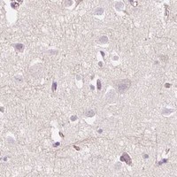


Comprehensive Human Protein Atlas IHC scoring per tissue (reliability: Approved — Secreted protein, tissue location of RNA and protein is expected to differ.). Rows are taken directly from the HPA tissue chart — click any row's HPA link to view the source.
| Tissue | Cell type | Level | Evidence | Source |
|---|---|---|---|---|
| Small intestine | Glandular cells | Medium | Protein (IHC) | HPA → |
| Tissue | Cell type | Level | Evidence | Source |
|---|---|---|---|---|
| Adipose tissue | Adipocytes | Not detected | Protein (IHC) | HPA → |
| Adrenal gland | Glandular cells | Not detected | Protein (IHC) | HPA → |
| Appendix | Glandular cells | Not detected | Protein (IHC) | HPA → |
| Bone marrow | Hematopoietic cells | Not detected | Protein (IHC) | HPA → |
| Breast | Adipocytes | Not detected | Protein (IHC) | HPA → |
For CCL25 chromogenic IHC on paraffin sections, interpret staining in light of its secretion, limited tissue distribution, and unreported antibody epitope (UniProt O15444; HPA tissue IHC).
A04077 has IHC images from paraffin-embedded mouse and rat large intestine (image captions) and lists human reactivity and IF as an application (catalog). No IF image is supplied (catalog).
A04077 will render with an IHC figure from paraffin-embedded mouse large intestine at 1:300 (image caption). Its applications include IHC and IF, its listed reactivity is human, and a second IHC caption shows rat large intestine at 1:300 (catalog; image captions).
Which to pick: Choose A04077 for paraffin-section IHC because its own images show mouse and rat large intestine; the fixative is unreported (image captions). For IF/ICC, A04077 lists IF and human reactivity, but the payload supplies no IF image or ICC claim (catalog). For cross-species work, the mouse and rat IHC images support only those pictured samples; the listed reactivity is human, so verify performance in the intended species (image captions; catalog).