CCM2 / Cerebral cavernous malformations 2 protein · IHC design guide

Design Immunohistochemistry for CCM2

Plan CCM2 chromogenic IHC around its general cytoplasmic tissue pattern (HPA tissue IHC). Use spermatogonia and tonsil germinal center cells as high-staining references, with liver cholangiocytes as a reported unstained cell population (HPA tissue IHC).

Evidence assembled Oct 2026 · For research use; verify linked source records and product datasheet before use
Immunohistochemistry protocol sheet for CCM2 (IHC for CCM2): expected localisation General cytoplasmic staining (HPA tissue IHC), antibody A01908, validated IHC image, and IHC protocol steps
Printable CCM2 IHC protocol sheet — expected localisation General cytoplasmic staining (HPA tissue IHC), antibody A01908, controls and protocol steps. Open the full CCM2 IHC guide →

CCM2 Immunohistochemistry Experimental Design Guide

Expected localisation, validated protocols, controls and antibodies — the at-a-glance facts below, then the full design guide.

Must know before staining
Expected localisation General cytoplasmic staining (HPA tissue IHC)
Staining pattern Cytoplasmic; high in spermatogonia and germinal center cells (HPA tissue IHC)
Antigen retrieval Citrate pH 6.0 HIER, 95–98 °C, 20 min (rule: cytoplasmic / membrane antigen)
Positive control ⓘ Testis+4 more · see all
Negative control ⓘ Liver
Important caveats
Reasons your staining may differ from the expected pattern.
Fixation Keep fixation consistent across paraffin sections. (standard IHC practice; not target-specific)
Caveat Brain RNA enrichment may not predict strongest IHC staining (HPA tissue IHC)
Regulation Expression regulation unreported (UniProt)
Isoform / epitope 4 isoforms; antibody epitope coverage is unreported (UniProt)
Section 1

Recommended CCM2 IHC & IF Protocols

Compare the catalog antibody’s IHC-P protocol with three published CCM2 chromogenic IHC protocols (PMC2640205; PMC7160551; PMC2856024).

Recommended immunohistochemistry (IHC-P) protocol parameters
SampleParaffin-embedded human liver tissue; fixative not specified (datasheet A01908)
FixationImage fixative and duration unreported (datasheet A01908); verify before use.
Sectioning4–5 µm sections on charged slides (standard)
DeparaffinisationXylene, graded ethanol series to water (standard)
Antigen retrievalHeat-induced epitope retrieval in citrate buffer, pH 6.0, 20 min at 95–98 °C (standard rule: cytoplasmic / membrane antigen)
Peroxidase block3% H2O2, 10 min, room temperature (standard)
Blocking10% normal serum of the secondary host, 30 min, room temperature (standard)
Primary antibodyRabbit anti-CCM2, 1:50-1:200 (datasheet A01908)
Primary incubationOvernight at 4 °C (standard)
DetectionHRP-polymer secondary, DAB chromogen 5–10 min (standard)
CounterstainHematoxylin, blue, dehydrate and mount (standard)
Expected resultCCM2-positive staining in spermatogonia cells of testis (HPA tissue IHC: High). HPA tissue profile: General cytoplasmic expression. No signal in the no-primary control.
💡Decision noteStart with citrate pH 6.0 heat retrieval at 95–98 °C for 20 min (page retrieval rule); adjust for the tissue and antibody used.
Section 2

What Is the Expected CCM2 Staining Pattern?

For paraffin-section IHC, expect mainly cytoplasmic CCM2 staining in spermatogonia and tonsillar germinal center cells, where HPA reports high staining; several glandular and cerebral-cortex cell populations show medium staining (HPA tissue IHC). HPA describes general cytoplasmic expression with Supported reliability, meaning medium consistency between staining and RNA data (HPA tissue IHC). CCM2 is cytoplasmic and has no transmembrane segment (UniProt Q9BSQ5).

What am I looking at on my slide?
Cytoplasmic signal in spermatogonia or tonsillar germinal center cells.This matches HPA's high-staining cell populations and its general cytoplasmic profile (HPA tissue IHC). Judge signal in the named cells, rather than treating every cell in the section as an expected positive (HPA tissue IHC).
Predominantly nuclear, surface-restricted, or extracellular deposit-like staining.These patterns do not match UniProt's cytoplasmic location or HPA's general cytoplasmic tissue-IHC profile (UniProt Q9BSQ5; HPA tissue IHC). Treat an isolated, strong wrong-compartment pattern as suspect and check controls before assigning it to CCM2 (general IHC practice).
Strong staining in liver cholangiocytes.HPA reports CCM2 as not detected in cholangiocytes; that finding does not make the whole liver a negative control (HPA tissue IHC). Recheck cell identification and consider antibody cross-reactivity or endogenous detection activity (general IHC practice).
Diffuse color across cells and surrounding tissue, obscuring cell boundaries.An indiscriminate deposit is harder to reconcile with HPA's cell-resolved cytoplasmic pattern (HPA tissue IHC). Assess a no-primary control for endogenous detection activity, then review blocking and detection conditions (general IHC practice).
No signal in spermatogonia or tonsillar germinal center cells.Absence in these HPA high-staining populations weakens confidence in a negative result elsewhere (HPA tissue IHC). Verify tissue identity and assay controls, then review retrieval, antibody dilution, and detection conditions (general IHC practice).
💡Expected CCM2 appearanceCall a result convincing when cytoplasmic staining is clear in HPA high-staining spermatogonia or tonsillar germinal center cells; diffuse deposit or strong nuclear-only staining is suspect (HPA tissue IHC; UniProt Q9BSQ5; general IHC practice).
How each factor affects the staining
Tissue and cell choiceTestis spermatogonia and tonsillar germinal center cells are high; cerebral-cortex neurons and several glandular populations are medium (HPA tissue IHC). Cerebellar granular-layer cells and hippocampal neurons are low, so weak signal there alone is a less decisive assay check (HPA tissue IHC).
Strength of IHC evidenceHPA rates the tissue pattern Supported, with medium consistency between antibody staining and RNA expression (HPA tissue IHC). Its listed antibodies HPA020273 and HPA021669 are IHC Supported (HPA antibodies). Interpret an unexpected pattern with controls and cell identity in view (general IHC practice).
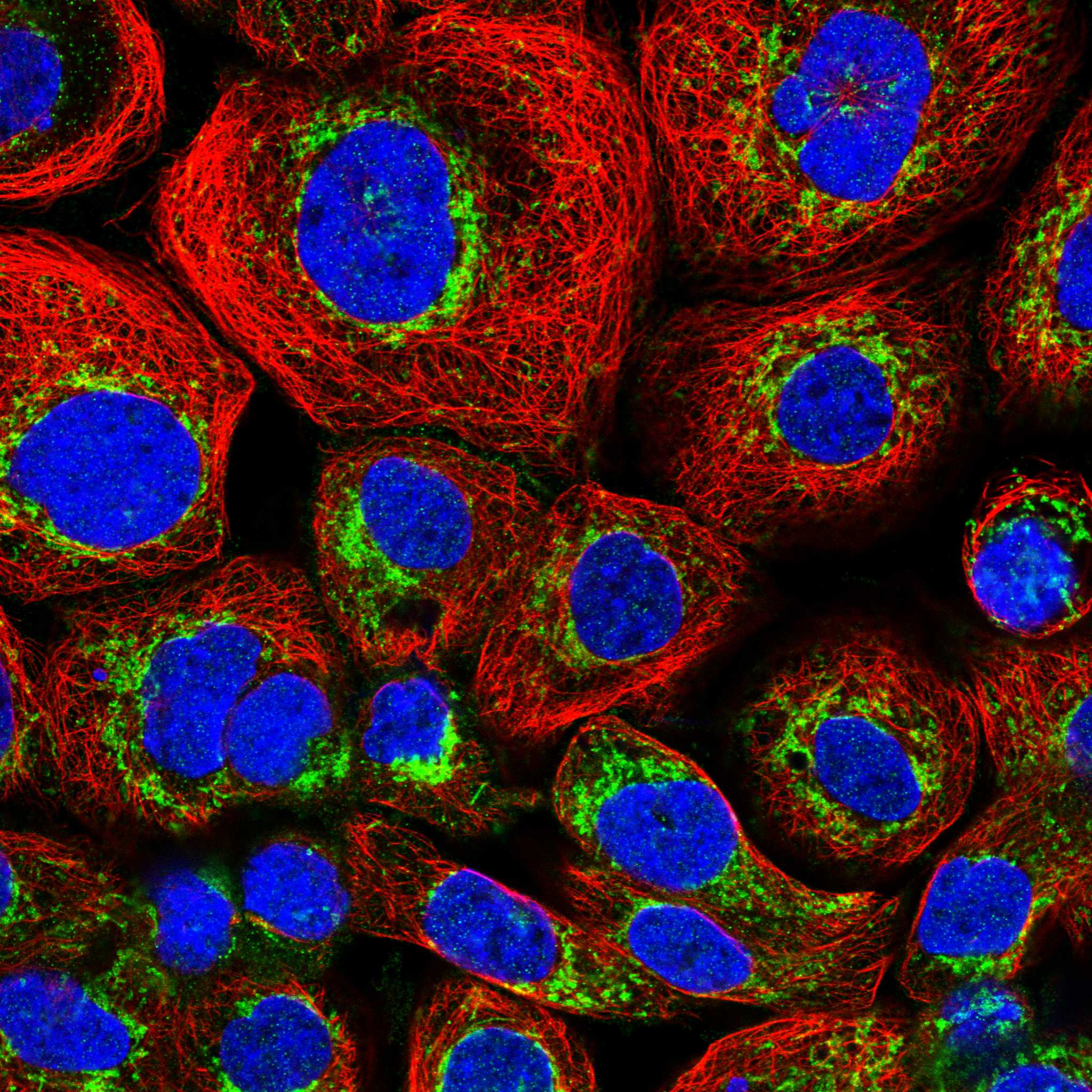
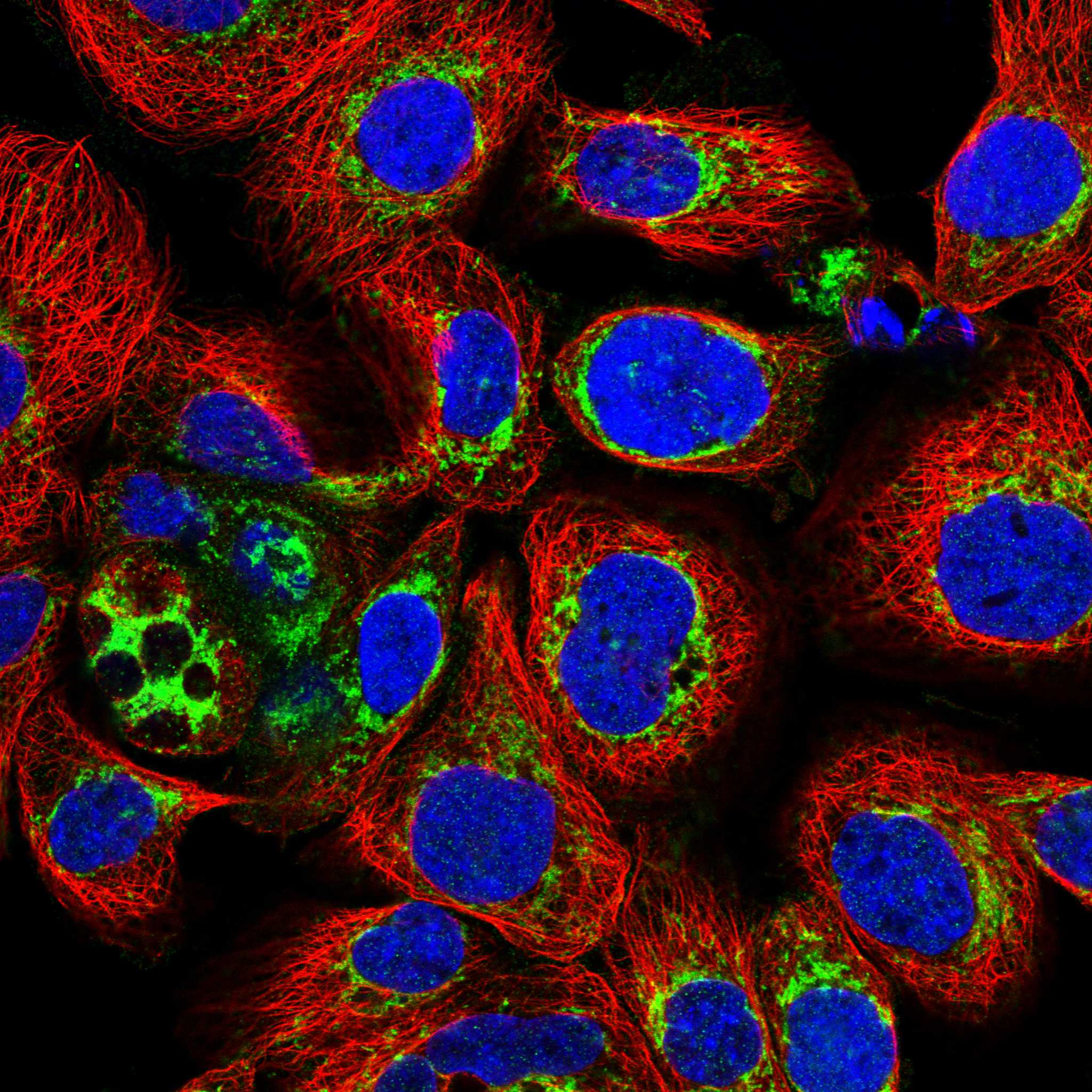
IF/ICC pattern?HPA reports an enhanced mitochondrial location in ICC-IF, while UniProt lists cytoplasm and HPA describes general cytoplasmic tissue-IHC staining (HPA subcellular; UniProt Q9BSQ5; HPA tissue IHC). Can punctate IF signal be expected? Yes in HPA's ICC-IF context; do not impose that subcellular resolution on chromogenic tissue IHC (HPA subcellular; general IHC practice).
Isoforms and epitopeUniProt lists four CCM2 isoforms and a PID domain at residues 59–248 (UniProt Q9BSQ5). The supplied sources do not identify the IHC antibody's epitope or establish isoform-specific staining. Do not infer which isoform produced a cell's signal from its intensity alone (UniProt Q9BSQ5; general IHC practice).
Processing and retrievalUniProt lists no signal peptide or propeptide and describes one 1–444 protein chain, with no transmembrane segment (UniProt Q9BSQ5). There is no supplied CCM2-specific retrieval or fixation-sensitivity finding. Select and assess retrieval using assay controls as general paraffin-IHC practice.
Why is my staining missing, weak or wrong?
SituationLikely causeNext action
Positive-control cells are unstained.The assay may have insufficient detectable signal; HPA reports high staining in spermatogonia and tonsillar germinal center cells (HPA tissue IHC; general IHC practice).Confirm that those cells are present, then check retrieval, the catalog antibody's IHC-P instructions, dilution, and detection controls (general IHC practice).
Color appears throughout the section.Diffuse deposit may reflect nonspecific binding or endogenous detection activity (general IHC practice).Compare a no-primary control; review blocking, antibody concentration, washes, and detection chemistry before interpreting cell staining (general IHC practice).
Signal is mainly nuclear.That distribution conflicts with cytoplasmic localisation reported by UniProt and HPA tissue IHC (UniProt Q9BSQ5; HPA tissue IHC).Check counterstain and compartment assignment, then compare positive and no-primary controls before calling CCM2 positive (general IHC practice).
Cholangiocytes stain strongly.HPA reports cholangiocytes as not detected; cross-reactivity or endogenous detection activity is possible (HPA tissue IHC; general IHC practice).Verify cell identity and inspect no-primary and positive-tissue controls; do not generalize the HPA cholangiocyte result to all liver cells (HPA tissue IHC; general IHC practice).
Only faint signal appears in a low-staining brain population.Low staining is reported in cerebellar granular-layer cells and hippocampal neurons (HPA tissue IHC).Assess a high-staining control population before declaring assay failure or interpreting the faint signal as an unexpected negative (HPA tissue IHC; general IHC practice).
Punctate ICC-IF differs from tissue-IHC appearance.HPA reports enhanced mitochondrial ICC-IF localisation but general cytoplasmic tissue-IHC expression (HPA subcellular; HPA tissue IHC).Interpret each application against its own HPA observation; use tissue cell identity and cytoplasmic distribution for this chromogenic IHC readout (HPA tissue IHC; general IHC practice).

Sample controls for CCM2 IHC & IF

🧪Run tonsil first and look for staining in germinal center cells (HPA: High in tonsil germinal center cells). Use liver cholangiocytes as the biological comparison (HPA: Not detected in liver cholangiocytes); on the tonsil slide, cells outside the scored germinal center population should show no specific chromogen above background, but HPA does not establish those cells as CCM2-negative.
Positive control tissue: Testis (Spermatogonia cells, HPA High)
Negative control tissue: Liver (HPA Not detected)
ICC-IF cell lines (HPA subcellular resource): HPA ICC-IF images show CCM2 in A-431, U-251MG, U2OS, MCF-7, with annotated localisation: Mitochondria (enhanced) (HPA subcellular).
Technical controls: Include no-primary (secondary-only) and matched immunoglobulin controls: host- and isotype-matched for a monoclonal primary, or normal IgG from the primary host species for a polyclonal primary (standard IHC practice). Confirm specificity with CCM2-knockout material if available, or peptide competition when the immunizing peptide is available; quench endogenous peroxidase and check background in tonsil inflammatory cells (standard chromogenic IHC practice).
⚠️Feasibility: A target-specific fixation window or fixation effect is unreported, and the selected A01908 paraffin-section liver caption does not state a fixative (selected-SKU caption: fixative unreported). That caption uses microwave retrieval in 10 mM PBS, pH 7.2, at 1:100, but does not establish that retrieval is required (selected-SKU caption). There is no supplied comparison showing that frozen sections or IF are easier; for IF interpretation, HPA reports enhanced mitochondrial localization in its ICC images, whereas UniProt annotates CCM2 as cytoplasmic (HPA subcellular; UniProt Q9BSQ5 subcellular).

HPA tissue IHC evidence for CCM2

Comprehensive Human Protein Atlas IHC scoring per tissue (reliability: Supported — Medium consistency between antibody staining and RNA expression data.). Rows are taken directly from the HPA tissue chart — click any row's HPA link to view the source.

Positive expression · recommended positive controls

TissueCell typeLevelEvidenceSource
Testis Spermatogonia cells High Protein (IHC) HPA →
Tonsil Germinal center cells High Protein (IHC) HPA →
Adrenal gland Glandular cells Medium Protein (IHC) HPA →
Appendix Glandular cells Medium Protein (IHC) HPA →
Cerebral cortex Neuronal cells Medium Protein (IHC) HPA →

Undetected expression · recommended negative controls

TissueCell typeLevelEvidenceSource
Liver Cholangiocytes Not detected Protein (IHC) HPA →
Section 3

Advanced CCM2 IHC Tips

These questions address CCM2 staining in paraffin sections with chromogenic IHC; the immunofluorescence entry covers the separate IF/ICC application.

Which retrieval conditions should I start with for CCM2 in paraffin sections?
Start with citrate buffer at pH 6.0 for heat induced antigen retrieval at 95–98 °C for 20 min (page IHC protocol). Allow sections to cool consistently, then compare staining with a control section processed in the same run; variable heating can change apparent cytoplasmic intensity (standard IHC practice; UniProt Q9BSQ5: cytoplasmic). If staining remains weak, a catalog image for SKU A01908 documents microwave retrieval in 10 mM PBS at pH 7.2 with antibody at 1:100 in paraffin embedded human liver (A01908 tissue IHC caption). Treat that image as an alternative retrieval example, and assess signal alongside background before changing the working method (standard IHC practice).
How should I assess whether fixation is reducing CCM2 staining?
Target specific fixation sensitivity is unknown because the supplied CCM2 evidence does not report a fixative or a controlled fixation comparison (A01908 tissue IHC caption: fixative not stated). Process matched sections with the same fixation history, section thickness, retrieval conditions and detection run when comparing CCM2 intensity (standard IHC practice). If archived blocks stain inconsistently, compare a control tissue across runs before changing citrate retrieval at pH 6.0, 95–98 °C for 20 min (page IHC protocol; standard IHC practice). Record fixation details where available, but do not infer a CCM2 specific fixation effect from tissue staining patterns or protein features (HPA tissue IHC; UniProt Q9BSQ5).
What cellular pattern should count as plausible CCM2 staining?
Look first for intracellular, predominantly cytoplasmic signal: UniProt annotates CCM2 in the cytoplasm, and tissue IHC reports general cytoplasmic expression (UniProt Q9BSQ5: cytoplasm; HPA tissue IHC: general cytoplasmic expression). A finely punctate pattern may merit investigation because the subcellular IF dataset assigns CCM2 an enhanced mitochondrial location, but that finding does not establish an IHC pattern for this antibody (HPA subcellular: mitochondria enhanced). Score cells against nearby morphology and a same run control, keeping nuclear only staining or a sharp extracellular rim separate from plausible intracellular signal (standard IHC practice; UniProt Q9BSQ5: no transmembrane segment). Document diffuse and punctate cytoplasmic patterns separately when both appear (standard IHC practice).
Could isoforms or phosphorylation explain a change in CCM2 IHC staining?
CCM2 has 4 annotated isoforms and a PID domain spanning residues 59–248, so an antibody's epitope determines which forms it can detect (UniProt Q9BSQ5: isoforms and PID domain). The record also lists phosphoserines at 15, 164, 384 and 393, plus phosphothreonine at 394; altered staining alone cannot identify a phosphorylation event (UniProt Q9BSQ5: modified residues). Check the antibody's stated immunogen or epitope against the isoforms before interpreting a lost signal, if that information is available (standard IHC practice; UniProt Q9BSQ5: alternative splicing). Keep retrieval and detection constant across compared sections so differences in staining remain interpretable (standard IHC practice).
How can IF help resolve uncertain CCM2 staining in tissue?
On the separate IF/ICC workflow, multiplex CCM2 with a marker for the cell type being assessed, such as a neuronal cell marker when examining cerebral cortex, where neuronal staining is reported (HPA tissue IHC: cerebral cortex neuronal cells, medium; standard IF practice). Choose fluorophores after checking the specimen's autofluorescence in each channel; a spectrally separated, longer wavelength channel can improve interpretation where background is strong (standard IF practice). Plan permeabilisation for an intracellular epitope, since CCM2 has no transmembrane segment and is annotated as cytoplasmic, while the antibody epitope's precise location remains unspecified (UniProt Q9BSQ5: topology and cytoplasm). Assess any puncta against the reported mitochondrial IF localisation without assuming that the paraffin section antibody reproduces it (HPA subcellular: mitochondria enhanced).
What should I check when CCM2 staining appears widespread or muddy?
Inspect a no primary control and the hematoxylin counterstain to distinguish deposited chromogen from tissue colour or nonspecific staining (standard chromogenic IHC practice). Ensure the routine peroxidase block is effective before DAB development, since endogenous enzyme activity can mimic a positive signal in chromogenic IHC (standard chromogenic IHC practice). Compare staining in a documented positive population with a lower signal population on the same run: HPA reports high staining in testis spermatogonia and no detected staining in liver cholangiocytes (HPA tissue IHC: testis and liver). If background persists, optimise blocking, antibody concentration and wash stringency while keeping retrieval at pH 6.0 initially (standard IHC practice; page IHC protocol).
How should I quantify CCM2 staining across paraffin sections? ⚠ ANSWER MARKED FOR VERIFICATION
Define the cell population and compartment before scoring, then report the percentage of positive cells and cytoplasmic intensity using the same threshold across slides (standard IHC practice; HPA tissue IHC: general cytoplasmic expression). An H score combines intensity categories with their positive cell percentages on a 0–300 scale; report the scoring rule alongside the result (standard IHC practice). For spatial counts, express positive cells per mm² of viable, evaluable tissue and exclude folds and necrotic areas (standard IHC practice). Normalise comparisons to the same cell type or anatomical region and document retrieval, detection and imaging settings across runs (standard IHC practice).
How can I distinguish genuine CCM2 signal from staining artefact?
A plausible result is intracellular staining in a relevant cell population, consistent with cytoplasmic CCM2 and the tissue IHC profile (UniProt Q9BSQ5: cytoplasm; HPA tissue IHC: general cytoplasmic expression). Compare the pattern with documented high staining in spermatogonia or germinal center cells, while remembering that HPA reports liver cholangiocytes as not detected (HPA tissue IHC: testis, tonsil and liver). Treat nuclear only deposits, tissue edge accentuation, necrotic regions and colour persisting in a no primary control as reasons to investigate artefact (standard chromogenic IHC practice; UniProt Q9BSQ5: cytoplasm). Check the peroxidase block when DAB signal appears in the control, and confirm a disputed pattern in independently processed sections (standard chromogenic IHC practice).
Boster reagents

Best CCM2 / Cerebral cavernous malformations 2 protein IHC Antibodies

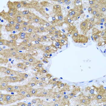
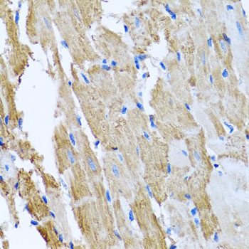
A01908 has real IHC images of paraffin-embedded human liver, rat heart, and mouse heart (catalog: IHC image captions); no IF image or IF application is listed (catalog: applications and images).

Real IHC data Immunohistochemistry of paraffin-embedded human liver using CCM2 antibody at dilution of 1:100 .Perform microwave antigen retrieval with 10 mM PBS buffer pH 7.2 before commencing with IHC staining protocol.
Anti-CCM2 Antibody
Cat # A01908

A01908 is listed for IHC in human, mouse, and rat samples (catalog: applications and reactivity). Its IHC images show paraffin-embedded human liver, rat heart, and mouse heart stained at 1:100 (catalog: IHC image captions).

Which to pick: Choose A01908 for tissue IHC in paraffin sections; its own images use microwave retrieval in 10 mM PBS, pH 7.2 (catalog: A01908 IHC image captions). A01908 is listed as reactive with human, mouse, and rat, but its clonality is unreported (catalog: reactivity and clone field). No IF/ICC choice is supported because A01908 has no listed IF application or IF image; its captions report paraffin embedding but do not report the fixative (catalog: applications, IF images, and IHC image captions).

Each figure is that product's own IHC / IF validation image from its datasheet.

References

  1. UniProt Consortium. UniProt entry Q9BSQ5 (CCM2_HUMAN, Cerebral cavernous malformations 2 protein).
  2. Human Protein Atlas. CCM2 tissue IHC expression (reliability: Supported).
  3. Human Protein Atlas. CCM2 subcellular location (ICC-IF): Localized to the mitochondria..
  4. Human Protein Atlas. CCM2 antibody validation summary (3 antibodies).
  5. A two-hit mechanism causes cerebral cavernous malformations: complete inactivation of CCM1, CCM2 or CCM3 in affected endothelial cells. Human molecular genetics 2009 — PMC2640205.
  6. Emerging roles of CCM genes during tumorigenesis with potential application as novel biomarkers across major types of cancers. Oncology reports 2020 — PMC7160551.
  7. Cerebral cavernous malformations proteins inhibit Rho kinase to stabilize vascular integrity. The Journal of experimental medicine 2010 — PMC2856024.
  8. Epicardial CCM2 Promotes Cardiac Development and Repair Via its Regulation on Cytoskeletal Reorganization. JACC. Basic to translational science 2024 — PMC10950406.
  9. PubMed PMID:14702039 — UniProt-cited evidence.
  10. PubMed PMID:15498874 — UniProt-cited evidence.
  11. PubMed PMID:12853948 — UniProt-cited evidence.