This website uses cookies to ensure you get the best experience on our website.


- Table of Contents
Plan CCNA1 chromogenic IHC in paraffin sections using ciliated-cell staining in bronchus and fallopian tube as tissue references (HPA tissue IHC). Compare the nuclear expectation (UniProt) with observed staining, and titrate the IHC-validated antibody at 1:50–1:200 (datasheet: A03889-3).

Expected localisation, validated protocols, controls and antibodies — the at-a-glance facts below, then the full design guide.
| Expected localisation | Nucleus expected (UniProt); ciliary rootlets observed (HPA tissue IHC) | |
| Staining pattern | Bronchial ciliated cell bodies and fallopian ciliary rootlets stain (HPA tissue IHC) | |
| Antigen retrieval | Tris-EDTA pH 9.0 HIER, 95–98 °C, 20 min (rule: nuclear antigen) | |
| Positive control | Bronchus+2 more · see all | |
| Negative control | Adipose tissue+4 more · see all |
| Fixation | Keep fixation consistent across sections. (standard IHC practice; not target-specific) | |
| Caveat | Nuclear expectation diverges from ciliary-rootlet staining (UniProt; HPA tissue IHC) | |
| Regulation | Very high expression in testis (UniProt) | |
| Isoform / epitope | 3 isoforms; no processing annotated (UniProt) |
The catalog antibody’s IHC-P protocol is paired with published CCNA1 staining protocols for gastric cancer and head and neck squamous cell carcinoma (PMC4598091; PMC3404904).
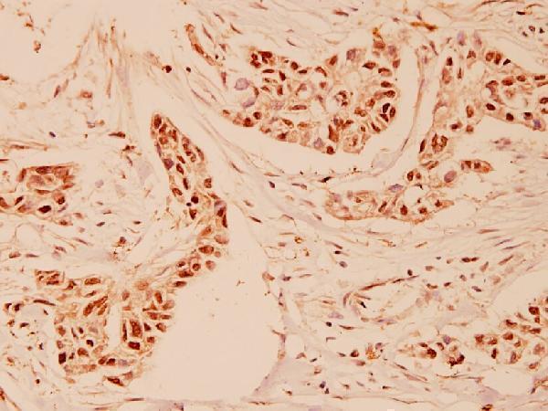
| Sample | Paraffin-embedded human breast carcinoma tissue; fixative not specified (datasheet A03889-3) |
| Fixation | Image fixative and duration unreported (datasheet A03889-3); verify before use. |
| Sectioning | 4–5 µm sections on charged slides (standard) |
| Deparaffinisation | Xylene, graded ethanol series to water (standard) |
| Antigen retrieval | Heat-induced epitope retrieval in Tris-EDTA buffer, pH 9.0, 20 min at 95–98 °C (standard rule: nuclear antigen) |
| Peroxidase block | 3% H2O2, 10 min, room temperature (standard) |
| Blocking | 10% normal serum of the secondary host, 30 min, room temperature (standard) |
| Primary antibody | Rabbit anti-CCNA1, 1:50-1:200 (datasheet A03889-3) |
| Primary incubation | Overnight at 4 °C (standard) |
| Detection | HRP-polymer secondary, DAB chromogen 5–10 min (standard) |
| Counterstain | Hematoxylin, blue, dehydrate and mount (standard) |
| Expected result | CCNA1-positive staining in ciliated cells (cell body) of bronchus (HPA tissue IHC: High). HPA tissue profile: Expression in mainly in testis. No signal in the no-primary control. |
CCNA1 is a nuclear protein with no transmembrane segment (UniProt P78396). In tissue IHC, expect low staining in testis pachytene spermatocytes and high staining in bronchial ciliated cell bodies; HPA also reports high staining at fallopian tube ciliary rootlets (HPA tissue IHC). Tissue IHC reliability is Enhanced, with medium consistency between staining and RNA expression (HPA tissue IHC). The rootlet observation needs care because it differs from the reported nuclear localisation (UniProt P78396; HPA subcellular ICC-IF).
| Nuclear staining in pachytene spermatocytes, even if faint (HPA tissue IHC; UniProt P78396). | This fits the reported low testis IHC signal and nuclear localisation (HPA tissue IHC; UniProt P78396). Assess the named cells rather than treating uniformly strong staining throughout testis as the expected pattern (HPA tissue IHC). |
| Strong staining in bronchial ciliated cell bodies (HPA tissue IHC). | The cell type and high signal match the tissue observation (HPA tissue IHC). Check whether signal is nuclear, since HPA's tissue entry specifies the cell body but does not resolve its subcellular pattern; nucleoplasm and nuclear bodies are approved ICC-IF locations (HPA tissue IHC; HPA subcellular ICC-IF). |
| Predominantly diffuse cytoplasmic staining, or staining confined to ciliary rootlets (HPA tissue IHC). | Diffuse cytoplasmic staining conflicts with reported nuclear localisation (UniProt P78396; HPA subcellular ICC-IF). Rootlet staining is reported as high in fallopian tube, but its apparent compartment differs from those localisation records; assess it separately before calling it specific CCNA1 (HPA tissue IHC; UniProt P78396; HPA subcellular ICC-IF). |
| Strong staining in cells listed as not detected, such as adipocytes or bone marrow hematopoietic cells (HPA tissue IHC). | This disagrees with the cited tissue observations and raises possible cross-reactivity or endogenous detection activity (HPA tissue IHC; standard IHC practice). Compare a reagent-omission control and the expected cell distribution before scoring it as CCNA1 (standard IHC practice). |
| No discernible staining in a selected positive tissue, or color spread across most of the section (HPA tissue IHC; standard IHC practice). | Absent signal in testis alone is inconclusive because pachytene spermatocyte staining is reported as low; bronchial ciliated cell bodies offer a reported high-signal comparison (HPA tissue IHC). Widespread color obscuring cell boundaries suggests background rather than a readable localisation pattern (standard IHC practice). |
| Tissue and cell selection (HPA tissue IHC) | Testis pachytene spermatocytes are reported low, bronchial ciliated cell bodies high, and several listed cell types not detected; use the specific cell population when judging whether a section behaves as expected (HPA tissue IHC). |
| Compartment and topology (UniProt P78396; HPA subcellular ICC-IF) | UniProt places CCNA1 in the nucleus and reports no transmembrane segment; approved ICC-IF locations are nucleoplasm and nuclear bodies (UniProt P78396; HPA subcellular ICC-IF). These records support a nuclear cross-check but do not resolve the tissue rootlet observation (HPA tissue IHC). |
| Antibody evidence by application (HPA antibodies; HPA tissue IHC) | HPA077614 has Enhanced IHC validation, whereas HPA060646 has Approved ICC status and no listed IHC status (HPA antibodies). The overall tissue reliability is Enhanced despite medium staining–RNA consistency; do not transfer an ICC approval to an IHC interpretation (HPA tissue IHC; HPA antibodies). |
| Isoforms and processing (UniProt P78396) | Three isoforms are listed, with no annotated signal peptide, propeptide or glycosylation sites (UniProt P78396). The supplied records give no epitope location or isoform-specific IHC pattern, so they cannot predict which isoform an individual stain detects (UniProt P78396). |
| IF/ICC cross-check (HPA subcellular ICC-IF) | What should an IF/ICC comparison show? Approved localisation is nucleoplasm and nuclear bodies, with images listed for PC-3, Rh30 and U2OS (HPA subcellular ICC-IF). Use that location as a comparison, while interpreting tissue IHC against its own cell-specific observations (HPA tissue IHC). |
| Situation | Likely cause | Next action |
|---|---|---|
| Testis section appears entirely negative (HPA tissue IHC). | The reported signal in pachytene spermatocytes is low, so a negative-looking field may miss the relevant cells or a subtle signal (HPA tissue IHC). | Confirm pachytene spermatocytes are present and inspect nuclei; compare a bronchial section containing ciliated cells, reported high, while checking the general IHC detection controls (HPA tissue IHC; standard IHC practice). |
| Ciliated cells stain, but only at fallopian tube ciliary rootlets (HPA tissue IHC). | High rootlet staining is reported in tissue IHC, yet nuclear localisation is reported by UniProt and ICC-IF; the records do not explain the difference (HPA tissue IHC; UniProt P78396; HPA subcellular ICC-IF). | Record rootlet and nuclear staining separately; compare appropriate reagent-omission controls and another reported positive cell population before interpreting the rootlet signal as specific (HPA tissue IHC; standard IHC practice). |
| Broad cytoplasmic signal obscures nuclei (UniProt P78396; HPA subcellular ICC-IF). | The pattern differs from reported nuclear localisation and may reflect nonspecific staining or background in the IHC workflow (UniProt P78396; HPA subcellular ICC-IF; standard IHC practice). | Review the counterstain and control section, then check blocking, antibody concentration and detection settings using general IHC practice; no CCNA1-specific dilution or retrieval condition is supplied (standard IHC practice). |
| Strong staining appears in listed negative cells (HPA tissue IHC). | Adipocytes and bone marrow hematopoietic cells are reported as not detected, so cross-reactivity or endogenous detection activity is possible (HPA tissue IHC; standard IHC practice). | Compare a primary-antibody-omission control and inspect whether color follows the expected cells and nuclear compartment; address endogenous detection activity if the control also develops color (standard IHC practice; HPA tissue IHC; UniProt P78396). |
| Brown color is widespread even where cell structure is hard to distinguish (standard IHC practice). | Diffuse chromogen can arise from general staining background or endogenous detection activity; the appearance alone cannot identify which (standard IHC practice). | Check reagent-omission controls, blocking and chromogen development, then reassess localisation against the counterstain and HPA's named cell populations (standard IHC practice; HPA tissue IHC). |
| A positive section loses signal after a workflow change (standard IHC practice). | Target-specific fixation effects are not established by the supplied assay evidence. Verify with a matched IHC source before attributing a result to fixation. | Compare matched sections and document the changed retrieval, antibody and detection steps; verify the assay controls before assigning the loss to CCNA1 biology (standard IHC practice). |


Comprehensive Human Protein Atlas IHC scoring per tissue (reliability: Enhanced — Medium consistency between antibody staining and RNA expression data.). Rows are taken directly from the HPA tissue chart — click any row's HPA link to view the source.
| Tissue | Cell type | Level | Evidence | Source |
|---|---|---|---|---|
| Bronchus | Ciliated cells (cell body) | High | Protein (IHC) | HPA → |
| Fallopian tube | Ciliated cells (ciliary rootlets) | High | Protein (IHC) | HPA → |
| Nasopharynx | Ciliated cells (cell body) | Medium | Protein (IHC) | HPA → |
| Tissue | Cell type | Level | Evidence | Source |
|---|---|---|---|---|
| Adipose tissue | Adipocytes | Not detected | Protein (IHC) | HPA → |
| Adrenal gland | Glandular cells | Not detected | Protein (IHC) | HPA → |
| Appendix | Glandular cells | Not detected | Protein (IHC) | HPA → |
| Bone marrow | Hematopoietic cells | Not detected | Protein (IHC) | HPA → |
| Breast | Adipocytes | Not detected | Protein (IHC) | HPA → |
Troubleshoot CCNA1 staining in paraffin sections by checking retrieval, nuclear localisation, controls and cell-specific scoring (UniProt P78396; HPA subcellular).
Real IHC data show human paraffin-embedded breast carcinoma tissue (A03889-3 image caption); IF/ICC data show PC-3 cells (PB9485 IF caption). Both antibodies list Human, Mouse, and Rat reactivity (catalog reactivity).
The rendered card is A03889-3, with IHC demonstrated on paraffin-embedded human breast carcinoma tissue at 1:100 (A03889-3 image caption). PB9485 has IF/ICC data from PC-3 cells but has no IHC card here (PB9485 IF caption; catalog applications).
Which to pick: Choose A03889-3 for paraffin-section IHC because its own image documents that preparation; the fixative is unreported (A03889-3 image caption). Choose PB9485 for IF/ICC because its cell image documents that application (PB9485 IF caption). Both list Human, Mouse, and Rat reactivity, while the supplied images document human samples only (catalog reactivity; A03889-3 image caption; PB9485 IF caption).