This website uses cookies to ensure you get the best experience on our website.


- Table of Contents
Plan CCNE2 chromogenic IHC in paraffin sections around its nuclear staining pattern, which appears in a varying fraction of tissue cells (HPA tissue IHC). This guide covers fixation, staining controls and scoring of positive nuclei.

Expected localisation, validated protocols, controls and antibodies — the at-a-glance facts below, then the full design guide.
| Expected localisation | Nuclear staining in a varying fraction of cells (HPA tissue IHC) | |
| Staining pattern | Nuclear staining in subsets of glandular and hematopoietic cells (HPA tissue IHC) | |
| Antigen retrieval | Tris-EDTA pH 9.0 HIER, 95–98 °C, 20 min (rule: nuclear antigen) | |
| Positive control | Appendix+4 more · see all | |
| Negative control | Adipose tissue+4 more · see all |
| Fixation | Keep fixation consistent across sections (standard IHC practice; not target-specific). Selected-image fixative and duration unreported (datasheet M04536); verify before use. | |
| Caveat | The fraction of positive nuclei varies between tissues (HPA tissue IHC) | |
| Regulation | Elevated in tumor-derived cells (UniProt) | |
| Isoform / epitope | 2 isoforms, Long and Short; epitope coverage is unknown (UniProt) |
The catalog antibody IHC-P protocol is accompanied by published CCNE2 protocols for paraffin sections (PMC8459753; PMC8656552).
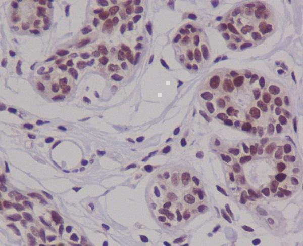
| Sample | Paraffin-embedded human breast carcinoma tissue; fixative not specified (datasheet M04536) |
| Fixation | Image fixative and duration unreported (datasheet M04536); verify before use. |
| Sectioning | 4–5 µm sections on charged slides (standard) |
| Deparaffinisation | Xylene, graded ethanol series to water (standard) |
| Antigen retrieval | Heat-induced epitope retrieval in Tris-EDTA buffer, pH 9.0, 20 min at 95–98 °C (standard rule: nuclear antigen) |
| Peroxidase block | 3% H2O2, 10 min, room temperature (standard) |
| Blocking | 10% normal serum of the secondary host, 30 min, room temperature (standard) |
| Primary antibody | Rabbit monoclonal (clone GHH-3) anti-CCNE2, 1:50 (datasheet M04536) |
| Primary incubation | Overnight at 4 °C (standard) |
| Detection | HRP-polymer secondary, DAB chromogen 5–10 min (standard) |
| Counterstain | Hematoxylin, blue, dehydrate and mount (standard) |
| Expected result | CCNE2-positive staining in glandular cells of appendix (HPA tissue IHC: Medium). HPA tissue profile: Nuclear expression in varying fraction of cells in several tissues. No signal in the no-primary control. |
CCNE2 should appear primarily in nuclei, in a varying fraction of cells rather than uniformly across a section (UniProt O96020: nucleus; HPA tissue IHC: nuclear expression). HPA reports medium staining in selected glandular, hematopoietic, glial, germinal center, trophoblastic and keratinocyte populations (HPA tissue IHC). Its tissue assessment is Enhanced, with medium consistency between antibody staining and RNA expression (HPA tissue IHC). CCNE2 has no transmembrane segment (UniProt O96020: topology).
| Discrete nuclear staining in some hematopoietic cells of bone marrow or germinal center cells of lymph node, with other cells unstained. | This fits the reported medium staining in those populations and the varying fraction of nuclear-positive cells (HPA tissue IHC). Record the positive fraction and intensity within the identified cell population; a uniformly positive field is not required by the HPA pattern (HPA tissue IHC). |
| Predominantly cytoplasmic or membrane-like staining, with little nuclear signal. | This conflicts with the recorded nuclear location and absence of a transmembrane segment (UniProt O96020: location and topology; HPA tissue IHC). Treat it as a possible staining artefact, then review morphology and controls before assigning a CCNE2 result (general IHC practice). |
| Strong staining in adipocytes or in an unrelated cell population while expected nuclei are negative. | HPA reports adipocytes as not detected in its listed adipose and breast samples (HPA tissue IHC). Check whether the signal follows tissue pigments, endogenous detection activity or antibody cross-reactivity; morphology and control sections help distinguish these possibilities (general IHC practice). |
| Diffuse color across tissue, including extracellular spaces, with no clear nuclear boundaries. | This does not resemble the reported nuclear pattern (HPA tissue IHC; UniProt O96020: nucleus). Review no-primary and detection controls, blocking, washing and chromogen development before interpreting weak nuclear color against the background (general IHC practice). |
| No signal in a section chosen as a positive comparator. | First confirm that the sampled cell population matches a reported positive one: HPA lists medium staining in bone marrow hematopoietic cells, not every cell in bone marrow (HPA tissue IHC). A negative field can reflect sampling or a staining-run problem; compare the control slide and run conditions before calling the specimen negative (general IHC practice). |
| Cell-cycle distribution | CCNE2 acts in late G1 and early S phase (UniProt O96020: function), and HPA reports a varying fraction of nuclear-positive cells (HPA tissue IHC). Interpret the fraction within each cell population rather than expecting every nucleus to stain. |
| Choice of tissue and cell population | HPA reports medium staining in appendix glandular cells, placenta trophoblastic cells and skin keratinocytes, but not detected staining in several other specified cell populations (HPA tissue IHC). Name the cell population when selecting and scoring a comparator. |
| Antibody evidence | CAB007825 has Enhanced IHC status and CAB019374 has Approved IHC status (HPA antibodies). The HPA tissue profile has Enhanced reliability with medium RNA–staining consistency (HPA tissue IHC); these ratings do not establish that every specimen or cell will stain. |
| Isoforms and epitope | UniProt lists Long and Short isoforms and a single annotated 1–404 chain (UniProt O96020: isoforms and processing). The supplied records give no antibody epitope or isoform coverage, so isoform-specific staining cannot be assigned from these data. |
| IF/ICC: is the nuclear pattern independently confirmed by cell images? | HPA supplies no main ICC-IF location, additional location or cell-line images for CCNE2 (HPA subcellular). Use the IHC nuclear pattern as an expectation only; this record cannot establish an IF/ICC staining pattern or protocol. |
| Situation | Likely cause | Next action |
|---|---|---|
| A putative positive section shows no nuclear staining. | The examined field may lack the relevant cells, or the staining run may have failed (HPA tissue IHC: varying positive fraction; general IHC practice). | Locate the reported cell population on the section, then inspect the positive control and the run's reagent and detection records before interpreting absence of signal (HPA tissue IHC; general IHC practice). |
| Color is concentrated outside nuclei. | The distribution disagrees with CCNE2's recorded nuclear location (UniProt O96020: nucleus; HPA tissue IHC). Nonspecific staining or misidentified cell boundaries are possible (general IHC practice). | Review the counterstain and morphology, compare a no-primary control, and score only clearly assigned nuclei (general IHC practice). |
| Many unexpected cells stain, including adipocytes. | HPA lists adipocytes as not detected in adipose tissue and breast samples (HPA tissue IHC). Unexpected color may arise from cross-reactivity or endogenous detection activity (general IHC practice). | Check the affected cell identity and control sections; compare staining with the reported positive and negative populations before attributing the color to CCNE2 (HPA tissue IHC; general IHC practice). |
| Weak nuclear color cannot be separated from diffuse background. | Background from the detection workflow can obscure a varying fraction of positive nuclei (general IHC practice; HPA tissue IHC: nuclear expression in varying fractions). | Inspect no-primary and detection controls; review blocking, washes and chromogen development, then reassess whether individual nuclei have distinct signal (general IHC practice). |
| A low-staining lung sample is treated as a failed positive control. | HPA reports low staining in lung alveolar cells, while UniProt cites elevated lung expression from one literature account (HPA tissue IHC; UniProt O96020: tissue specificity). The records address different evidence and do not guarantee strong IHC staining. | Use the HPA cell-specific IHC observation to judge that slide; choose a reported medium-staining population when a clearer IHC comparator is needed (HPA tissue IHC). |
| Two sections differ in the fraction of stained nuclei. | HPA describes a varying positive fraction, and CCNE2 has a late-G1/early-S cell-cycle role (HPA tissue IHC; UniProt O96020: function). Neither source establishes a target-specific fixation effect. | Compare the same identified cell population, record positive fraction and intensity, and check run controls before treating the difference as biological (HPA tissue IHC; general IHC practice). |

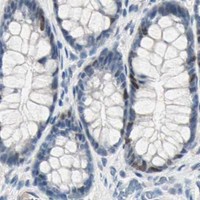


Comprehensive Human Protein Atlas IHC scoring per tissue (reliability: Enhanced — Medium consistency between antibody staining and RNA expression data.). Rows are taken directly from the HPA tissue chart — click any row's HPA link to view the source.
| Tissue | Cell type | Level | Evidence | Source |
|---|---|---|---|---|
| Appendix | Glandular cells | Medium | Protein (IHC) | HPA → |
| Bone marrow | Hematopoietic cells | Medium | Protein (IHC) | HPA → |
| Caudate | Glial cells | Medium | Protein (IHC) | HPA → |
| Cerebellum | Cells in molecular layer | Medium | Protein (IHC) | HPA → |
| Cerebral cortex | Glial cells | Medium | Protein (IHC) | HPA → |
| Tissue | Cell type | Level | Evidence | Source |
|---|---|---|---|---|
| Adipose tissue | Adipocytes | Not detected | Protein (IHC) | HPA → |
| Adrenal gland | Glandular cells | Not detected | Protein (IHC) | HPA → |
| Breast | Adipocytes | Not detected | Protein (IHC) | HPA → |
| Cervix | Glandular cells | Not detected | Protein (IHC) | HPA → |
| Endometrium | Cells in endometrial stroma | Not detected | Protein (IHC) | HPA → |
Troubleshoot CCNE2 staining in paraffin sections by checking retrieval, nuclear localisation, antibody specificity and cell level scoring before interpreting chromogenic signal.
Anti-CCNE2 antibodies cover IHC and IF/ICC in human and mouse samples (catalog: M04536); a rat-reactive option is also listed (catalog: A04536-3). M04536 has IHC and IF image captions (catalog: M04536).
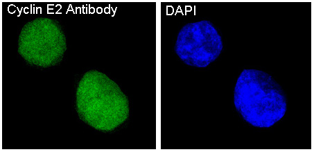
The rendered card is M04536, with IHC shown in paraffin-embedded human breast carcinoma (M04536 IHC image caption). It also lists IF/ICC for human and mouse and has an IF image caption for HeLa cells (catalog: M04536 applications, reactivity and IF image caption).
Which to pick: For tissue IHC, choose M04536: its own image caption documents paraffin-embedded human breast carcinoma, while the fixative is unreported (M04536 IHC image caption). For IF/ICC, M04536 lists both applications and has an IF image caption for HeLa cells (catalog: M04536 applications and IF image caption). For rat samples, consider polyclonal A04536-3, which lists rat reactivity and IHC and IF applications (catalog: A04536-3); its payload provides no IHC image caption.