This website uses cookies to ensure you get the best experience on our website.


- Table of Contents
Plan CCNK IHC-P around its broad nuclear tissue pattern (HPA tissue IHC). Compare high staining in placental trophoblasts with undetected staining in adipocytes, and optimize the catalog antibody within 1:50–1:200 (HPA tissue IHC; datasheet A06254).

Expected localisation, validated protocols, controls and antibodies — the at-a-glance facts below, then the full design guide.
| Expected localisation | Nuclear staining across tissues (HPA tissue IHC) | |
| Staining pattern | Broad nuclear staining in tissue cells (HPA tissue IHC) | |
| Antigen retrieval | Citrate pH 6.0 HIER, heat-mediated (datasheet A06254) | |
| Positive control | Adrenal gland+4 more · see all | |
| Negative control | Adipose tissue+2 more · see all |
| Fixation | Matched tissue-IHC evidence does not establish the fixation claim. Validate the specimen-specific method before use. (selected-SKU IHC image A06254) | |
| Caveat | Adipocytes may lack detectable staining (HPA tissue IHC) | |
| Regulation | Widely expressed; highest in testis (UniProt) | |
| Isoform / epitope | 4 isoforms; epitope effects unknown (UniProt) |
The catalog antibody has a datasheet IHC-P protocol (datasheet A06254). The published endometrial tissue protocol below uses a different retrieval buffer (PMC11898804 methods).
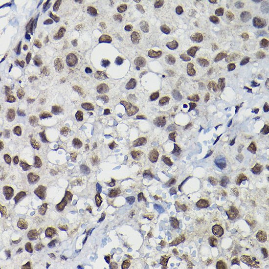
| Sample | Paraffin-embedded human breast cancer tissue; fixative not specified (datasheet A06254) |
| Fixation | Image fixative and duration unreported (datasheet A06254); verify before use. |
| Sectioning | 4–5 µm sections on charged slides (standard) |
| Deparaffinisation | Xylene, graded ethanol series to water (standard) |
| Antigen retrieval | Heat retrieval: Citrate pH 6.0 (datasheet A06254); 20 min, 95–100 °C (standard) |
| Peroxidase block | 3% H2O2, 10 min, room temperature (standard) |
| Blocking | 10% normal serum of the secondary host, 30 min, room temperature (standard) |
| Primary antibody | Rabbit anti-CCNK, 1:50-1:200 (datasheet A06254) |
| Primary incubation | Overnight at 4 °C (standard) |
| Detection | HRP-polymer secondary, DAB chromogen 5–10 min (standard) |
| Counterstain | Hematoxylin, blue, dehydrate and mount (standard) |
| Expected result | CCNK-positive staining in glandular cells of adrenal gland (HPA tissue IHC: High). HPA tissue profile: Ubiquitous nuclear expression. No signal in the no-primary control. |
CCNK should appear predominantly in nuclei in paraffin-section IHC (UniProt O75909: nucleus; HPA: ubiquitous nuclear expression). Expect staining across many cell types, with high signal reported in adrenal, epididymal, and parathyroid glandular cells, cerebral cortical glia, and placental trophoblasts (HPA: tissue IHC). HPA rates the tissue pattern Supported, with medium consistency between antibody staining and RNA data (HPA: reliability). CCNK has no transmembrane segment (UniProt O75909: topology).
| Clear nuclear staining in adrenal glandular cells or placental trophoblasts, with nuclei identifiable by counterstain. | This matches high staining in those cells (HPA: tissue IHC) and CCNK's nuclear location (UniProt O75909). Compare cells within the same section: HPA reports cell-specific levels, so a uniformly dark field is less informative than identifiable stained nuclei (general IHC practice). |
| Strong, convincing cytoplasmic or membrane staining dominates while nuclei remain weak. | Treat this as a localisation mismatch requiring investigation: CCNK is nuclear (UniProt O75909), and HPA describes ubiquitous nuclear tissue staining (HPA: tissue IHC). Review controls and detection conditions before scoring that signal as CCNK; morphology alone cannot identify the cause (general IHC practice). |
| Adipocytes, liver cholangiocytes, or splenic red-pulp cells stain strongly. | These particular cell populations were not detected in the supplied HPA tissue observations (HPA: tissue IHC). Suspect cross-reactivity or endogenous detection activity, and assess a no-primary control (general IHC practice). Do not call the entire adipose, liver, or spleen section negative. |
| Brown signal coats stroma, cytoplasm, or most of the section without crisp nuclear boundaries. | Diffuse background prevents a confident nuclear call (UniProt O75909: nucleus; general IHC practice). Compare a no-primary control and inspect blocking, washes, detection reagent, and counterstain (general IHC practice). HPA tissue levels cannot establish which workflow step caused the background. |
| No nuclear signal appears in adrenal glandular cells or placental trophoblasts. | Those are useful expected-positive cell populations because HPA reports high staining there (HPA: tissue IHC). Check section integrity, the catalog antibody's IHC-P instructions, and detection controls before interpreting absence as biology (general IHC practice). HPA's Supported rating is not a guarantee for every specimen. |
| Tissue and cell selection | HPA reports high staining in five named cell populations, medium staining in appendix, breast, and bronchus, and low staining in several other populations (HPA: tissue IHC). Select and score the specified cells, since a whole-tissue label can obscure these differences. |
| IHC evidence strength | The tissue profile is Supported, with medium agreement between antibody staining and RNA expression (HPA: reliability). HPA000645 has no supplied IHC status; HPA077073 is Supported for IHC (HPA: antibody validation). Interpret discordant staining with this distinction in view. |
| Isoforms and epitope coverage | Four CCNK isoforms are listed (UniProt O75909: isoforms). Their presence alone does not show which isoforms the catalog antibody recognises; an epitope map would be needed to make that claim. Do not explain a cell-level staining difference by isoform choice without supporting evidence. |
| Processing and nuclear context | UniProt lists a single 1–580 CCNK chain, with no signal peptide, propeptide, or transmembrane segment (UniProt O75909: processing and topology). These annotations support a nuclear interpretation; they provide no basis here for predicting a secreted or surface staining pattern. |
| Antigen retrieval | Use the catalog antibody's IHC-P retrieval instructions when available and judge the result against expected nuclear staining (general IHC practice; UniProt O75909: nucleus). No supplied source establishes CCNK-specific fixation sensitivity or predicts which retrieval condition will recover signal. |
| IF/ICC: where should CCNK appear? | HPA reports enhanced nucleoplasmic localisation and Enhanced ICC validation for HPA000645 and HPA077073 (HPA: subcellular location; HPA: antibody validation). Expect nucleoplasmic fluorescence. These ICC findings support localisation interpretation here; IF/ICC procedure belongs in its separate guide. |
| Situation | Likely cause | Next action |
|---|---|---|
| Expected-positive nuclei are blank. | The assay may have failed, or the sampled cells may be unsuitable; HPA reports high staining in adrenal glandular cells and placental trophoblasts (HPA: tissue IHC). | Verify the cell identity and section, run an appropriate positive control, and follow the catalog antibody's IHC-P retrieval and dilution instructions (general IHC practice). Do not infer a CCNK-specific fixation effect from this result. |
| Nuclear signal is faint throughout a control section. | Detection conditions may be weak, although HPA's high categories describe particular cell populations rather than every cell in a tissue (HPA: tissue IHC; general IHC practice). | Confirm that the expected cells are present; review the catalog IHC-P dilution, detection reagents, and development time while retaining a no-primary control (general IHC practice). |
| Cytoplasm or cell borders stain more strongly than nuclei. | The dominant compartment conflicts with CCNK's nuclear annotation and HPA's nuclear tissue profile (UniProt O75909: subcellular location; HPA: tissue IHC). | Check morphology against the counterstain, compare a no-primary control, and reassess antibody specificity before scoring the pattern as CCNK (general IHC practice). |
| Brown staining appears in expected-negative cell populations. | Adipocytes, cholangiocytes, and splenic red-pulp cells are listed as not detected (HPA: tissue IHC); apparent signal may reflect cross-reactivity or endogenous detection activity (general IHC practice). | Identify the stained cell type precisely, examine a no-primary control, and check the relevant endogenous-activity block for the detection chemistry (general IHC practice). |
| Diffuse precipitate obscures nuclear boundaries. | Background or excessive development can prevent compartment scoring (general IHC practice); HPA's nuclear profile does not explain a diffuse deposit (HPA: tissue IHC). | Inspect no-primary and reagent controls, washing, chromogen development, and counterstain; repeat only after the nuclei can be distinguished (general IHC practice). |
| A test tissue disagrees with an expected intensity category. | HPA reports a Supported profile with medium antibody–RNA consistency, and intensity categories apply to specified cells (HPA: reliability; HPA: tissue IHC). | Record the exact cell population and nuclear fraction, compare an expected-positive control in the same run, and describe the discrepancy without assigning an unsupported tissue-wide CCNK status (general IHC practice). |


Comprehensive Human Protein Atlas IHC scoring per tissue (reliability: Supported — Medium consistency between antibody staining and RNA expression data.). Rows are taken directly from the HPA tissue chart — click any row's HPA link to view the source.
| Tissue | Cell type | Level | Evidence | Source |
|---|---|---|---|---|
| Adrenal gland | Glandular cells | High | Protein (IHC) | HPA → |
| Cerebral cortex | Glial cells | High | Protein (IHC) | HPA → |
| Epididymis | Glandular cells | High | Protein (IHC) | HPA → |
| Parathyroid gland | Glandular cells | High | Protein (IHC) | HPA → |
| Placenta | Trophoblastic cells | High | Protein (IHC) | HPA → |
CCNK is predominantly nuclear, so evaluate chromogenic staining by cell type and compartment alongside matched controls (UniProt O75909; HPA tissue IHC).
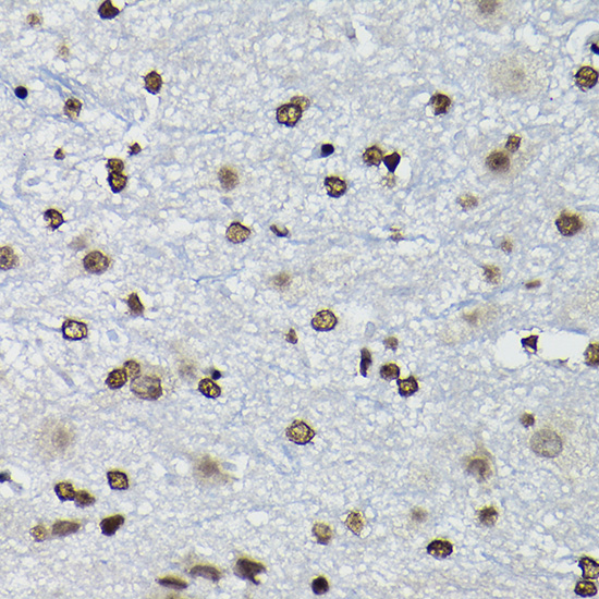
The catalog antibody has real IHC images from paraffin-embedded human breast cancer and mouse spinal cord (A06254 image captions). IF/ICC image data are unreported (catalog: no IF images).
A06254 has IHC images of paraffin-embedded human breast cancer and mouse spinal cord, each stained at 1:100 (A06254 image captions). Its listed applications are IHC and WB, and its listed reactivity is Human, Mouse, and Rat (catalog: A06254 applications and reactivity).
Which to pick: Choose A06254 for tissue IHC: it is a rabbit polyclonal antibody with paraffin-section IHC images in human and mouse tissue (catalog: A06254 host; A06254 image captions). For cross-species work, Human, Mouse, and Rat are listed as reactive, but the supplied IHC images cover Human and Mouse only (catalog: A06254 reactivity; A06254 image captions). No IF/ICC application or image is listed, so there is no supported IF/ICC pick here; the IHC captions specify paraffin embedding and citrate retrieval, while the fixative is unreported (catalog: A06254 applications and IF images; A06254 image captions).