This website uses cookies to ensure you get the best experience on our website.


- Table of Contents
Plan chromogenic paraffin-section IHC around granular cytoplasmic staining, with high signal in bronchial ciliated cells and late spermatids (HPA tissue IHC). Start with 2–5 μg/ml of the IHC-validated antibody (datasheet A05058-1), and interpret staining cautiously because agreement with RNA expression is low (HPA tissue IHC).

Expected localisation, validated protocols, controls and antibodies — the at-a-glance facts below, then the full design guide.
| Expected localisation | Granular cytoplasm in tissue sections (HPA tissue IHC) | |
| Staining pattern | Granular cytoplasm; high in ciliated cells and late spermatids (HPA tissue IHC) | |
| Antigen retrieval | EDTA pH 8.0 HIER, heat-mediated (datasheet A05058-1) | |
| Positive control | Bronchus+4 more · see all | |
| Negative control | Adipose tissue+1 more · see all |
| Fixation | Keep fixation consistent across sections (standard IHC practice; not target-specific) | |
| Caveat | Tissue staining has low agreement with RNA expression (HPA tissue IHC) | |
| Regulation | Ubiquitination promotes degradation (UniProt) | |
| Isoform / epitope | 2 isoforms; epitope coverage is unknown (UniProt) |
The catalog antibody uses EDTA pH 8.0 retrieval (datasheet A05058-1). One published CCP110 tissue-section protocol provides a citrate-based alternative (PMC9996364).
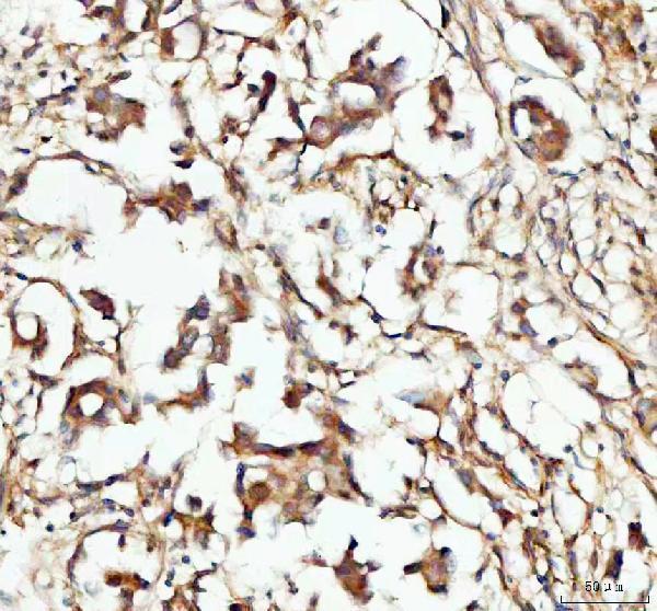
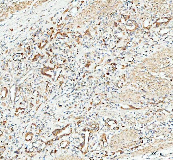
| Sample | Paraffin-embedded human breast cancer tissue; fixative not specified (datasheet A05058-1) |
| Fixation | Image fixative and duration unreported (datasheet A05058-1); verify before use. |
| Sectioning | 4–5 µm sections on charged slides (standard) |
| Deparaffinisation | Xylene, graded ethanol series to water (standard) |
| Antigen retrieval | Heat retrieval: EDTA pH 8.0 (datasheet A05058-1); 20 min, 95–100 °C (standard) |
| Peroxidase block | 3% H2O2, 10 min, room temperature (standard) |
| Blocking | 10% goat serum (datasheet A05058-1) |
| Primary antibody | Rabbit anti-CCP110, 2-5μg/ml (datasheet A05058-1) |
| Primary incubation | Overnight at 4 °C (datasheet A05058-1) |
| Detection | HRP-conjugated secondary, DAB chromogen (datasheet A05058-1) |
| Counterstain | Hematoxylin, blue, dehydrate and mount (standard) |
| Expected result | CCP110-positive staining in ciliated cells (cell body) of bronchus (HPA tissue IHC: High). HPA tissue profile: Granular cytoplasmic expression in most tissues, including testis. No signal in the no-primary control. |
CCP110 localizes to centrioles, centrosomes and cilium basal bodies and has no transmembrane segment (UniProt O43303 localization and topology). In paraffin-section IHC, expect granular cytoplasmic staining, especially in bronchial ciliated-cell bodies and testicular late spermatids (HPA tissue IHC). HPA rates the tissue staining Approved but reports low consistency between antibody staining and RNA expression; interpret intensity with that limitation (HPA tissue IHC).
| Granular cytoplasmic stain is strong in bronchial ciliated-cell bodies or elongated or late spermatids (HPA tissue IHC). | This matches HPA's High observations in those cell populations (HPA tissue IHC). Small cytoplasmic granules fit CCP110's centrosomal and centriolar localization, although chromogenic IHC alone cannot identify an individual centriole (UniProt O43303 localization; general IHC practice). |
| A sharp nuclear, surface-membrane or extracellular pattern dominates instead of cytoplasmic granules (HPA tissue IHC; UniProt O43303 topology). | Treat the compartment mismatch as possible artefact: UniProt places CCP110 at centrosomal structures and reports no transmembrane segment (UniProt O43303 localization and topology). Compare the pattern with a known-positive section and the negative reagent control before scoring it (general IHC practice). |
| Adipocytes or smooth muscle cells stain strongly while expected positive cells do not (HPA tissue IHC). | HPA reports CCP110 as Not detected in adipocytes and smooth muscle cells (HPA tissue IHC). A signal confined to those cells warrants investigation of cross-reactivity or endogenous detection activity; morphology alone cannot distinguish the two (general IHC practice). |
| Brown precipitate is widespread across tissue compartments, obscuring cell borders and the expected granular pattern (HPA tissue IHC). | Diffuse background cannot be scored as CCP110-positive cells (general IHC practice). Review blocking, wash stringency, antibody concentration and chromogen development, then compare with a section processed without primary antibody (general IHC practice). |
| No convincing signal appears in bronchial ciliated-cell bodies or testicular late spermatids (HPA tissue IHC). | These are HPA High reference populations, so a blank result leaves assay performance unresolved (HPA tissue IHC). Check section identity and positive-control performance before interpreting another tissue as negative; HPA's RNA–staining consistency is low (HPA tissue IHC; general IHC practice). |
| Cell population and tissue | HPA records High staining in bronchial ciliated-cell bodies and late spermatids, Medium in several other listed cell populations, and Not detected in adipocytes and smooth muscle cells (HPA tissue IHC). Score the identified cell population rather than assigning one intensity to an entire section (general IHC practice). |
| Subcellular resolution | UniProt places CCP110 at centrioles, centrosomes and basal bodies (UniProt O43303 localization). HPA describes tissue IHC as granular cytoplasmic (HPA tissue IHC). A chromogenic granule can support this pattern, but cannot establish its precise organelle without higher-resolution localization (general IHC practice). |
| Ciliation state | CCP110 is removed from the mother centriole during initiation of ciliogenesis and remains at the daughter centriole in ciliated cells (UniProt O43303 localization). Do not require identical centriole-level placement across every cell; assess the tissue IHC pattern at the resolution available (general IHC practice). |
| Antibody evidence and isoforms | HPA lists antibody HPA039402 as IHC Approved and reports low consistency between antibody staining and RNA expression (HPA tissue IHC; HPA antibody record). UniProt lists two isoforms, but the supplied evidence does not establish whether this antibody detects them equally (UniProt O43303 isoforms). |
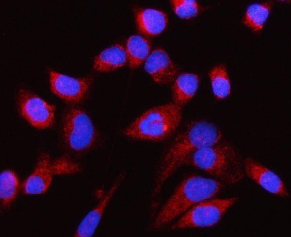
| IF/ICC: what localization is reported? | HPA reports an approved centriolar-satellite localization in ICC-IF (HPA subcellular). That observation can guide localization checks, while this section's primary readout remains granular cytoplasmic staining in paraffin-section IHC (HPA tissue IHC). |
| Situation | Likely cause | Next action |
|---|---|---|
| The positive-control section has no stain. | The assay may have failed, or the expected cell population may be absent from the section (general IHC practice). | Confirm bronchial ciliated-cell bodies or testicular late spermatids are present, then check antibody dilution, retrieval, detection reagents and run controls (HPA tissue IHC; general IHC practice). |
| Granules are too faint to score. | Weak detection or a cell population with lower observed staining may explain the result (general IHC practice; HPA tissue IHC). | Compare with HPA High reference cells before adjusting antibody dilution or detection conditions; HPA reports Medium staining in several other listed populations (HPA tissue IHC; general IHC practice). |
| Nuclei or cell surfaces dominate the stain. | The pattern conflicts with CCP110's centrosomal localization and lack of a transmembrane segment (UniProt O43303 localization and topology). | Review morphology, compare a known-positive section and inspect the no-primary control before assigning CCP110 positivity (general IHC practice). |
| The whole section has diffuse brown background. | Nonspecific antibody binding, incomplete washing or excessive chromogen development can obscure a granular signal (general IHC practice). | Review blocking and washes, titrate the primary antibody or shorten chromogen development; judge improvement against the no-primary control (general IHC practice). |
| Adipocytes or smooth muscle cells appear strongly positive. | Both are reported Not detected by HPA; cross-reactivity or endogenous detection activity is possible (HPA tissue IHC; general IHC practice). | Compare no-primary and appropriate detection controls, then reassess whether the expected positive cell population shows granular cytoplasmic staining (general IHC practice; HPA tissue IHC). |
| A tissue looks negative despite otherwise working controls. | Cell composition and staining level vary across HPA's listed tissues; antibody staining also has low consistency with RNA expression (HPA tissue IHC). | Score the relevant cells separately, document the control result and report the finding as an IHC observation without inferring absent CCP110 protein from a single negative section (general IHC practice; HPA tissue IHC). |
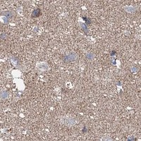


Comprehensive Human Protein Atlas IHC scoring per tissue (reliability: Approved — Low consistency between antibody staining and RNA expression data.). Rows are taken directly from the HPA tissue chart — click any row's HPA link to view the source.
| Tissue | Cell type | Level | Evidence | Source |
|---|---|---|---|---|
| Bronchus | Ciliated cells (cell body) | High | Protein (IHC) | HPA → |
| Testis | Elongated or late spermatids | High | Protein (IHC) | HPA → |
| Adrenal gland | Glandular cells | Medium | Protein (IHC) | HPA → |
| Appendix | Glandular cells | Medium | Protein (IHC) | HPA → |
| Bone marrow | Hematopoietic cells | Medium | Protein (IHC) | HPA → |
Troubleshoot CCP110 staining in paraffin sections by checking retrieval, controls, compartment, and cell type before comparing staining intensity across samples.
Anti-CCP110 antibody A05058-1 has human paraffin-section IHC and HELA-cell IF images (catalog image captions); listed reactivity covers human, mouse and rat (catalog reactivity).
A05058-1 is listed for IHC, with images of paraffin-embedded human breast cancer, colorectal adenocarcinoma and testicular germ cell tumor sections (catalog applications; A05058-1 IHC image captions). A05058-1 is also listed for IF/ICC, with an IF image in HELA cells (catalog applications; A05058-1 IF image caption).
Which to pick: For tissue IHC, choose A05058-1: its own paraffin-section captions document EDTA retrieval at pH 8.0 and 2 μg/ml primary antibody; the fixative is unreported (A05058-1 IHC image captions). For IF/ICC, A05058-1 is the listed option, with a HELA-cell IF image at 5 μg/ml (catalog applications; A05058-1 IF image caption). For mouse or rat samples, A05058-1 lists reactivity with both species, while the supplied images show human samples; clone type is unreported (catalog reactivity; A05058-1 image captions; catalog clone field).