This website uses cookies to ensure you get the best experience on our website.


- Table of Contents
Plan CCR10 paraffin IHC around cytoplasmic staining in several tissues, including immune cells (HPA tissue IHC). This guide pairs the catalog antibody’s documented IHC conditions with HPA tissue controls and its low staining–RNA consistency warning (datasheet A04731-1; HPA tissue IHC).

Expected localisation, validated protocols, controls and antibodies — the at-a-glance facts below, then the full design guide.
| Expected localisation | Cytoplasmic staining (HPA tissue IHC); membrane expected (UniProt) | |
| Staining pattern | Cytoplasmic signal in several tissues, including immune cells (HPA tissue IHC) | |
| Antigen retrieval | Citrate pH 6 HIER, heat-mediated (datasheet A04731-1) | |
| Positive control | Adrenal gland+4 more · see all | |
| Negative control | Adipose tissue+4 more · see all |
| Fixation | Keep fixation consistent across sections (standard IHC practice; not target-specific) | |
| Caveat | Staining has low consistency with RNA expression (HPA tissue IHC) | |
| Regulation | Expression varies by tissue (UniProt) | |
| Isoform / epitope | No annotated isoforms; epitope side may affect access (UniProt) |
The catalog antibody uses citrate pH 6 heat retrieval (datasheet A04731-1). Four published CCR10 IHC protocols provide additional sample-specific conditions (PMC8218397; PMC6862375; PMC4490744; PMC3522182).
| Sample | Paraffin-embedded human oesophagus squama cancer tissue; fixative not specified (datasheet A04731-1) |
| Fixation | Image fixative and duration unreported (datasheet A04731-1); verify before use. |
| Sectioning | 4–5 µm sections on charged slides (standard) |
| Deparaffinisation | Xylene, graded ethanol series to water (standard) |
| Antigen retrieval | Heat retrieval: Citrate pH 6, 20 min (datasheet A04731-1) |
| Peroxidase block | 3% H2O2, 10 min, room temperature (standard) |
| Blocking | 10% goat serum (datasheet A04731-1) |
| Primary antibody | Rabbit anti-CCR10, 1μg/ml (datasheet A04731-1) |
| Primary incubation | Overnight at 4 °C (datasheet A04731-1) |
| Detection | Streptavidin-biotin complex (SABC), DAB chromogen (datasheet A04731-1) |
| Counterstain | Hematoxylin, blue, dehydrate and mount (standard) |
| Expected result | CCR10-positive staining in glandular cells of adrenal gland (HPA tissue IHC: Medium). HPA tissue profile: Cytoplasmic expression in several tissues, including immune cells. No signal in the no-primary control. |
CCR10 is a seven-pass cell-membrane receptor (UniProt P46092 topology and subcellular location). In paraffin-section IHC, HPA reports mainly cytoplasmic staining across several tissues, including immune cells, with medium staining in lung macrophages and colon endothelial cells (HPA tissue IHC). Treat this observed pattern cautiously: HPA rates its tissue IHC reliability Uncertain because antibody staining has low consistency with RNA expression (HPA tissue IHC).
| Medium cytoplasmic staining in lung macrophages or colon endothelial cells (HPA tissue IHC). | This resembles the reported IHC pattern in those cell types (HPA tissue IHC). Record cell identity and compartment separately; cytoplasmic staining on a section does not, by itself, resolve whether CCR10 is at the plasma membrane (UniProt P46092 subcellular location; HPA tissue IHC). |
| Predominantly nuclear staining, with little cell-associated cytoplasmic or membrane signal. | A nuclear pattern conflicts with CCR10's annotated cell-membrane location and HPA's cytoplasmic tissue-IHC profile (UniProt P46092 subcellular location; HPA tissue IHC). Consider nonspecific detection or an interpretation error; compare the staining with a control section before calling it CCR10 (general IHC practice). |
| Strong staining in adipocytes or oral-mucosa squamous epithelium. | These cell types were not detected in the supplied HPA tissue IHC observations (HPA tissue IHC). Investigate antibody cross-reactivity or endogenous detection activity as possibilities (general IHC practice), while allowing for the reported uncertainty of the HPA tissue profile (HPA tissue IHC). |
| Similar color across cells, extracellular spaces, and the counterstained section. | Uniform diffuse color lacks the cell-specific distribution needed to interpret a positive IHC result (general IHC practice). Check background with a no-primary control and reassess blocking, antibody concentration, washing, and detection conditions before scoring compartments (general IHC practice). |
| No visible signal in lung macrophages or colon endothelial cells. | These are reported medium-staining populations, so their absence prompts a technical check, but is not definitive evidence of assay failure (HPA tissue IHC; reliability Uncertain). Confirm the expected cells are present, then examine retrieval, antibody incubation, and detection controls (general IHC practice). |
| Compartment and epitope (UniProt P46092 topology). | CCR10 has 7 transmembrane segments and both extracellular and cytoplasmic regions (UniProt P46092 topology). The supplied record does not locate the antibody epitope, so topology alone cannot predict its staining pattern or establish a retrieval condition (UniProt P46092 topology; supplied evidence). |
| Choice of comparison tissue (HPA tissue IHC). | Lung macrophages and colon endothelial cells were scored Medium; adipocytes and oral-mucosa squamous epithelial cells were Not detected (HPA tissue IHC). Use these as comparisons, while treating discordance cautiously because HPA rates tissue-IHC reliability Uncertain (HPA tissue IHC). |
| Antibody evidence (HPA antibodies; HPA tissue IHC). | The listed antibody with an IHC assessment, CAB011684, is rated Uncertain; the other two have no IHC assessment in the supplied HPA antibody list (HPA antibodies). A single stained section therefore cannot resolve antibody specificity, especially given the uncertain tissue profile (HPA antibodies; HPA tissue IHC). |
| IF/ICC Q&A: What compartment should I expect (HPA subcellular; UniProt P46092)? | HPA calls the endoplasmic reticulum the approved main ICC-IF location, whereas UniProt annotates cell membrane (HPA subcellular; UniProt P46092 subcellular location). These assay-specific observations should be reported separately; the supplied HPA antibodies with ICC assessments are rated Uncertain (HPA antibodies). |
| Situation | Likely cause | Next action |
|---|---|---|
| Reported medium-staining cells are present, but the IHC section is blank (HPA tissue IHC). | The result could reflect a detection or incubation problem; HPA's uncertain tissue profile also limits how strongly the expected signal can be assumed (general IHC practice; HPA tissue IHC). | Check that a detection control worked, verify antibody incubation and section handling, and review retrieval conditions as general IHC checks. Do not assign a CCR10-specific retrieval requirement from these sources (general IHC practice; supplied evidence). |
| Color is concentrated in nuclei rather than the reported cytoplasmic profile (HPA tissue IHC). | A nuclear-dominant result is discordant with both the reported tissue pattern and UniProt cell-membrane annotation; nonspecific detection is a possibility (HPA tissue IHC; UniProt P46092 subcellular location; general IHC practice). | Compare with a no-primary control, inspect the counterstain, and score only cell-associated signal that can be distinguished from background (general IHC practice). |
| Adipocytes or oral-mucosa squamous cells stain strongly despite a Not detected HPA call (HPA tissue IHC). | Cross-reactivity or endogenous detection activity may account for unexpected color, although an uncertain reference profile cannot conclusively classify the specimen (general IHC practice; HPA tissue IHC). | Check a no-primary control and any relevant endogenous-activity control, then reassess whether staining follows recognizable cells and compartments (general IHC practice). |
| Background obscures the boundary between stained cells and surrounding tissue. | Excess detection signal, insufficient blocking, or inadequate washing can make chromogenic staining difficult to localize (general IHC practice). | Compare control sections, optimize blocking and washing, and adjust primary-antibody concentration within a validated workflow; rescore only after individual cells are distinguishable (general IHC practice). |
| A sample shows cytoplasmic staining without a clear membrane rim (HPA tissue IHC). | HPA describes a mainly cytoplasmic tissue-IHC profile, despite UniProt's cell-membrane annotation; the observations alone do not identify the source of the difference (HPA tissue IHC; UniProt P46092 subcellular location). | Document the observed compartment and cell type rather than requiring a continuous membrane outline; weigh the result against controls and HPA's Uncertain reliability rating (HPA tissue IHC; general IHC practice). |
| ICC-IF shows an endoplasmic-reticulum pattern that appears different from the IHC section (HPA subcellular; HPA tissue IHC). | HPA reports approved endoplasmic-reticulum localization in ICC-IF and a mainly cytoplasmic tissue-IHC profile; these are observations from different assays (HPA subcellular; HPA tissue IHC). | Interpret each image in its own assay context, noting the listed ICC antibody assessments are Uncertain; do not use the IF pattern alone to validate chromogenic tissue staining (HPA antibodies; HPA subcellular; general IHC practice). |


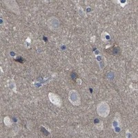
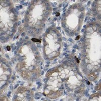
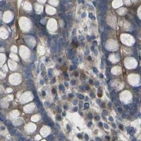
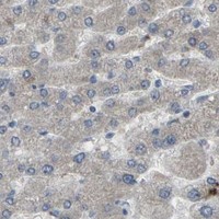


Comprehensive Human Protein Atlas IHC scoring per tissue (reliability: Uncertain — Low consistency between antibody staining and RNA expression data.). Rows are taken directly from the HPA tissue chart — click any row's HPA link to view the source.
| Tissue | Cell type | Level | Evidence | Source |
|---|---|---|---|---|
| Adrenal gland | Glandular cells | Medium | Protein (IHC) | HPA → |
| Cerebellum | Cells in granular layer | Medium | Protein (IHC) | HPA → |
| Colon | Endothelial cells | Medium | Protein (IHC) | HPA → |
| Duodenum | Glandular cells | Medium | Protein (IHC) | HPA → |
| Heart muscle | Cardiomyocytes | Medium | Protein (IHC) | HPA → |
| Tissue | Cell type | Level | Evidence | Source |
|---|---|---|---|---|
| Adipose tissue | Adipocytes | Not detected | Protein (IHC) | HPA → |
| Caudate | Glial cells | Not detected | Protein (IHC) | HPA → |
| Fallopian tube | Glandular cells | Not detected | Protein (IHC) | HPA → |
| Oral mucosa | Squamous epithelial cells | Not detected | Protein (IHC) | HPA → |
| Soft tissue | Fibroblasts | Not detected | Protein (IHC) | HPA → |
Use the catalog antibody’s paraffin-section workflow as the IHC starting point, and interpret CCR10 staining by cell type, compartment, and controls.
The catalog antibody has real IHC images from human paraffin sections of oesophageal squamous cancer, prostatic cancer and tonsil; IF images are absent (A04731-1 image captions; catalog applications).
A04731-1 will render with an IHC image from a human paraffin section of oesophageal squamous cancer (A04731-1 image caption). Its other IHC captions show human prostatic cancer and tonsil sections; the catalog lists Human, Mouse and Rat reactivity and IHC and WB applications (A04731-1 image captions; catalog applications and reactivity).
Which to pick: Choose A04731-1 for paraffin-section tissue IHC: its own captions document citrate retrieval at pH 6 for 20 minutes and primary antibody at 1 μg/ml overnight at 4°C (A04731-1 image captions). The fixative is unreported, and IF/ICC is not listed or illustrated, so there is no supported IF/ICC pick here (A04731-1 image captions; catalog applications and images). For cross-species planning, A04731-1 is a rabbit polyclonal antibody listed as reactive with Human, Mouse and Rat, although the supplied IHC images show human tissue only (catalog host, clonality and reactivity; A04731-1 image captions).