This website uses cookies to ensure you get the best experience on our website.


- Table of Contents
Plan CCR3 paraffin-section IHC around the mainly membranous tissue pattern reported by HPA (HPA tissue IHC). Compare controls and interpret staining cautiously because HPA rates its tissue staining reliability as uncertain (HPA tissue IHC).

Expected localisation, validated protocols, controls and antibodies — the at-a-glance facts below, then the full design guide.
| Expected localisation | Mainly membranous in tissue (HPA tissue IHC) | |
| Staining pattern | Mainly membranous staining in several tissues (HPA tissue IHC) | |
| Antigen retrieval | Citrate pH 6.0 HIER, 95–98 °C, 20 min (rule: cytoplasmic / membrane antigen) | |
| Positive control | Adrenal gland+4 more · see all | |
| Negative control | Adipose tissue+4 more · see all |
| Fixation | Keep fixation consistent across sections. (standard IHC practice; not target-specific) | |
| Caveat | Presumed off-target staining; reliability uncertain (HPA tissue IHC) | |
| Regulation | Expression regulation not specified (UniProt) | |
| Isoform / epitope | 2 isoforms; epitope coverage is unknown (UniProt) |
The catalog antibody’s IHC-P protocol is followed by three published CCR3 immunohistochemistry workflows: frozen mouse brain, paraffin-embedded bovine corpus luteum, and human prostate cancer tissue microarrays.
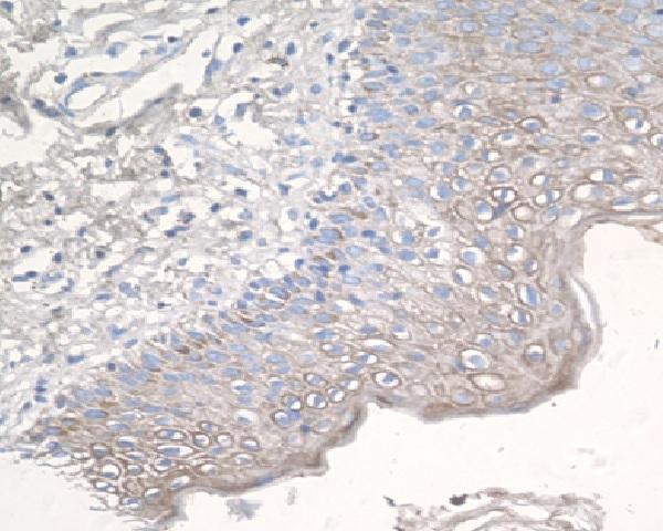
| Sample | Paraffin-embedded human epidemal cells of skin tissue; fixative not specified (datasheet M01748) |
| Fixation | Image fixative and duration unreported (datasheet M01748); verify before use. |
| Sectioning | 4–5 µm sections on charged slides (standard) |
| Deparaffinisation | Xylene, graded ethanol series to water (standard) |
| Antigen retrieval | Heat-induced epitope retrieval in citrate buffer, pH 6.0, 20 min at 95–98 °C (standard rule: cytoplasmic / membrane antigen) |
| Peroxidase block | 3% H2O2, 10 min, room temperature (standard) |
| Blocking | 10% normal serum of the secondary host, 30 min, room temperature (standard) |
| Primary antibody | Rabbit monoclonal (clone ECE-3) anti-CCR3, 1:50 (datasheet M01748) |
| Primary incubation | Overnight at 4 °C (standard) |
| Detection | HRP-polymer secondary, DAB chromogen 5–10 min (standard) |
| Counterstain | Hematoxylin, blue, dehydrate and mount (standard) |
| Expected result | CCR3-positive staining in glandular cells of adrenal gland (HPA tissue IHC: Medium). HPA tissue profile: Mainly membranous expression in several tissues. No signal in the no-primary control. |
CCR3 is a seven-transmembrane receptor assigned to the cell membrane, so a credible positive cell should show a membranous signal (UniProt P51677 topology; UniProt P51677 subcellular location). Eosinophils are the clearest cell-type expectation; UniProt reports only trace amounts in neutrophils and monocytes (UniProt P51677 tissue specificity). HPA describes mainly membranous tissue staining, but rates its tissue IHC profile Uncertain because staining and RNA data have low consistency and presumed off-target binding was observed (HPA tissue IHC).
| A crisp membrane-associated signal appears in cells identified as eosinophils by morphology. | This fits the receptor's location and strongest stated cell-type expectation (UniProt P51677 subcellular location and tissue specificity). Judge the signal alongside the tissue context and controls; HPA's overall tissue IHC reliability is Uncertain (HPA tissue IHC). |
| Signal is predominantly nuclear or fills the cytoplasm without a discernible membrane component. | That distribution conflicts with the assigned cell-membrane location and seven-transmembrane topology (UniProt P51677 subcellular location and topology). Treat it as suspect and check detection background before calling the cell positive (general IHC practice). |
| Strong staining appears in an unexpected cell population, especially across many cell types. | Investigate nonspecific binding or endogenous detection activity (general IHC practice). Cell identity alone is not decisive: HPA reports medium staining in several epithelial or glandular populations, while rating its tissue IHC profile Uncertain (HPA tissue IHC). |
| A diffuse chromogenic haze obscures cell borders throughout the section. | This cannot establish the membrane pattern expected for CCR3 (UniProt P51677 subcellular location; HPA tissue IHC profile). Compare a no-primary control, then review blocking, washes and detection conditions (general IHC practice). |
| There is no signal in a specimen with independently established CCR3-positive cells. | First confirm that those cells are present and that the detection controls worked (general IHC practice). HPA tissue examples alone are a weak positive-control standard because its tissue IHC reliability is Uncertain (HPA tissue IHC). |
| Topology and compartment | CCR3 has seven transmembrane segments and is assigned to the cell membrane (UniProt P51677 topology and subcellular location). Score a membrane-associated pattern rather than using total cellular color as the sole criterion (general IHC practice). |
| Cell population | UniProt places CCR3 in eosinophils and reports trace amounts in neutrophils and monocytes (UniProt P51677 tissue specificity). Cell morphology therefore matters when interpreting a stained inflammatory infiltrate (general IHC practice). |
| Tissue-level evidence | HPA reports mainly membranous staining and examples of medium epithelial or glandular staining, but flags low agreement with RNA expression and presumed off-target binding (HPA tissue IHC). Its tissue positives should be interpreted cautiously. |
| Antibody validation | The listed tissue IHC antibody CAB003795 is rated Uncertain; the separate antibody HPA069514 has Supported ICC evidence but no listed IHC rating (HPA antibodies). ICC support does not establish IHC-P performance for another antibody (general assay interpretation). |
| IF/ICC applicability | For the separate IF/ICC guide: HPA assigns an approved plasma-membrane location and lists images from RT-4 and U2OS (HPA subcellular ICC-IF). This location supports an IF interpretation, without changing the Uncertain tissue IHC assessment (HPA tissue IHC). |
| Isoforms and epitope coverage | UniProt lists two CCR3 isoforms (UniProt P51677 isoforms). The supplied record does not establish which isoform an IHC antibody recognizes; check the antibody's stated epitope before interpreting a negative result as absence of all CCR3 isoforms (general IHC practice). |
| Situation | Likely cause | Next action |
|---|---|---|
| No chromogen appears in an independently confirmed positive control. | A failed detection step or unsuitable assay conditions are possibilities (general IHC practice); HPA tissue examples alone do not resolve this because their IHC reliability is Uncertain (HPA tissue IHC). | Verify the control contains the expected cells, check the detection reagents, and review the IHC-P antibody instructions, including any specified retrieval and dilution (general IHC practice). |
| Staining is mostly nuclear or uniformly cytoplasmic. | The pattern disagrees with CCR3's assigned membrane location (UniProt P51677 subcellular location). Background or nonspecific staining may be contributing (general IHC practice). | Compare no-primary and appropriate tissue controls; assess whether any clear membrane-associated signal remains after background is accounted for (general IHC practice). |
| Many unrelated cell populations stain at similar strength. | Nonspecific binding or endogenous detection activity may contribute (general IHC practice). HPA also reports presumed off-target binding in its uncertain tissue IHC assessment (HPA tissue IHC). | Inspect no-primary controls, review blocking and detection chemistry, and require a plausible cellular distribution before scoring the section (general IHC practice; UniProt P51677 tissue specificity). |
| The whole section has diffuse color and cell borders are hard to resolve. | Background can obscure a membrane-associated result (general IHC practice; UniProt P51677 subcellular location). | Check no-primary staining, wash performance and detection conditions; score only cells whose membrane pattern can be distinguished from local background (general IHC practice). |
| An epithelial population stains, but eosinophils are not identified. | HPA records some medium epithelial staining, yet rates the overall tissue IHC profile Uncertain (HPA tissue IHC). The observation alone cannot confirm cell-specific CCR3 expression. | Identify cells by morphology, compare suitable controls, and report the epithelial pattern as an observation rather than assigning CCR3 specificity from color alone (general IHC practice; HPA tissue IHC). |
| IF/ICC shows a membrane signal while paraffin IHC is inconclusive. | HPA's approved plasma-membrane ICC-IF location and its Uncertain tissue IHC rating are distinct assessments (HPA subcellular ICC-IF; HPA tissue IHC). | Interpret each application with its own controls and antibody validation; do not transfer an ICC result to the IHC-P interpretation (HPA antibodies; general assay interpretation). |
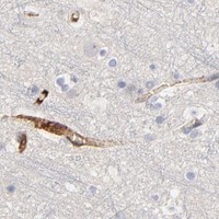


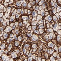


Comprehensive Human Protein Atlas IHC scoring per tissue (reliability: Uncertain — Low consistency between antibody staining and RNA expression data. Presumed off target binding observed and disregarded.). Rows are taken directly from the HPA tissue chart — click any row's HPA link to view the source.
| Tissue | Cell type | Level | Evidence | Source |
|---|---|---|---|---|
| Adrenal gland | Glandular cells | Medium | Protein (IHC) | HPA → |
| Esophagus | Squamous epithelial cells | Medium | Protein (IHC) | HPA → |
| Liver | Hepatocytes | Medium | Protein (IHC) | HPA → |
| Nasopharynx | Respiratory epithelial cells | Medium | Protein (IHC) | HPA → |
| Oral mucosa | Squamous epithelial cells | Medium | Protein (IHC) | HPA → |
| Tissue | Cell type | Level | Evidence | Source |
|---|---|---|---|---|
| Adipose tissue | Adipocytes | Not detected | Protein (IHC) | HPA → |
| Appendix | Glandular cells | Not detected | Protein (IHC) | HPA → |
| Breast | Adipocytes | Not detected | Protein (IHC) | HPA → |
| Caudate | Glial cells | Not detected | Protein (IHC) | HPA → |
| Cerebellum | Cells in granular layer | Not detected | Protein (IHC) | HPA → |
Troubleshoot CCR3 chromogenic IHC in paraffin sections by checking retrieval, compartment, cell identity and controls before interpreting staining.
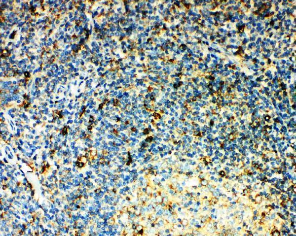
Both catalog antibodies have human tissue IHC images: paraffin-embedded skin cells for M01748 and tonsil for PA2176 (respective IHC captions). M01748 also lists mouse/rat reactivity and IF/ICC applications (M01748 catalog).
M01748 renders with its paraffin-embedded human skin-cell IHC image and lists IHC, ICC and IF applications with human, mouse and rat reactivity (M01748 catalog; IHC caption). PA2176 renders with its human tonsil IHC(P) image and lists IHC with human reactivity (PA2176 catalog; IHC caption).
Which to pick: For tissue IHC, choose PA2176 if its human tonsil IHC(P) example matches your sample, or M01748 if its paraffin-embedded human skin example is more relevant (respective IHC captions). For IF/ICC or mouse/rat samples, M01748 is the listed option: it is rabbit monoclonal clone ECE-3 and lists those applications and species (M01748 catalog). The M01748 caption reports paraffin embedding, while the PA2176 caption reports IHC(P); neither reports the fixative (respective IHC captions).