This website uses cookies to ensure you get the best experience on our website.


- Table of Contents
Plan CCT3 staining in paraffin sections using the mainly cytoplasmic lymphoid tissue pattern as a reference (HPA tissue IHC). Start with the IHC-validated antibody at 0.5–1 µg/mL (datasheet PB9926) and compare staining across cell types (HPA tissue IHC).

Expected localisation, validated protocols, controls and antibodies — the at-a-glance facts below, then the full design guide.
| Expected localisation | Mainly cytoplasmic in lymphoid tissues (HPA tissue IHC) | |
| Staining pattern | Lymphoid tissue: mainly cytoplasmic staining (HPA tissue IHC) | |
| Antigen retrieval | Citrate pH 6 HIER, heat-mediated (datasheet PB9926) | |
| Positive control | Adrenal gland+4 more · see all | |
| Negative control | None in HPA (detected in all 45 tissues); use no-primary + isotype controls |
| Fixation | Keep fixation conditions consistent across sections (standard IHC practice; not target-specific) | |
| Caveat | Staining intensity varies across tissues and cell types (HPA tissue IHC) | |
| Regulation | No expression regulator annotated (UniProt) | |
| Isoform / epitope | 2 isoforms; check epitope coverage across variants (UniProt) |
The catalog antibody’s IHC-P protocol (datasheet: PB9926) is accompanied by published CCT3 tissue IHC protocols from PMC13514722 and PMC5958781.
| Sample | Paraffin-embedded human lung cancer tissue; fixative not specified (datasheet PB9926) |
| Fixation | Image fixative and duration unreported (datasheet PB9926); verify before use. |
| Sectioning | 4–5 µm sections on charged slides (standard) |
| Deparaffinisation | Xylene, graded ethanol series to water (standard) |
| Antigen retrieval | Heat retrieval: Citrate pH 6, 20 min (datasheet PB9926) |
| Peroxidase block | 3% H2O2, 10 min, room temperature (standard) |
| Blocking | 10% goat serum (datasheet PB9926) |
| Primary antibody | Rabbit anti-CCT3, 0.5-1μg/ml (datasheet PB9926) |
| Primary incubation | Overnight at 4 °C (datasheet PB9926) |
| Detection | Streptavidin-biotin complex (SABC), DAB chromogen (datasheet PB9926) |
| Counterstain | Hematoxylin, blue, dehydrate and mount (standard) |
| Expected result | CCT3-positive staining in glandular cells of adrenal gland (HPA tissue IHC: High). HPA tissue profile: Mainly cytoplasmic expression in lymphoid tissues. No signal in the no-primary control. |
CCT3 is cytoplasmic and has no transmembrane segment (UniProt P49368). In paraffin sections, expect mainly cytoplasmic staining where tissue expression is observed, including glandular cells in breast and duodenum, glial cells in cerebral cortex, and cervical squamous epithelial cells (HPA: High). HPA rates its tissue IHC evidence Approved, with medium consistency between staining and RNA expression (HPA: tissue IHC).
| Cytoplasmic signal in breast or duodenal glandular cells, glia, or cervical squamous cells. | This fits the reported compartment and cell patterns (UniProt P49368: Cytoplasm; HPA: High in those cells). Compare cells within the same section; HPA's High rating does not require identical intensity in every cell. |
| Nuclear staining dominates while the expected cytoplasmic signal is absent. | That pattern conflicts with the annotated location (UniProt P49368: Cytoplasm). Treat it as questionable; inspect morphology and controls before assigning CCT3 positivity. Nuclear counterstain alone is not target staining (general IHC practice). |
| Strong staining appears chiefly in ovarian stroma or smooth muscle cells. | These are reported as Low, not negative (HPA: ovarian stroma and smooth muscle). Unexpectedly strong signal warrants a specificity check; the tissue profile alone cannot prove cross-reactivity or identify its source. |
| Brown color covers many cell types, extracellular areas, or blank spaces without a cellular pattern. | This does not resemble the mainly cytoplasmic tissue pattern (HPA: tissue IHC). Consider background from detection reagents, endogenous enzyme activity, or nonspecific binding; use controls to distinguish them (general IHC practice). |
| A breast glandular compartment expected to stain shows no detectable signal. | Breast glandular cells are rated High (HPA: tissue IHC), so check that the relevant cells are present. A blank result may reflect staining workflow failure or sample variation; one section does not overturn the HPA pattern. |
| Compartment and topology | CCT3 is annotated in the cytoplasm, with no transmembrane segment (UniProt P49368). Judge paraffin IHC by cellular cytoplasmic staining; a crisp surface outline alone is not the expected tissue pattern. |
| Tissue and cell context | HPA rates glandular cells High in adrenal gland, breast, duodenum, endometrium, and epididymis; glia High in caudate and cerebral cortex (HPA: tissue IHC). Low entries remain possible weak positives, not validated negatives. |
| Strength of pattern evidence | Tissue IHC is Approved but has medium consistency with RNA data (HPA: tissue IHC). HPA006543 is IHC Approved; the supplied antibody list gives no IHC status for HPA074177 (HPA: antibodies). Interpret isolated discordant staining cautiously. |
| Isoforms and epitope coverage | UniProt lists 2 isoforms but supplies no epitope or isoform coverage for the antibody (UniProt P49368). Isoform status therefore cannot explain a weak section or justify predicting a different compartment. |
| Antigen retrieval and detection | Retrieval conditions, blocking, and detection can affect paraffin IHC background and visibility (general IHC practice). No source here reports CCT3-specific fixation sensitivity or an optimal retrieval condition; assess workflow controls without attributing a result to CCT3 epitope masking. |
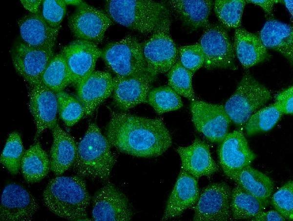
| IF/ICC Q: Should fluorescence match IHC exactly? | HPA approves cytosol as the main ICC-IF location and vesicles and plasma membrane as additional locations (HPA: subcellular). That informs an IF check; the tissue IHC expectation remains mainly cytoplasmic (HPA: tissue IHC). |
| Situation | Likely cause | Next action |
|---|---|---|
| No signal in a breast section's glandular cells. | Those cells are rated High (HPA: tissue IHC); an absent signal may reflect missing target cells, section quality, or an IHC workflow problem (general IHC practice). | Confirm glandular cells on the counterstain and run a known-positive section alongside reagent controls. Review the validated IHC workflow before changing conditions; no CCT3-specific fixation effect is documented here. |
| Nuclei stain strongly but cytoplasm stays clear. | A predominantly nuclear result conflicts with CCT3's annotated cytoplasmic location (UniProt P49368). Counterstain or nonspecific signal may be mistaken for chromogen (general IHC practice). | Distinguish counterstain color from chromogen, inspect a reagent control, and repeat the comparison in an HPA High cell compartment. Do not score nuclear-only color as expected CCT3 staining. |
| Uniform brown haze obscures cell borders. | Diffuse color lacks the mainly cytoplasmic distribution reported for CCT3 (HPA: tissue IHC). Endogenous detection activity or nonspecific binding can produce such background (general IHC practice). | Check a no-primary control and the blocking and detection steps. Judge a repeated stain only where cell boundaries and cytoplasm remain readable (general IHC practice). |
| Ovarian stroma or smooth muscle looks stronger than a chosen positive compartment. | Both cell types are listed Low (HPA: tissue IHC). The mismatch raises a specificity question, although HPA levels do not define a guaranteed intensity ratio between individual sections. | Compare sections processed together and inspect no-primary controls. Reassess cell identity and morphology before calling the unexpected staining CCT3-specific (general IHC practice). |
| Stain is strongest only at tissue edges or in damaged areas. | An edge-dominant pattern is not the reported cellular pattern (HPA: tissue IHC). Uneven reagent exposure or section damage can complicate interpretation (general IHC practice). | Score intact interior tissue with identifiable cells and compare the control section. Repeat staining if an intact cytoplasmic compartment cannot be assessed (general IHC practice). |
| A Low tissue is blank and is being used as a negative control. | Low is a reported staining level, not absence of CCT3 (HPA: esophageal and vaginal squamous cells, ovarian stroma, smooth muscle, chondrocytes). HPA lists no negative tissue entries in this payload. | Use reagent controls to assess background and a High cell compartment to assess detection. Do not label a blank Low sample a validated biological negative (HPA: tissue IHC; general IHC practice). |



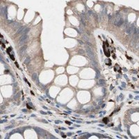


Comprehensive Human Protein Atlas IHC scoring per tissue (reliability: Approved — Medium consistency between antibody staining and RNA expression data.). Rows are taken directly from the HPA tissue chart — click any row's HPA link to view the source.
| Tissue | Cell type | Level | Evidence | Source |
|---|---|---|---|---|
| Adrenal gland | Glandular cells | High | Protein (IHC) | HPA → |
| Breast | Glandular cells | High | Protein (IHC) | HPA → |
| Caudate | Glial cells | High | Protein (IHC) | HPA → |
| Cerebral cortex | Glial cells | High | Protein (IHC) | HPA → |
| Cervix | Squamous epithelial cells | High | Protein (IHC) | HPA → |
| Tissue | Cell type | Level | Evidence | Source |
|---|---|---|---|---|
| None in HPA: CCT3 is detected in all 45 scored tissues. Use a no-primary (secondary-only) and an isotype control instead. | ||||
Troubleshoot chromogenic CCT3 staining in paraffin sections by checking retrieval, compartment, tissue context, and controls before comparing staining scores.
The catalog includes CCT3 staining in human paraffin sections and A431 cells (PB9926 and M05920 image captions); PB9926 also lists mouse and rat reactivity (catalog: PB9926 reactivity).
PB9926 has IHC images from human lung, mammary and intestinal cancer, and placenta paraffin sections, plus IF images from A431 cells (PB9926 image captions). M05920 has IF images from A431 cells and lists human reactivity (M05920 image captions; catalog: M05920 reactivity).
Which to pick: Choose PB9926 for tissue IHC: its rabbit antibody has a listed IHC application and 0.5–1 μg/ml IHC dilution, with paraffin section images; the fixative is unreported (catalog: PB9926 applications and dilution; PB9926 IHC captions). For IF/ICC, PB9926 has A431 images and a listed 2 μg/ml IF dilution, while mouse monoclonal M05920 (clone 12H4) has A431 IF images and a captioned 5 μg/ml concentration (PB9926 and M05920 IF captions; catalog: M05920 clone). For mouse or rat samples, PB9926 lists reactivity with both species, although its pictured IHC sections are human; M05920 lists human reactivity only (catalog: PB9926 and M05920 reactivity; PB9926 IHC captions).