This website uses cookies to ensure you get the best experience on our website.


- Table of Contents
Plan CCT5 chromogenic IHC around cytoplasmic staining, most abundant in seminiferous duct cells (HPA tissue IHC). This guide covers paraffin-section staining with the catalog antibody (datasheet PB9928) and interpretation of CNS nuclear positivity, which HPA disregards as off target (HPA tissue IHC).

Expected localisation, validated protocols, controls and antibodies — the at-a-glance facts below, then the full design guide.
| Expected localisation | General cytoplasmic staining (HPA tissue IHC) | |
| Staining pattern | Cytoplasmic staining, most abundant in seminiferous duct cells (HPA tissue IHC) | |
| Antigen retrieval | Citrate pH 6 HIER, heat-mediated (datasheet PB9928) | |
| Positive control | Caudate+4 more · see all | |
| Negative control | Esophagus+4 more · see all |
| Fixation | Keep paraffin-section fixation consistent (standard IHC practice; not target-specific); Matched tissue-IHC evidence does not establish the fixation claim. Validate the specimen-specific method before use. (selected-SKU IHC image PB9928) | |
| Caveat | CNS nuclear positivity may be off target (HPA tissue IHC) | |
| Regulation | No regulation annotated (UniProt) | |
| Isoform / epitope | 2 isoforms; epitope impact unresolved (UniProt) |
The catalog antibody’s IHC-P protocol is accompanied by four published CCT5 IHC protocols (PMC5609992; PMC11409311; PMC13014677; PMC13260463).
| Sample | Paraffin-embedded human intestinal cancer tissues; fixative not specified (datasheet PB9928) |
| Fixation | Image fixative and duration unreported (datasheet PB9928); verify before use. |
| Sectioning | 4–5 µm sections on charged slides (standard) |
| Deparaffinisation | Xylene, graded ethanol series to water (standard) |
| Antigen retrieval | Heat retrieval: Citrate pH 6, 20 min (datasheet PB9928) |
| Peroxidase block | 3% H2O2, 10 min, room temperature (standard) |
| Blocking | 10% goat serum (datasheet PB9928) |
| Primary antibody | Rabbit anti-CCT5, 0.5-1μg/ml (datasheet PB9928) |
| Primary incubation | Overnight at 4 °C (datasheet PB9928) |
| Detection | Streptavidin-biotin complex (SABC), DAB chromogen (datasheet PB9928) |
| Counterstain | Hematoxylin, blue, dehydrate and mount (standard) |
| Expected result | CCT5-positive staining in glial cells of caudate (HPA tissue IHC: High). HPA tissue profile: General cytoplasmic expression, most abundant in seminiferous duct cells. No signal in the no-primary control. |
CCT5 is expected mainly in the cytoplasm, consistent with its UniProt location and lack of a transmembrane segment (UniProt P48643: subcellular location, topology). HPA reports general cytoplasmic IHC expression, most abundant in seminiferous duct cells, with high staining in late spermatids and several other cell populations (HPA tissue IHC: Enhanced). Interpret CNS nuclear positivity cautiously: HPA regards it as off target (HPA tissue IHC: reliability description).
| Cytoplasmic staining in late spermatids, placental cytotrophoblasts, or lymph node germinal center cells. | This fits the reported high staining in those cell populations (HPA tissue IHC: High). Score the named cells and cytoplasmic compartment together; staining elsewhere in the section does not by itself establish a CCT5-positive result (HPA tissue IHC; general IHC interpretation). |
| Predominantly nuclear staining, especially in CNS cells. | This conflicts with the expected cytoplasmic location (UniProt P48643: subcellular location; HPA tissue IHC: profile). HPA specifically disregards CNS nuclear positivity as off target, so exclude it from CCT5 scoring even if the nuclear signal is strong (HPA tissue IHC: reliability description). |
| Strong staining in cells reported as not detected, such as cardiomyocytes or esophageal squamous cells. | The cell-level pattern disagrees with HPA tissue IHC (HPA tissue IHC: Not detected). Consider cross-reactivity or endogenous detection activity, then compare a known-positive cell population and appropriate detection controls before attributing the signal to CCT5 (general IHC interpretation). |
| Diffuse chromogen across cells, stroma, or much of the section without clear cytoplasmic boundaries. | A widespread haze is difficult to reconcile with a cell-resolved cytoplasmic pattern (HPA tissue IHC: profile). Background from detection or blocking is a possible explanation; interpret intensity only after a control section shows that the haze is separate from specific staining (general IHC practice). |
| No cytoplasmic signal in a known-positive population, such as testis late spermatids. | An absent signal disagrees with the reported high staining in that population (HPA tissue IHC: Testis, High). First check that the relevant cells are present and identifiable; then check the antibody, retrieval, detection, and control performance without assuming that CCT5 is absent (general IHC practice). |
| Cell population and compartment | HPA reports broad cytoplasmic expression but different cell-level intensities: late spermatids and placental cytotrophoblasts are High, whereas cardiomyocytes are Not detected (HPA tissue IHC). Select and score the named cells rather than treating an entire organ as uniformly positive or negative. |
| Antibody validation | HPA labels the tissue IHC profile Enhanced and records Enhanced IHC validation for HPA002238 and HPA005958; CAB006271 is Supported (HPA antibodies; HPA tissue IHC). These labels support the reported pattern but do not validate every antibody or every staining condition. |
| Isoforms and epitope | UniProt lists two CCT5 isoforms and a chain spanning residues 2–541, with no signal peptide or propeptide (UniProt P48643: isoforms, processing). The supplied evidence gives no antibody epitope, so isoform recognition and epitope-dependent staining cannot be predicted from this record. |
| IF/ICC Q: What localization is reported? | In sperm ICC-IF, HPA approves the mid piece as the main location and the principal piece and annulus as additional locations (HPA subcellular: sperm). This sperm-specific IF observation does not replace the cytoplasmic tissue IHC pattern or establish an IF protocol for paraffin sections. |
| Fixation sensitivity | Target-specific sensitivity to fixation or antigen retrieval is unreported in the supplied UniProt and HPA evidence. Retrieval may be optimized as a general IHC workflow step, but no CCT5-specific retrieval condition or fixation effect can be inferred (general IHC practice). |
| Situation | Likely cause | Next action |
|---|---|---|
| Known-positive cells are present, but chromogenic signal is absent. | The assay may have failed to reveal antigen; the supplied evidence does not identify a CCT5-specific fixation or retrieval failure. | Confirm cell identity in a high-staining population such as late spermatids (HPA tissue IHC: Testis, High). Check the IHC-validated antibody, retrieval and detection steps against assay controls; adjust one general workflow variable at a time (general IHC practice). |
| CNS nuclei stain more strongly than cytoplasm. | HPA identifies CNS nuclear positivity as off target (HPA tissue IHC: reliability description). | Do not score those nuclei as CCT5. Reassess cytoplasmic staining in an HPA positive population, and compare antibody and detection controls before accepting any CNS result (HPA tissue IHC; general IHC practice). |
| Cardiomyocytes or esophageal squamous cells show convincing signal. | These cells are reported as Not detected (HPA tissue IHC); cross-reactivity or endogenous detection activity is possible (general IHC interpretation). | Compare cell-specific staining with a known-positive section and run appropriate reagent controls. Review whether the signal follows cell boundaries and the expected cytoplasmic compartment (UniProt P48643: cytoplasm; general IHC practice). |
| Stain covers the section as a diffuse haze. | Excess nonspecific binding or detection background can obscure a discrete cellular pattern (general IHC practice). | Inspect controls, blocking and wash steps, then reassess antibody and detection reagent concentrations within the assay workflow (general IHC practice). Accept a pattern only when named cells can be distinguished from local background (HPA tissue IHC). |
| Signal appears restricted to membranes or extracellular material. | That distribution conflicts with CCT5's reported cytoplasmic location and lack of a transmembrane segment (UniProt P48643: subcellular location, topology). | Treat the distribution as suspect; check morphology and detection controls, then compare the same run with an HPA high cell population for intracellular staining (HPA tissue IHC; general IHC practice). |
| A weakly stained section seems to contradict a negative call. | HPA reports general cytoplasmic expression alongside cell populations scored Low or Not detected; organ-wide judgments can obscure those distinctions (HPA tissue IHC). | Record the exact cell population, compartment and intensity. Compare like cells with HPA's cell-level categories and keep weak background separate from interpretable cytoplasmic staining (HPA tissue IHC; general IHC interpretation). |


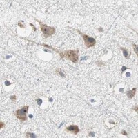


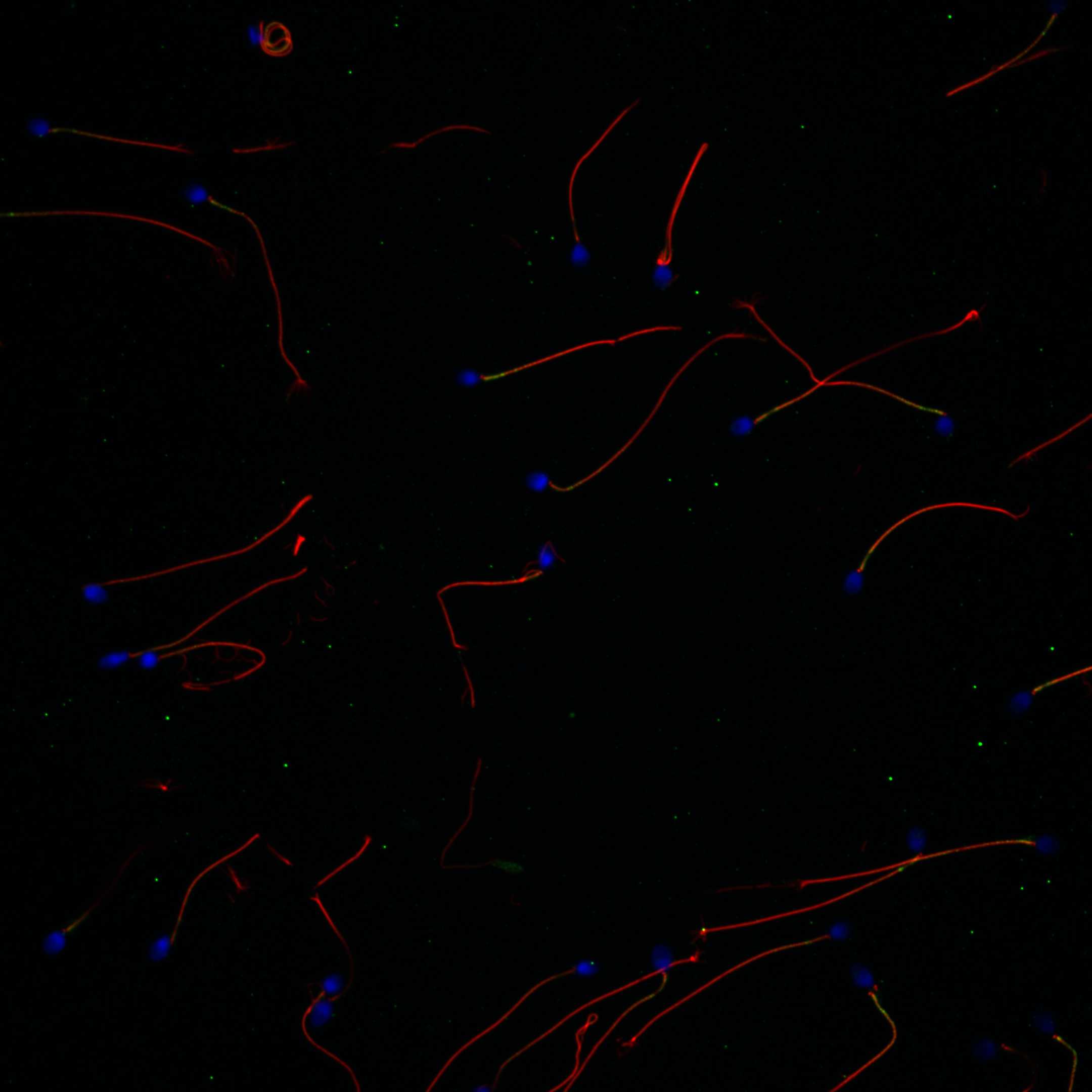
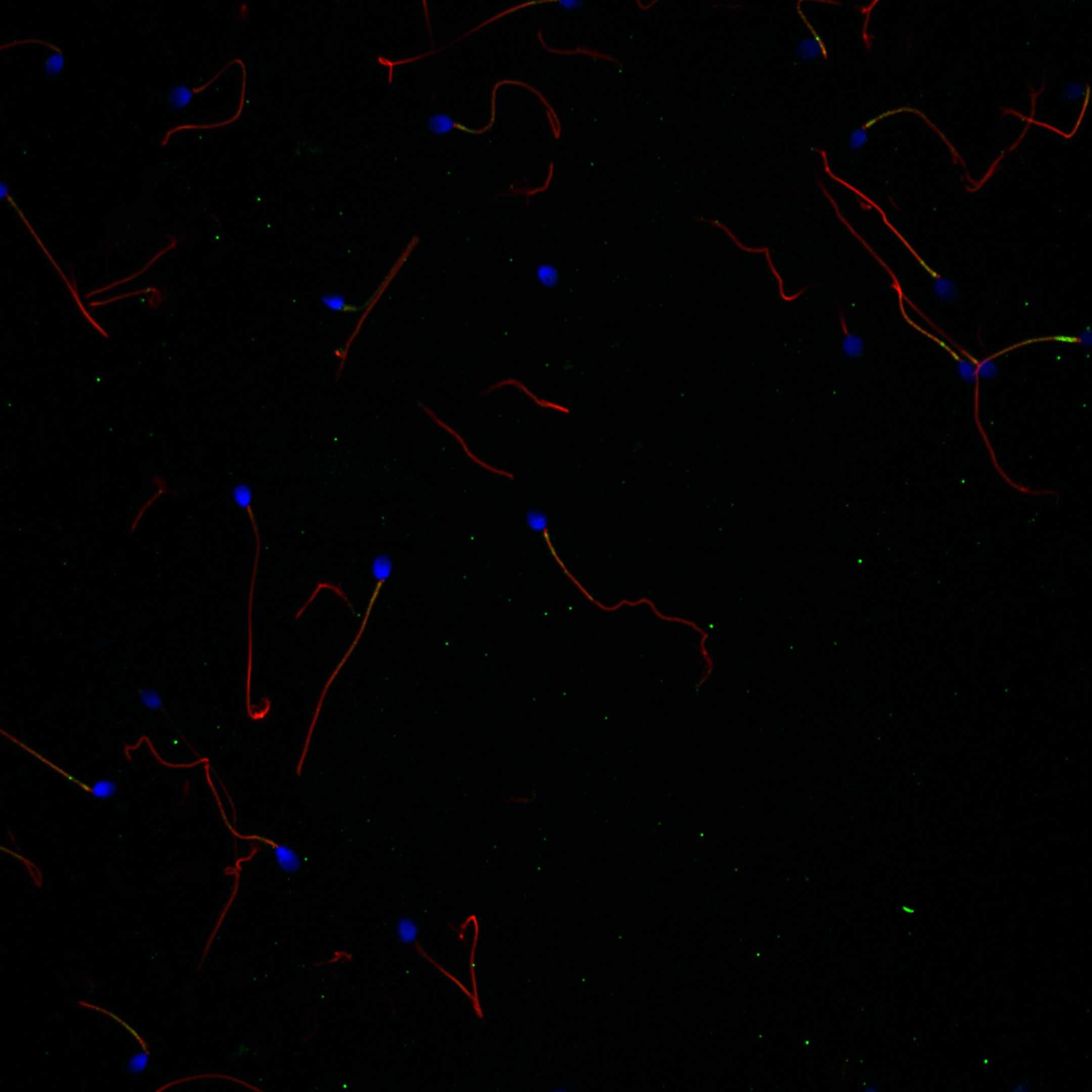
Comprehensive Human Protein Atlas IHC scoring per tissue (reliability: Enhanced — High consistency between antibody staining and RNA expression data. Nuclear positivity in CNS viewed as off target and disregarded.). Rows are taken directly from the HPA tissue chart — click any row's HPA link to view the source.
| Tissue | Cell type | Level | Evidence | Source |
|---|---|---|---|---|
| Caudate | Glial cells | High | Protein (IHC) | HPA → |
| Fallopian tube | Glandular cells | High | Protein (IHC) | HPA → |
| Lymph node | Germinal center cells | High | Protein (IHC) | HPA → |
| Parathyroid gland | Glandular cells | High | Protein (IHC) | HPA → |
| Placenta | Cytotrophoblasts | High | Protein (IHC) | HPA → |
| Tissue | Cell type | Level | Evidence | Source |
|---|---|---|---|---|
| Esophagus | Squamous epithelial cells | Not detected | Protein (IHC) | HPA → |
| Heart muscle | Cardiomyocytes | Not detected | Protein (IHC) | HPA → |
| Liver | Cholangiocytes | Not detected | Protein (IHC) | HPA → |
| Smooth muscle | Smooth muscle cells | Not detected | Protein (IHC) | HPA → |
| Vagina | Squamous epithelial cells | Not detected | Protein (IHC) | HPA → |
Troubleshoot CCT5 staining in paraffin sections by checking retrieval, tissue handling, cytoplasmic localisation and cell type before interpreting chromogenic signal.
PB9928 has IHC images from paraffin sections of human intestinal cancer tissue and mouse and rat ovary, plus an IF image from U20S cells (catalog image captions).
PB9928 is listed for IHC and IF, with human, mouse and rat reactivity (catalog applications and reactivity). Its IHC captions show paraffin sections of human intestinal cancer tissue and mouse and rat ovary; its IF caption shows U20S cells (catalog image captions).
Which to pick: For tissue IHC, choose PB9928: its images document paraffin sections with citrate retrieval at pH 6 for 20 minutes (PB9928 IHC captions); the fixative is unreported (PB9928 IHC captions). For IF/ICC, PB9928 is listed for both applications and has an IF image from U20S cells (catalog applications; PB9928 IF caption). For work across human, mouse and rat, PB9928 lists reactivity in all three and has an IHC image for each species; its host is rabbit, while clonality is unreported (catalog reactivity and host; PB9928 IHC captions; catalog clone field).