CD164 / Sialomucin core protein 24 · IHC design guide

Design Immunohistochemistry for CD164

Plan CD164 chromogenic IHC in paraffin sections using the granular cytoplasmic tissue pattern as a reference (HPA tissue IHC). Compare staining across consistently fixed sections, and consider isoform-specific expression when interpreting intensity (UniProt).

Evidence assembled Oct 2026 · For research use; verify linked source records and product datasheet before use
Immunohistochemistry protocol sheet for CD164 (IHC for CD164): expected localisation Granular cytoplasm in most tissues (HPA tissue IHC), antibody A05879, validated IHC image, and IHC protocol steps
Printable CD164 IHC protocol sheet — expected localisation Granular cytoplasm in most tissues (HPA tissue IHC), antibody A05879, controls and protocol steps. Open the full CD164 IHC guide →

CD164 Immunohistochemistry Experimental Design Guide

Expected localisation, validated protocols, controls and antibodies — the at-a-glance facts below, then the full design guide.

Must know before staining
Expected localisation Granular cytoplasm in most tissues (HPA tissue IHC)
Staining pattern Granular cytoplasmic staining; high in glandular cells (HPA tissue IHC)
Antigen retrieval Tris-EDTA pH 9.0 HIER, heat-mediated (datasheet A05879)
Positive control ⓘ Appendix+4 more · see all
Negative control ⓘ Vagina
Important caveats
Reasons your staining may differ from the expected pattern.
Fixation Matched tissue-IHC evidence does not establish the fixation claim. Validate the specimen-specific method before use. (selected-SKU IHC image A05879)
Caveat Isoform-specific expression may change staining intensity (UniProt)
Regulation Isoform 1 higher in bone metastases (UniProt)
Isoform / epitope 5 isoforms; check extracellular vs cytoplasmic epitope (UniProt)
Section 1

Recommended CD164 IHC & IF Protocols

The catalog antibody's IHC-P protocol (datasheet: A05879) is accompanied by four published CD164 IHC protocols (PMC6089154; PMC5589567; PMC7610312; PMC11121609).

Recommended immunohistochemistry (IHC-P) protocol parameters
SampleParaffin-embedded human Squamous cell carcinoma of lung tissue; fixative not specified (datasheet A05879)
FixationImage fixative and duration unreported (datasheet A05879); verify before use.
Sectioning4–5 µm sections on charged slides (standard)
DeparaffinisationXylene, graded ethanol series to water (standard)
Antigen retrievalHeat retrieval: Tris-EDTA pH 9.0 (datasheet A05879); 20 min, 95–100 °C (standard)
Peroxidase block3% H2O2, 10 min, room temperature (standard)
Blocking10% normal serum of the secondary host, 30 min, room temperature (standard)
Primary antibodyRabbit anti-CD164, 1:100-1:300 (datasheet A05879)
Primary incubationOvernight at 4 °C (standard)
DetectionHRP-polymer secondary, DAB chromogen 5–10 min (standard)
CounterstainHematoxylin, blue, dehydrate and mount (standard)
Expected resultCD164-positive staining in glandular cells of appendix (HPA tissue IHC: High). HPA tissue profile: Granular cytoplasmic expression in most tissues. No signal in the no-primary control.
💡Decision noteStart with heat-mediated Tris-EDTA, pH 9.0 (datasheet: A05879); compare the published EDTA and citrate conditions if retrieval needs optimization (PMC6089154; PMC5589567; PMC11121609).
Section 2

What Is the Expected CD164 Staining Pattern?

CD164 is observed as granular cytoplasmic staining in most tissues, consistent with its endosome and lysosome membrane locations; cell surface staining is also plausible from its transmembrane topology (HPA tissue IHC; UniProt Q04900 location and topology). Expect stronger glandular staining in appendix, cervix, gallbladder, rectum and stomach, with medium staining in bone marrow hematopoietic cells (HPA tissue IHC). HPA rates the tissue pattern Approved, with medium agreement between staining and RNA data (HPA tissue IHC).

What am I looking at on my slide?
Granular cytoplasmic staining in glandular cells, especially in the listed high-staining tissues (HPA tissue IHC).This matches HPA's predominant tissue pattern. Compare cells within the same section: a positive result is cell-associated and granular, while neighboring structures can provide context for background (HPA tissue IHC; general IHC practice).
Cell-surface staining accompanies intracellular granules (UniProt Q04900 location; HPA tissue IHC).A surface component is compatible with CD164's extracellular residues 24–162 and membrane-spanning residues 163–183; the HPA tissue IHC profile chiefly describes granular cytoplasm (UniProt Q04900 topology; HPA tissue IHC).
Predominantly nuclear staining, without the expected granular cellular pattern (HPA tissue IHC).Nuclear staining is unsupported by the supplied location records; consider an artefact or off-target signal. Recheck the compartment in an HPA high-staining tissue before scoring it as CD164 (UniProt Q04900 location; HPA tissue IHC).
Strong staining in vaginal squamous epithelial cells (HPA: Not detected).This conflicts with the HPA tissue observation and raises concern for cross-reactivity or endogenous chromogenic activity. Check a detection-only control and the expected positive cells before interpreting it (HPA tissue IHC; general IHC practice).
Diffuse tissue-wide color or no signal in appendix glandular cells (HPA: High).Diffuse color obscures cell-specific interpretation; absent signal in an expected positive suggests an assay failure. Read background controls and verify that the positive-control section stained as expected (HPA tissue IHC; general IHC practice).
💡Expected CD164 appearanceCall a section positive when glandular cells show distinct granular cytoplasmic staining, typically strong in appendix, cervix, gallbladder, rectum or stomach; diffuse color or dominant nuclear signal is suspect (HPA tissue IHC; UniProt Q04900 location).
How each factor affects the staining
Compartment and topologyCD164 is listed at endosome, lysosome and cell membranes and as secreted; interpret granular intracellular staining alongside any surface component (UniProt Q04900 location and topology; HPA tissue IHC).
Tissue and cell selectionHPA reports High glandular staining in five listed tissues, Medium hematopoietic staining in bone marrow, and no detection in vaginal squamous epithelium (HPA tissue IHC).
Isoforms and processingUniProt lists 5 isoforms, a signal peptide at residues 1–23, and a mature chain at 24–197; staining cannot identify an isoform without epitope-specific evidence (UniProt Q04900).
Glycosylation and epitopeUniProt lists 10 glycosylation sites in the extracellular region. The supplied record does not identify this antibody's epitope, so it cannot establish an epitope-specific retrieval requirement (UniProt Q04900 topology and glycosylation).
Antibody evidenceHPA lists HPA010636, a rabbit polyclonal antibody, as Approved for IHC; that rating and medium RNA agreement warrant comparison with tissue controls, not an Enhanced-validation claim (HPA antibodies; HPA tissue IHC).
IF/ICC Q: What pattern is supported?A: HPA summarizes the location as membrane but supplies no ICC-IF image-bearing cell lines or main-location detail; interpret IF/ICC localization with that limit and UniProt's intracellular membrane locations (HPA subcellular; UniProt Q04900 location).
Why is my staining missing, weak or wrong?
SituationLikely causeNext action
Expected-positive glandular cells are blank in appendix or stomach (HPA: High).Target-specific fixation effects are not established by the supplied assay evidence. Verify with a matched IHC source before attributing a result to fixation.Check section integrity and positive-control staining, then review the antibody, retrieval and detection steps under the chosen IHC procedure (general IHC practice).
All tissue structures acquire diffuse chromogen, masking cell boundaries (general IHC practice).Excess background can arise from insufficient blocking or detection-system background; diffuse staining is unlike HPA's granular cellular profile (general IHC practice; HPA tissue IHC).Inspect a detection-only control and review blocking, washing and chromogen development before assigning CD164 positivity (general IHC practice).
Signal appears mainly in nuclei (HPA tissue IHC; UniProt Q04900 location).Neither source lists the nucleus as a CD164 location; predominant nuclear signal may be nonspecific (HPA tissue IHC; UniProt Q04900 location).Compare an expected-positive glandular section and a detection control; score only a reproducible, compartment-consistent signal (HPA tissue IHC; general IHC practice).
Vaginal squamous cells stain strongly (HPA: Not detected).The result disagrees with HPA's cell-type observation; cross-reactivity or endogenous detection activity is possible (HPA tissue IHC; general IHC practice).Assess a detection-only control and compare high-staining glandular tissue before treating the squamous signal as CD164 (HPA tissue IHC; general IHC practice).
Prostate glandular cells stain weakly (HPA: Low).Weak staining can fit the supplied normal-tissue observation; UniProt's prostate-cancer expression statement does not set a normal-gland intensity threshold (HPA tissue IHC; UniProt Q04900 tissue specificity).Use an HPA high-staining glandular tissue to assess assay performance; interpret prostate staining by its own cell pattern and controls (HPA tissue IHC; general IHC practice).
Surface staining is present with granular cytoplasmic staining (UniProt Q04900 location; HPA tissue IHC).CD164 has a transmembrane segment and endosomal and lysosomal locations, so both distributions are biologically plausible (UniProt Q04900 location and topology).Record each compartment and its cell type; compare the granular component with HPA tissue observations rather than rejecting a controlled surface signal (HPA tissue IHC; UniProt Q04900 location).

Sample controls for CD164 IHC & IF

🧪Run appendix first: its glandular cells should stain strongly (HPA: High in appendix glandular cells). Run vagina as the negative tissue (HPA: Not detected in vaginal squamous epithelial cells); on the appendix slide, non-glandular cells with little or no chromogen are candidate internal negatives, but verify their status because the HPA row identifies only glandular cells.
Positive control tissue: Appendix (Glandular cells, HPA High)
Negative control tissue: Vagina (HPA Not detected)
ICC-IF cell lines (HPA subcellular resource): HPA carries no ICC-IF cell line for CD164; derive a cell-line control from the positive tissue's cell type (Glandular cells) and confirm it by RNA or western blot first.
Technical controls: Include a no-primary (secondary-only) section, a concentration-matched isotype control appropriate to the primary antibody’s host species and clonality, and CD164-knockout tissue if available; a peptide-block control is suitable only when the antibody’s immunizing peptide is available. Check endogenous peroxidase activity in the appendix section before interpreting chromogenic signal, and compare glandular staining with the control sections (HPA: High in appendix glandular cells).
⚠️Feasibility: No target-specific fixation window or fixation effect is reported in the supplied evidence; the selected A05879 paraffin-section caption does not state the fixative (caption: fixative unreported). Its reported IHC conditions use Tris-EDTA retrieval at pH 9.0, primary antibody at 1:200 overnight at 4°C, and secondary antibody at 1:200 for 45 minutes at room temperature (A05879 caption); retrieval dependence and whether frozen-section IHC or IF would be easier remain unreported. In appendix, glandular luminal material or background chromogen can complicate scoring, so assess staining in intact glandular cells against the controls (HPA: High in appendix glandular cells).

HPA tissue IHC evidence for CD164

Comprehensive Human Protein Atlas IHC scoring per tissue (reliability: Approved — Medium consistency between antibody staining and RNA expression data.). Rows are taken directly from the HPA tissue chart — click any row's HPA link to view the source.

Positive expression · recommended positive controls

TissueCell typeLevelEvidenceSource
Appendix Glandular cells High Protein (IHC) HPA →
Cervix Glandular cells High Protein (IHC) HPA →
Gallbladder Glandular cells High Protein (IHC) HPA →
Rectum Glandular cells High Protein (IHC) HPA →
Stomach Glandular cells High Protein (IHC) HPA →

Undetected expression · recommended negative controls

TissueCell typeLevelEvidenceSource
Vagina Squamous epithelial cells Not detected Protein (IHC) HPA →
Section 3

Advanced CD164 IHC Tips

Troubleshoot CD164 staining in paraffin sections by checking retrieval, controls, cellular distribution, and scoring before interpreting chromogenic signal.

Which retrieval condition should I start with for CD164 in paraffin sections?
Use heat-mediated Tris-EDTA at pH 9.0 for antigen retrieval on paraffin sections (datasheet A05879). The selected tissue image used this buffer before overnight primary incubation at 4°C with antibody diluted 1:200 (caption A05879). If staining is weak, first compare a known positive section processed alongside the sample and confirm that heating and cooling were consistent (general IHC practice; HPA: high expression in appendix glandular cells). Only then compare another retrieval buffer as a fallback, keeping antibody dilution and detection constant (general IHC practice). Excessive retrieval can damage morphology and complicate interpretation of granular cytoplasmic staining (general IHC practice; HPA: granular cytoplasmic profile).
How should I assess whether fixation is affecting CD164 staining?
The selected image documents paraffin sections but does not state a fixative, so CD164 sensitivity to any particular fixation condition is unknown (caption A05879). Record fixative, fixation duration, specimen thickness, and processing history for each section before comparing staining intensity (general IHC practice). Test sections with different known fixation histories using the same pH 9.0 retrieval and 1:200 primary dilution, with a control section in each run (datasheet A05879; general IHC practice). Judge changes against morphology and staining in the expected cell population, since an intensity difference alone cannot establish a CD164-specific fixation effect (general IHC practice; HPA: high expression in appendix glandular cells).
Where should convincing CD164 staining appear in chromogenic sections?
Expect predominantly granular cytoplasmic staining across many tissues, as described for tissue IHC (HPA: granular cytoplasmic expression in most tissues). Membrane-associated signal can also be biologically plausible because CD164 has an extracellular segment at residues 24–162, a transmembrane segment at 163–183, and a cytoplasmic tail at 184–197 (UniProt Q04900 topology). Endosomal and lysosomal membrane localisation provides a basis for punctate intracellular signal, while secretion means diffuse extracellular material requires cautious interpretation (UniProt Q04900 subcellular localisation). Compare staining with a matched no-primary control and cell morphology; isolated nuclear staining does not fit the supplied localisation evidence (general IHC practice; UniProt Q04900 subcellular localisation).
Could epitope location or isoforms explain different CD164 staining patterns?
Check the catalog antibody’s stated immunogen or epitope before comparing sections, because the supplied tissue caption does not locate its binding site (caption A05879). CD164 has 5 annotated isoforms and 10 listed glycosylation sites within its extracellular region, so isoform coverage and epitope accessibility require antibody-specific evidence (UniProt Q04900 isoforms, glycosylation, and topology). Isoforms 1 and 3 occur in hematopoietic and non-hematopoietic tissues, whereas isoform 2 has a different listed tissue distribution (UniProt Q04900 tissue specificity). A difference in chromogenic intensity cannot by itself identify an isoform or prove glycosylation-dependent masking; compare antibody documentation and matched controls before assigning either explanation (general IHC practice).
How can I adapt CD164 localisation checks to multiplex IF?
Treat IF/ICC as a separate assay: the supplied catalog image documents paraffin-section IHC, and no IF/ICC validation is supplied here (caption A05879; provided evidence). Multiplex CD164 with a marker identifying the cell population being assessed, such as a glandular epithelial marker when examining glandular cells (HPA: high expression in appendix glandular cells; general IF practice). Select a fluorophore channel after checking tissue autofluorescence and single-stain controls, favoring a spectrally separated channel with adequate signal (general IF practice). For an extracellular epitope at residues 24–162, compare staining without permeabilisation; assessment of the cytoplasmic tail at 184–197 or intracellular compartments requires a compatible permeabilisation step (UniProt Q04900 topology and subcellular localisation; general IF practice).
What should I check when CD164 DAB staining looks diffuse?
Run no-primary and known positive controls to distinguish detection background from the expected granular cytoplasmic pattern (general IHC practice; HPA: granular cytoplasmic profile). Block endogenous peroxidase before chromogenic detection and inspect unstained tissue pigment, especially where brown material could be mistaken for DAB (general IHC practice). The selected image used primary antibody at 1:200 overnight at 4°C and secondary antibody at 1:200 for 45 minutes at room temperature; use those values as documented starting conditions (caption A05879). If background persists, vary blocking, washing, or antibody concentration one factor at a time while preserving the same retrieval and control sections (general IHC practice; datasheet A05879).
How should I score CD164 staining across samples? ⚠ ANSWER MARKED FOR VERIFICATION
Define the eligible cell population and compartment before scoring, since tissue IHC describes granular cytoplasmic expression and UniProt also lists membrane-associated compartments (HPA: tissue IHC profile; UniProt Q04900 subcellular localisation). Report the percentage of positive eligible cells and an intensity-based H-score, using consistent thresholds, fields, and exposure to chromogen (general IHC quantification practice). If spatial abundance matters, report positive cells per mm² of viable tissue and normalize counts to the measured viable area, excluding folds and necrosis (general IHC quantification practice). Stratify glandular and other cell populations rather than pooling them, and retain a common reference section across staining runs (HPA: cell-specific tissue IHC results; general IHC practice).
How do I distinguish credible CD164 signal from artefact?
Look for staining in intact cells with a plausible granular cytoplasmic or membrane-associated distribution, supported by the tissue IHC profile and annotated compartments (HPA: granular cytoplasmic profile; UniProt Q04900 subcellular localisation). High staining in appendix glandular cells offers a reference, whereas vaginal squamous epithelial cells are listed as not detected in the HPA tissue dataset (HPA: appendix high; HPA: vagina not detected). Treat nuclear-only signal, uniform staining along section edges, and deposits confined to necrosis as suspect until matched controls resolve them (UniProt Q04900 subcellular localisation; general IHC practice). Compare no-primary controls and peroxidase-blocked sections when brown signal could reflect detection background or endogenous enzyme activity (general IHC practice).
Boster reagents

Best CD164 / Sialomucin core protein 24 IHC Antibodies

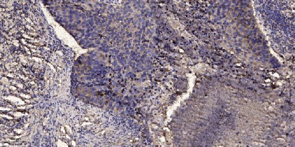
A05879 has real IHC data from paraffin-embedded human lung squamous cell carcinoma (IHC image caption); IF is listed without a figure (catalog applications; catalog IF images). Human, mouse and rat reactivity is listed (catalog reactivity).

Real IHC data Immunohistochemical analysis of paraffin-embedded human Squamous cell carcinoma of lung. 1, Antibody was diluted at 1:200 (4° overnight). 2, Tris-EDTA, pH9.0 was used for antigen retrieval. 3, Secondary antibody was diluted at 1:200 (room temperature, 45min).
Anti-Sialomucin core protein 24 CD164 Antibody
Cat # A05879

A05879 will render with an IHC figure from paraffin-embedded human lung squamous cell carcinoma (IHC image caption). Its listed applications include IHC and IF, and its listed reactivity covers human, mouse and rat (catalog applications; catalog reactivity).

Which to pick: Choose A05879 for paraffin-section IHC: its figure uses a 1:200 primary dilution and Tris-EDTA retrieval at pH 9.0; the fixative is unreported (IHC image caption). A05879 is also listed for IF, but ICC validation and an IF figure are unreported (catalog applications; catalog IF images). For mouse or rat samples, A05879 lists reactivity with both species, although its IHC figure shows human tissue only; the antibody is polyclonal (catalog reactivity; IHC image caption; catalog dilution_raw).

Each figure is that product's own IHC / IF validation image from its datasheet.

References

  1. UniProt Consortium. UniProt entry Q04900 (MUC24_HUMAN, Sialomucin core protein 24).
  2. Human Protein Atlas. CD164 tissue IHC expression (reliability: Approved).
  3. Human Protein Atlas. CD164 subcellular location (ICC-IF): Membrane.
  4. Human Protein Atlas. CD164 antibody validation summary (1 antibodies).
  5. CD164 promotes tumor progression and predicts the poor prognosis of bladder cancer. Cancer medicine 2018 — PMC6089154.
  6. CD164 promotes lung tumor-initiating cells with stem cell activity and determines tumor growth and drug resistance via Akt/mTOR signaling. Oncotarget 2017 — PMC5589567.
  7. RP11‑619L19.2 promotes colon cancer development by regulating the miR‑1271‑5p/CD164 axis. Oncology reports 2020 — PMC7610312.
  8. Distinctiveness of Femoral and Acetabular Mesenchymal Stem and Progenitor Populations in Patients with Primary and Secondary Hip Osteoarthritis Due to Developmental Dysplasia. International journal of molecular sciences 2024 — PMC11121609.
  9. PubMed PMID:1478919 — UniProt-cited evidence.
  10. PubMed PMID:9763543 — UniProt-cited evidence.
  11. PubMed PMID:10878358 — UniProt-cited evidence.