This website uses cookies to ensure you get the best experience on our website.


- Table of Contents
Use bone marrow hematopoietic cells or colonic enterocyte microvilli as positive IHC references (HPA tissue IHC). This guide covers expected staining patterns and a catalog antibody starting dilution of 1:100 (HPA tissue IHC; datasheet: 1:100).

Expected localisation, validated protocols, controls and antibodies — the at-a-glance facts below, then the full design guide.
| Expected localisation | Marrow/spleen cytoplasm; colon/rectum membrane (HPA tissue IHC) | |
| Staining pattern | Marrow/spleen subsets: cytoplasmic; enterocyte microvilli: membranous (HPA tissue IHC) | |
| Antigen retrieval | Citrate pH 6.0 HIER, 95–98 °C, 20 min (rule: cytoplasmic / membrane antigen) | |
| Positive control | Appendix+4 more · see all | |
| Negative control | Adipose tissue+4 more · see all |
| Fixation | Keep formalin fixation consistent across paraffin sections. (standard IHC practice; not target-specific) | |
| Caveat | Only a variable subset of mature neutrophils expresses CD177 (UniProt) | |
| Regulation | TNF, fMLP or CXCL8 increase surface CD177 (UniProt) | |
| Isoform / epitope | 3 isoforms; check the mature ectodomain epitope (UniProt) |
The catalog antibody’s IHC-P protocol is followed by four published CD177 IHC protocols (PMC3734037; PMC10941067; PMC6476150; PMC10641157).
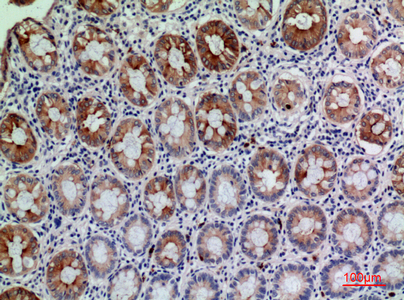
| Sample | Paraffin-embedded human colon tissue; fixative not specified (datasheet A03958) |
| Fixation | Image fixative and duration unreported (datasheet A03958); verify before use. |
| Sectioning | 4–5 µm sections on charged slides (standard) |
| Deparaffinisation | Xylene, graded ethanol series to water (standard) |
| Antigen retrieval | Heat-induced epitope retrieval in citrate buffer, pH 6.0, 20 min at 95–98 °C (standard rule: cytoplasmic / membrane antigen) |
| Peroxidase block | 3% H2O2, 10 min, room temperature (standard) |
| Blocking | 10% normal serum of the secondary host, 30 min, room temperature (standard) |
| Primary antibody | Rabbit anti-CD177, 1:100 (datasheet A03958) |
| Primary incubation | Overnight at 4 °C (standard) |
| Detection | HRP-polymer secondary, DAB chromogen 5–10 min (standard) |
| Counterstain | Hematoxylin, blue, dehydrate and mount (standard) |
| Expected result | CD177-positive staining in enterocytes - Microvilli of appendix (HPA tissue IHC: High). HPA tissue profile: Cytoplasmic expression in a subset of cells in bone marrow and spleen. Membranous expression in colon and rectum. No signal in the no-primary control. |
CD177 is a GPI-anchored surface protein with no transmembrane segment; it also occurs in neutrophil granule membranes and can be shed (UniProt Q8N6Q3 topology, subcellular location). In IHC, expect staining in a subset of marrow hematopoietic cells and at intestinal enterocyte microvilli; HPA also reports cytoplasmic staining in a subset of splenic cells (HPA: Enhanced tissue IHC).
| Distinct staining marks only some marrow hematopoietic cells; nearby cells remain pale. | A mixed result fits CD177 expression in a subset of mature neutrophils. Expression begins at the metamyelocyte stage, and the positive fraction varies between people; score the fraction and compartment rather than expecting every granulocyte to stain (UniProt Q8N6Q3 tissue specificity). |
| Colon or rectum shows a crisp apical enterocyte border. | This matches HPA's high microvillar staining and reported membranous expression in these tissues (HPA: colon and rectum tissue IHC). Read the epithelial border separately from leukocytes in the same section so their different expected patterns are not conflated (HPA: tissue IHC; UniProt Q8N6Q3 tissue specificity). |
| Staining is predominantly nuclear, without the expected cell surface or cytoplasmic pattern. | A nuclear pattern does not match the supplied CD177 locations and should prompt an artefact check (UniProt Q8N6Q3 subcellular location; HPA: tissue IHC). Review the counterstain and detection controls before calling those nuclei CD177 positive (standard IHC practice). |
| Strong signal appears in adipocytes or another cell type HPA lists as undetected. | That result conflicts with the specified negative cell population, though it does not by itself prove cross-reactivity (HPA: adipose tissue, adipocytes, not detected). Check antibody specificity and endogenous detection activity with appropriate controls; assess each named cell population rather than declaring an entire tissue negative (standard IHC practice; HPA: tissue IHC). |
| Diffuse colour covers tissue and blank spaces, or a known-positive marrow section has no specific signal. | Diffuse deposition obscures compartment scoring and suggests background; an absent signal in a known-positive section leaves the run uninterpretable (standard IHC practice). Marrow hematopoietic cells provide an HPA high-staining comparator, while their subset pattern means a small sampled field can miss positive cells (HPA: bone marrow tissue IHC; UniProt Q8N6Q3 tissue specificity). |
| Cell mixture and maturation | CD177 appears from the metamyelocyte stage onward and labels only a variable subset of mature neutrophils. Field selection and the proportion of those cells therefore affect the apparent positive fraction (UniProt Q8N6Q3 tissue specificity). |
| Compartment and processing | CD177 has a signal peptide, a processed mature chain and a GPI-anchor annotation. Neutrophils store it mainly in secondary and tertiary granules; surface expression can rise after stimulation, and CD177 can be shed (UniProt Q8N6Q3 processing, keywords, subcellular location). These facts support more than one plausible cellular pattern, without predicting how paraffin processing changes it. |
| Tissue-specific appearance | HPA reports cytoplasmic expression in subsets of marrow and splenic cells, but membranous expression in colon and rectum. Its listed high signal in enterocyte microvilli gives a separate epithelial pattern to recognize (HPA: Enhanced tissue IHC). |
| Antibody evidence | HPA marks tissue IHC reliability Enhanced and lists HPA041820, HPA046601 and HPA077640 as IHC Enhanced (HPA: tissue IHC and antibody validation). That supports the observed tissue pattern; it does not supply target-specific antigen retrieval, fixation sensitivity or IF performance. |
| Antigen retrieval and detection controls | Retrieval conditions, blocking and chromogenic detection should be assessed with positive and negative controls for the chosen IHC assay (standard IHC practice). The supplied HPA and UniProt records do not specify a CD177 retrieval condition or show that fixation changes CD177 signal. |
| IF/ICC Q: Is an image-backed localisation available? | A: HPA lists no main subcellular location and no cell lines with ICC-IF images; its listed antibodies have no ICC validation entry (HPA: subcellular record and antibody validation). Treat the UniProt locations as biological context, not an IF-validated staining pattern (UniProt Q8N6Q3 subcellular location). |
| Situation | Likely cause | Next action |
|---|---|---|
| No specific staining in a marrow positive control | The run may have failed, or the examined field may contain too few CD177-positive cells (standard IHC practice; UniProt Q8N6Q3 tissue specificity). | Check that hematopoietic cells and multiple fields were examined, then review the assay's retrieval, antibody and detection steps against its own validated conditions (HPA: bone marrow high; standard IHC practice). |
| Uniform nuclear staining dominates | Nuclear localisation is unsupported by the supplied CD177 patterns; counterstain or nonspecific detection may be contributing (UniProt Q8N6Q3 subcellular location; HPA: tissue IHC; standard IHC practice). | Compare the nuclear colour with the counterstain and a no-primary control, and require the expected cellular compartment before scoring positive (standard IHC practice; HPA: tissue IHC). |
| Diffuse chromogen obscures cell borders | Background can arise from detection chemistry or insufficient blocking and washing (standard IHC practice). | Inspect a no-primary control, review blocking and wash steps, and score only resolved cellular staining after background is controlled (standard IHC practice). |
| Adipocytes stain strongly | HPA lists adipocytes in adipose tissue as not detected, so that cell-specific result warrants a specificity or endogenous-activity check (HPA: adipose tissue IHC; standard IHC practice). | Confirm cell identity, compare a no-primary control and the IHC-validated antibody's pattern, then avoid assigning CD177 positivity until the unexpected signal is resolved (standard IHC practice; HPA: antibody validation). |
| Every marrow granulocyte appears positive | That uniform pattern conflicts with the reported variable positive subset; cell identity, staining threshold or background may be misread (UniProt Q8N6Q3 tissue specificity; standard IHC practice). | Recheck morphology and controls, and record the positive fraction with its scoring threshold across several fields (standard IHC practice; UniProt Q8N6Q3 tissue specificity). |
| Colon or rectum lacks an apical border despite tissue colour | Diffuse colour does not reproduce HPA's enterocyte-microvillar pattern and cannot establish epithelial CD177 staining (HPA: colon and rectum tissue IHC). | Inspect border preservation and a no-primary control; compare the compartment with an HPA high-staining reference pattern before calling the section positive (HPA: tissue IHC; standard IHC practice). |
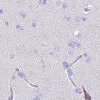
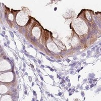


Comprehensive Human Protein Atlas IHC scoring per tissue (reliability: Enhanced — High consistency between antibody staining and RNA expression data.). Rows are taken directly from the HPA tissue chart — click any row's HPA link to view the source.
| Tissue | Cell type | Level | Evidence | Source |
|---|---|---|---|---|
| Appendix | Enterocytes - Microvilli | High | Protein (IHC) | HPA → |
| Bone marrow | Hematopoietic cells | High | Protein (IHC) | HPA → |
| Cervix | Squamous epithelial cells | High | Protein (IHC) | HPA → |
| Colon | Enterocytes - Microvilli | High | Protein (IHC) | HPA → |
| Oral mucosa | Squamous epithelial cells | High | Protein (IHC) | HPA → |
| Tissue | Cell type | Level | Evidence | Source |
|---|---|---|---|---|
| Adipose tissue | Adipocytes | Not detected | Protein (IHC) | HPA → |
| Adrenal gland | Glandular cells | Not detected | Protein (IHC) | HPA → |
| Breast | Adipocytes | Not detected | Protein (IHC) | HPA → |
| Bronchus | Respiratory epithelial cells | Not detected | Protein (IHC) | HPA → |
| Caudate | Glial cells | Not detected | Protein (IHC) | HPA → |
Troubleshoot CD177 staining in paraffin sections by checking retrieval, cell identity, staining compartment and controls before scoring chromogenic signal.
A03958 has a human paraffin-section IHC image (IHC image caption: human colon) and is listed for IF and for human and mouse reactivity (catalog: applications and reactivity).
A03958 will render with a human colon paraffin-section IHC image at 1:100 (IHC image caption). The catalog lists A03958 for IHC and IF in human and mouse, but provides no IF image (catalog: applications, reactivity, image alts).
Which to pick: Choose A03958 for tissue IHC when a human paraffin-section example is useful; its own caption documents human colon at 1:100, with the fixative unreported (IHC image caption). A03958 is also the listed IF option, though the catalog provides no IF/ICC image (catalog: applications, image alts). For mouse work, A03958 is the only listed choice because its reactivity includes mouse; it is a rabbit polyclonal antibody, while its IHC image shows human tissue only (catalog: reactivity and host; dilution_raw: polyclonal; IHC image caption).