This website uses cookies to ensure you get the best experience on our website.


- Table of Contents
Plan CD1B chromogenic IHC in paraffin sections using the catalog antibody’s documented tonsil protocol (datasheet A02158-1). Assess lysosomal staining with appropriate controls, bearing in mind its low consistency with RNA expression (HPA tissue IHC).

Expected localisation, validated protocols, controls and antibodies — the at-a-glance facts below, then the full design guide.
| Expected localisation | Lysosomal staining in several tissues (HPA tissue IHC) | |
| Staining pattern | Lysosomal staining in glandular cells (HPA tissue IHC) | |
| Antigen retrieval | Citrate pH 6 HIER, heat-mediated (datasheet A02158-1) | |
| Positive control | Adipose tissue+4 more · see all | |
| Negative control | Bone marrow+4 more · see all |
| Fixation | Keep fixation consistent across sections (standard IHC practice; not target-specific) | |
| Caveat | Staining shows low consistency with RNA (HPA tissue IHC) | |
| Regulation | Expression regulation is not specified (UniProt) | |
| Isoform / epitope | 2 isoforms; epitope differences unknown; check sidedness (UniProt) |
Compare the catalog antibody’s citrate pH 6 HIER protocol (datasheet A02158-1) with the published CD1B IHC protocols listed below (PMC7055113; PMC6553556; PMC13553427; PMC11629625).
| Sample | Paraffin-embedded human tonsil tissue; fixative not specified (datasheet A02158-1) |
| Fixation | Image fixative and duration unreported (datasheet A02158-1); verify before use. |
| Sectioning | 4–5 µm sections on charged slides (standard) |
| Deparaffinisation | Xylene, graded ethanol series to water (standard) |
| Antigen retrieval | Heat retrieval: Citrate pH 6, 20 min (datasheet A02158-1) |
| Peroxidase block | 3% H2O2, 10 min, room temperature (standard) |
| Blocking | 10% goat serum (datasheet A02158-1) |
| Primary antibody | Rabbit anti-CD1B, 1μg/ml (datasheet A02158-1) |
| Primary incubation | Overnight at 4 °C (datasheet A02158-1) |
| Detection | Streptavidin-biotin complex (SABC), DAB chromogen (datasheet A02158-1) |
| Counterstain | Hematoxylin, blue, dehydrate and mount (standard) |
| Expected result | CD1B-positive staining in adipocytes of adipose tissue (HPA tissue IHC: Medium). HPA tissue profile: Lysosomal expression in several tissues. No signal in the no-primary control. |
CD1B can appear at the cell surface and in intracellular compartments as it traffics through endosomes and lysosomes (UniProt P29016 topology and subcellular location). In paraffin-section IHC, HPA reports lysosomal staining in several tissues, including medium staining in adipocytes and selected glandular cells (HPA tissue IHC). Treat these as observed patterns, not universal expectations: HPA rates its IHC profile Approved but reports low consistency with RNA expression (HPA tissue IHC).
| Discrete intracellular granules or a vesicular pattern in adipocytes or glandular cells, with possible cell-edge staining. | This is compatible with HPA’s lysosomal IHC profile and its medium staining in adipocytes and selected glandular cells (HPA tissue IHC). A cell-edge component is biologically plausible because CD1B has an extracellular domain and one transmembrane segment (UniProt P29016 topology). Score the compartments separately. |
| Strong, uniform nuclear staining with little cytoplasmic or membrane-associated signal. | A nuclear-dominant pattern conflicts with the reported membrane, endosome and lysosome locations (UniProt P29016 subcellular location) and HPA’s lysosomal IHC profile (HPA tissue IHC). Consider nonspecific staining or an imaging/scoring error; this pattern alone does not establish CD1B positivity. |
| Prominent staining in skeletal-muscle myocytes or bone-marrow hematopoietic cells. | HPA reports CD1B as not detected in those specific cell populations (HPA tissue IHC). Recheck cell identity and compare controls; unexpected signal could reflect cross-reactivity or endogenous chromogen-generating activity (general IHC practice). A different staining result does not, by itself, identify the cause. |
| Diffuse chromogen across cells, stroma and slide background, without a defined cellular compartment. | This does not resemble the reported lysosomal tissue pattern (HPA tissue IHC). Inspect the no-primary control and review blocking, washes and detection chemistry for background (general IHC practice). Diffuse signal cannot be scored as CD1B solely because the section contains an HPA-reported positive cell type. |
| No staining in an adipocyte or glandular-cell population selected as a positive comparator. | HPA reports medium staining in adipocytes from adipose tissue or breast and in selected glandular cells (HPA tissue IHC). Verify that the relevant cells are present, then assess the antibody and detection run with controls (general IHC practice). HPA’s Approved rating and low RNA–staining consistency limit how strongly one negative section can be interpreted (HPA tissue IHC). |
| Compartment and topology | CD1B spans the membrane at residues 304–324, with residues 18–303 extracellular and 325–333 cytoplasmic (UniProt P29016 topology). Its trafficking between the surface, endosomes and lysosomes permits both cell-edge and intracellular staining (UniProt P29016 subcellular location). The record does not locate the catalog antibody’s epitope. |
| Choice of tissue and cell population | HPA reports medium staining in adipocytes and several glandular-cell populations, but no detection in skeletal-muscle myocytes and bone-marrow hematopoietic cells (HPA tissue IHC). Use the named cell population when comparing sections; a whole-tissue label can conceal mixed cells and focal staining (general IHC interpretation). |
| Strength of the tissue evidence | The tissue profile is Approved, yet HPA explicitly notes low consistency between antibody staining and RNA expression; the listed antibody HPA021824 is IHC Approved, without an IHC Enhanced designation in the supplied record (HPA tissue IHC; HPA antibody validation). Interpret an unexpected distribution cautiously and retain controls. |
| Antigen retrieval and detection | Target-specific fixation effects are not established by the supplied assay evidence. Verify with a matched IHC source before attributing a result to fixation. |
| Situation | Likely cause | Next action |
|---|---|---|
| A presumed positive section has no chromogen. | The expected cell population may be absent, or the IHC run may have failed (general IHC practice); HPA’s positive calls apply to named cells and staining levels (HPA tissue IHC). | Locate the named cells, inspect a run control and confirm primary-antibody and detection steps (general IHC practice). Avoid calling the tissue CD1B-negative until the run is valid. |
| The no-primary control is stained. | Endogenous detection activity or nonspecific chromogen deposition is possible (general IHC practice). This control cannot demonstrate CD1B binding. | Address the relevant endogenous activity, blocking or wash issue, then repeat the control alongside the primary-antibody section (general IHC practice). |
| Signal fills nuclei or appears uniformly across unrelated cell types. | The pattern conflicts with CD1B’s reported membrane and intracellular trafficking locations (UniProt P29016) and HPA’s lysosomal IHC profile (HPA tissue IHC). | Check counterstain, focus, control sections and background before scoring; require a defined cellular compartment in the named cell population (general IHC practice). |
| Skeletal-muscle myocytes or bone-marrow hematopoietic cells stain prominently. | Those cell populations are listed as not detected (HPA tissue IHC); possible explanations include mistaken cell identification, nonspecific antibody binding or detection background (general IHC practice). | Confirm the stained cell type and compare no-primary and tissue controls. Report the unexpected pattern instead of treating it as established CD1B expression. |
| A positive cell population shows only faint or scattered granules. | HPA’s medium staining examples describe observed levels, not a guaranteed intensity for every section (HPA tissue IHC). Section quality and run conditions can complicate interpretation (general IHC practice). | Score the fraction of relevant cells, intensity and compartment together; compare the same run’s controls and document uncertainty when the pattern is below a clear positive call (general IHC practice). |
| Q: Should ICC-IF Golgi or vesicle staining dictate the paraffin-section IHC call? | HPA’s ICC-IF summary places CD1B mainly in Golgi apparatus and vesicles, with additional cytosol, whereas its tissue IHC summary describes lysosomal expression (HPA subcellular ICC-IF; HPA tissue IHC). | A: Use the tissue IHC pattern and cell population for this chromogenic section. Treat the ICC-IF observations as context, not an IHC protocol or a requirement to assign every IHC granule to the Golgi. |
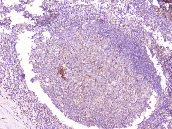
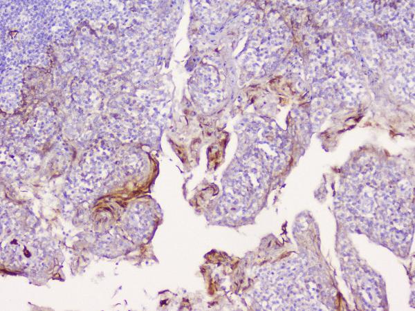
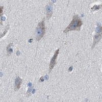


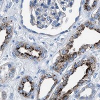


Comprehensive Human Protein Atlas IHC scoring per tissue (reliability: Approved — Low consistency between antibody staining and RNA expression data.). Rows are taken directly from the HPA tissue chart — click any row's HPA link to view the source.
| Tissue | Cell type | Level | Evidence | Source |
|---|---|---|---|---|
| Adipose tissue | Adipocytes | Medium | Protein (IHC) | HPA → |
| Adrenal gland | Glandular cells | Medium | Protein (IHC) | HPA → |
| Appendix | Glandular cells | Medium | Protein (IHC) | HPA → |
| Breast | Adipocytes | Medium | Protein (IHC) | HPA → |
| Bronchus | Respiratory epithelial cells | Medium | Protein (IHC) | HPA → |
| Tissue | Cell type | Level | Evidence | Source |
|---|---|---|---|---|
| Bone marrow | Hematopoietic cells | Not detected | Protein (IHC) | HPA → |
| Caudate | Glial cells | Not detected | Protein (IHC) | HPA → |
| Epididymis | Glandular cells | Not detected | Protein (IHC) | HPA → |
| Ovary | Ovarian stroma cells | Not detected | Protein (IHC) | HPA → |
| Skeletal muscle | Myocytes | Not detected | Protein (IHC) | HPA → |
Troubleshoot CD1B staining in paraffin sections by checking retrieval, compartment, cell identity and controls before interpreting chromogenic signal.
One human-reactive antibody has real IHC data from a paraffin-embedded tonsil section (catalog image: A02158-1). No IF/ICC data are provided (catalog: applications, image alts).
A02158-1 is listed for human IHC (catalog: applications, reactivity). Its IHC image shows CD1b staining in a paraffin-embedded human tonsil section (catalog image: A02158-1).
Which to pick: Choose A02158-1 for human paraffin-section IHC: it is rabbit polyclonal (catalog: host, dilution_raw), and its own image documents staining after citrate retrieval (catalog image: A02158-1). The fixative is unreported (catalog image: A02158-1). No listed SKU has IF/ICC validation or nonhuman reactivity, so neither use has a supported pick here (catalog: applications, reactivity, IF image alts).