This website uses cookies to ensure you get the best experience on our website.
- Table of Contents
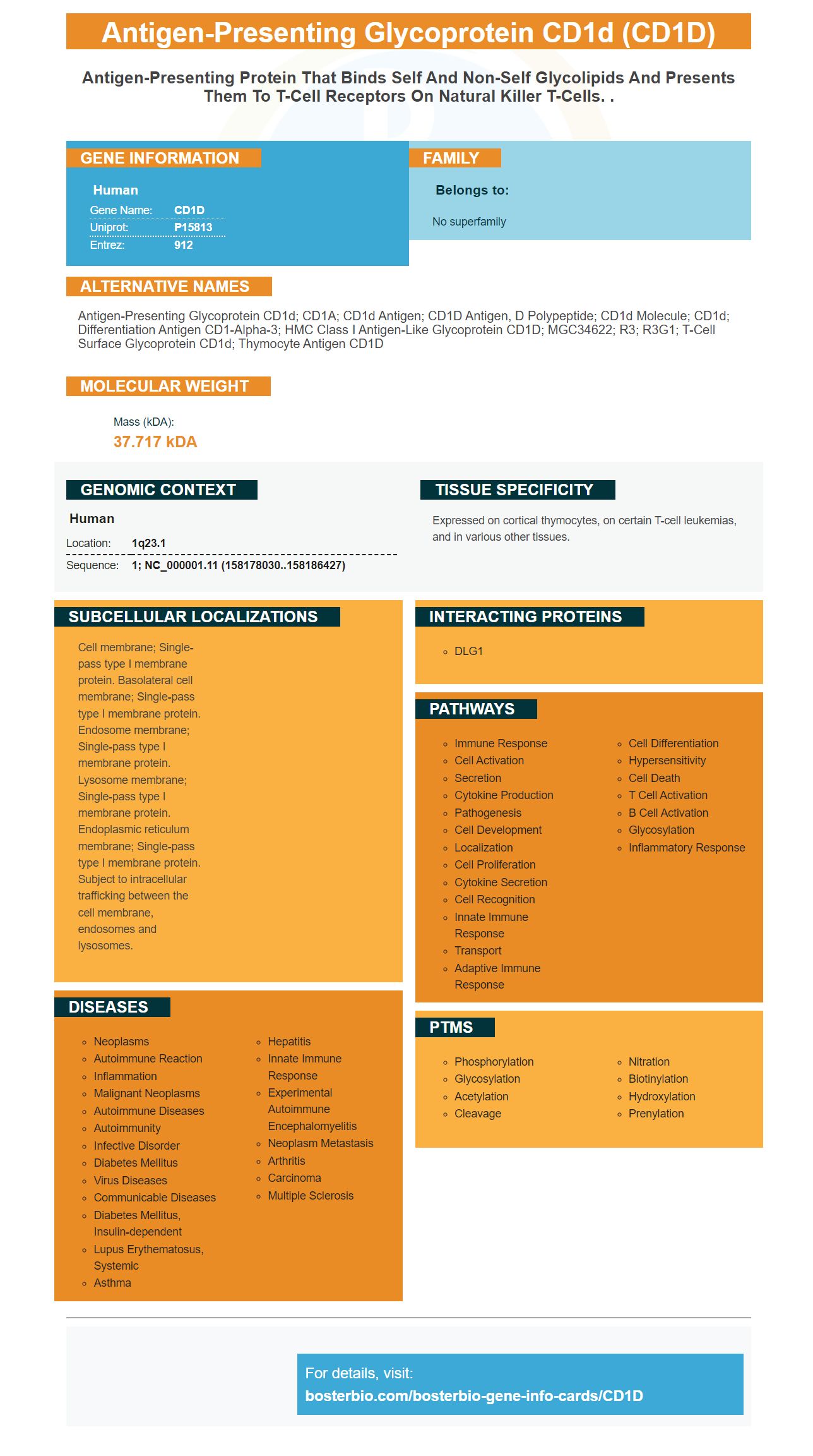
Facts about Antigen-presenting glycoprotein CD1d.
Human | |
---|---|
Gene Name: | CD1D |
Uniprot: | P15813 |
Entrez: | 912 |
Belongs to: |
---|
No superfamily |
antigen-presenting glycoprotein CD1d; CD1A; CD1d antigen; CD1D antigen, d polypeptide; CD1d molecule; CD1d; differentiation antigen CD1-alpha-3; HMC class I antigen-like glycoprotein CD1D; MGC34622; R3; R3G1; T-cell surface glycoprotein CD1d; thymocyte antigen CD1D
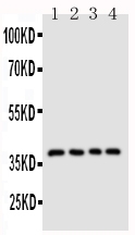
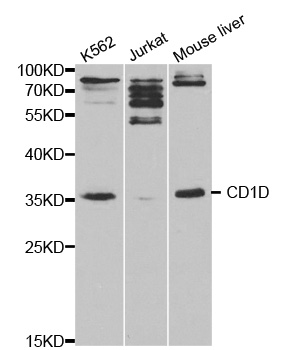
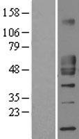
Mass (kDA):
37.717 kDA
Human | |
---|---|
Location: | 1q23.1 |
Sequence: | 1; NC_000001.11 (158178030..158186427) |
Expressed on cortical thymocytes, on certain T-cell leukemias, and in various other tissues.
Cell membrane; Single-pass type I membrane protein. Basolateral cell membrane; Single-pass type I membrane protein. Endosome membrane; Single-pass type I membrane protein. Lysosome membrane; Single-pass type I membrane protein. Endoplasmic reticulum membrane; Single-pass type I membrane protein. Subject to intracellular trafficking between the cell membrane, endosomes and lysosomes.
Boster Bio has the best CD1D antibody. Their high-affinity antibody has been extensively discussed in the research community in the past 25 years. Furthermore, Boster antibodies are tested on Western Blotting, Immunohistochemistry, and ELISA, making them a trusted choice in your research project. This article will provide information about Boster Bio as well as his career.
A range of B cell malignancies include hairy cell, variant hairy, and lymphoma of the splenic margin zone. The CD1D marker is a lot of expression. These diseases are usually incurable and make them ideal targets for treatment. In this study, CD1d expression was detected on the BMDCs' surfaces from hCD1d-KI mouse, but not on the surfaces of WT mice. These responses were confirmed by blocking with mAbs that are specific for human CD1d.
The expression of CD1D on B cells is constantly changing and requires further investigation to determine the exact pathways involved. The evidence from studies on the experimental side suggests that CD1D cells and iNKT cells play an important role in the development of humoral immunity. It isn't clear what role CD1d and the iNKT cells are playing in enhancing the humoral immune system. These immune cells are involved in the production of powerful adjuvant activity and assist in the development of therapeutic protocols to increase the effectiveness of vaccines.
The hCD1dKI mice are a useful model to study human INKT cells in live. The hCD1d–KI mouse is equipped with an equivalent amount of iNKT cells to normal human lymphocytes. This mouse model may prove beneficial in further studies of the iNKT cells that populate peripheral tissues. Moreover, hCD1dKI mice offer an efficient and precise method to compare the proliferative potential of mature CD4+ cells with DN INKT.
We conducted flow cytometry analysis on mouse lymphoma cells in FACS buffer with 1.1 percent BSA and 0.1 percent FCS. We utilized CD1d tetramers that were loaded with PBS57 from the NIH Tetramer Facility. The cells were stained using Foxp3/Transcription factor staining buffer and fixed cells were analysed using the fixable viability stain. For analysis we utilized BD Biosciences flow cytometers as well as FlowJo software.
Our results indicated that mice that lack the CD1D marker possess a broader range of IgA antibodies. The IgA repertoire of mice without CD1d is normal, however the IgA repertoire has been altered. This finding suggests that the deficiency of CD1d in mice can lead to changes in the microbiota which alters the IgA repertoire. This study concludes that CD1D expression in the intestinal lining is vital for determining how IgA antibodies are made and used.
The immunization of mice using alum with NPKLH led to the formation of high affinity AFCs that were monitored for up to seven days after transfer. It was difficult to assess the AFCs beyond day 14 due to the low number. Even after day 14 high affinity AFCs were still produced, even after the elimination of Clec9A targeted APCs.
The spleens of mice were removed on the seventh day and sera was collected via retroorbital eye bleedings. The recipients were vaccinated with 100 mg NP-KLH that was emulsified in CFA one week prior to transfer of the cells. After 30 days the T cells of the recipients had gone. Secondary immune responses were measured using pooled B cells from the primary recipient.
The activated B cell antigen-binding were collected from mice and mKO2lo/ mice. They were stained with CD86 and assessed for the presence or absence of CXCR4. Flow cytometric gating of GC zone division was observed on day 7. Antigen-specific GC B cells from mice with IL21r were analyzed to determine the phenotype of the light zone in IL21r-/ mice.
Histology of the NP–BSA protein shows that NP–BSA binds with ECs in blood vessels. While BSA is not a carbohydrate-specific but it does colocalize with MRs and TfRs. It is therefore a useful biomarker to NP-BSA. A NP with a higher carbohydrate specificity than its protein counterpart is more suitable for use in vascular applications.
The final contrast agent has an extremely narrow distribution as well as a negative surface charge of -26 mV up to -30mV. Supplements I and II provide information on how to calculate the maximum Gd tessellation. The effects of this toxic compound on the kidneys and lung are still not known. However, the NPBSA-Gd contrast agents could be used as EC-targeted contrast agents. Further research is needed to verify their potential for diagnostic use.
PMID: 2467814 by Calabi F., et al. Two classes of CD1 genes.
PMID: 2463622 by Balk S.P., et al. Isolation and characterization of a cDNA and gene coding for a fourth CD1 molecule.