This website uses cookies to ensure you get the best experience on our website.


- Table of Contents
Plan chromogenic CD1E IHC on paraffin sections using the IHC-validated antibody at 1:100 (datasheet). Interpret cytoplasmic and membranous staining with caution because the tissue-IHC profile has uncertain reliability (HPA tissue IHC).

Expected localisation, validated protocols, controls and antibodies — the at-a-glance facts below, then the full design guide.
| Expected localisation | Cytoplasmic and membranous tissue staining (HPA tissue IHC) | |
| Staining pattern | Several cell types, including immune cells; cytoplasmic/membranous (HPA tissue IHC) | |
| Antigen retrieval | Citrate pH 6.0 HIER, 95–98 °C, 20 min (rule: cytoplasmic / membrane antigen) | |
| Positive control | Epididymis+3 more · see all | |
| Negative control | Adipose tissue+4 more · see all |
| Fixation | Keep tissue fixation consistent across sections. (standard IHC practice; not target-specific) | |
| Caveat | Tissue staining has low consistency with RNA expression (HPA tissue IHC) | |
| Regulation | Maturation alters CD1e distribution (UniProt) | |
| Isoform / epitope | 13 isoforms; check extracellular/cytoplasmic epitopes after cleavage (UniProt) |
The catalog antibody’s IHC-P protocol is accompanied by published medulloblastoma multiplex staining (PMC10716463) and paraffin-embedded tumor tissue chromogenic staining (PMC10373563).
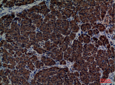
| Sample | Paraffin-embedded human pancreas tissue; fixative not specified (datasheet A06184) |
| Fixation | Image fixative and duration unreported (datasheet A06184); verify before use. |
| Sectioning | 4–5 µm sections on charged slides (standard) |
| Deparaffinisation | Xylene, graded ethanol series to water (standard) |
| Antigen retrieval | Heat-induced epitope retrieval in citrate buffer, pH 6.0, 20 min at 95–98 °C (standard rule: cytoplasmic / membrane antigen) |
| Peroxidase block | 3% H2O2, 10 min, room temperature (standard) |
| Blocking | 10% normal serum of the secondary host, 30 min, room temperature (standard) |
| Primary antibody | Rabbit anti-CD1E, 1:100 (datasheet A06184) |
| Primary incubation | Overnight at 4 °C (standard) |
| Detection | HRP-polymer secondary, DAB chromogen 5–10 min (standard) |
| Counterstain | Hematoxylin, blue, dehydrate and mount (standard) |
| Expected result | CD1E-positive staining in glandular cells of epididymis (HPA tissue IHC: Medium). HPA tissue profile: Cytoplasmic and membranous expression in several different cell types, including immune cells. No signal in the no-primary control. |
CD1E staining should be assessed by cell type and compartment: UniProt places the membrane form in the Golgi and endosomal pathway and the processed soluble form in the lysosome lumen (UniProt P15812). HPA reports cytoplasmic and membranous tissue staining, but rates its IHC evidence uncertain because staining and RNA expression have low consistency (HPA tissue IHC).
| Granular cytoplasmic staining in tonsil germinal center cells or lymph node non-germinal center cells. | This is compatible with reported medium IHC staining and intracellular CD1E trafficking; morphology alone cannot identify a Golgi, endosome, or lysosome (HPA tissue IHC; UniProt P15812). |
| Apparent nuclear staining dominates, without a convincing cytoplasmic pattern. | Treat this as suspect in IHC: HPA supports Golgi localisation by ICC-IF, while its additional nucleolar localisation is uncertain; review controls before assigning CD1E (HPA subcellular). |
| Strong staining appears in bronchial respiratory epithelium or adipocytes. | Investigate nonspecific antibody binding or endogenous chromogenic activity: HPA reports these specific cell populations as not detected, although its overall IHC reliability is uncertain (HPA tissue IHC). |
| Color spreads across tissue compartments, including cells expected to lack staining. | A diffuse pattern does not resolve CD1E localisation; compare a no-primary control, blocking, and detection conditions using general IHC practice, then reassess the cell-specific result (HPA tissue IHC). |
| No staining appears in a well-preserved tonsil or lymph node section. | Check the assay before calling CD1E absent: HPA reports medium staining in specified cells of these tissues, but its uncertain IHC reliability limits any single-section conclusion (HPA tissue IHC). |
| Cell type and tissue context | HPA reports medium staining in tonsil germinal center cells, lymph node non-germinal center cells, epididymal glandular cells, and testicular Leydig cells; lung macrophages are low (HPA tissue IHC). |
| Membrane topology and processing | CD1E has a transmembrane segment at residues 305–325; UniProt also describes a cleaved soluble form in mature dendritic-cell lysosomes, so membrane-only scoring could miss a compatible intracellular pattern (UniProt P15812). |
| Cell state and compartment | UniProt places CD1E predominantly in the trans-Golgi network of immature dendritic cells and its soluble form in the lysosome lumen of mature dendritic cells; routine chromogenic IHC cannot prove either organelle from color alone (UniProt P15812). |
| Antibody and isoform limits | HPA rates one antibody's IHC result uncertain and lists another without an IHC rating; UniProt lists 13 isoforms, but these records do not establish which forms an antibody detects (HPA antibodies; UniProt P15812). |
| IF/ICC Q: What localisation can its separate guide assess? | A: HPA supports a mainly Golgi signal in ICC-IF images from JURKAT and REH; its additional nucleolar assignment is uncertain. That IF evidence does not upgrade tissue IHC reliability (HPA subcellular; HPA tissue IHC). |
| Situation | Likely cause | Next action |
|---|---|---|
| Positive-control tissue has no convincing signal. | The expected cell population may be missed, or an IHC workflow step may have failed; HPA reports medium staining only in specified cells and rates its IHC evidence uncertain (HPA tissue IHC). | Locate those cells on the counterstained section; check the catalog antibody's IHC-P instructions, retrieval, detection reagents, and a concurrent positive control using general IHC practice. |
| Signal is mainly nuclear or nucleolar. | This conflicts with the supported Golgi assignment; HPA labels its additional nucleolar ICC-IF localisation uncertain, and that finding does not validate nuclear tissue IHC (HPA subcellular). | Compare cell morphology and no-primary controls; repeat with an independently validated IHC antibody if available, and avoid scoring nuclear color alone as CD1E. |
| Strong color appears in HPA-undetected cell populations. | Nonspecific binding or endogenous detection activity is possible when bronchial respiratory epithelial cells or adipocytes stain strongly despite their HPA not-detected calls (HPA tissue IHC). | Run no-primary and detection controls; review blocking and detection conditions as general IHC practice, then report the exact stained cell population rather than calling the whole tissue positive. |
| A weak signal appears only in lung macrophages. | Weak macrophage staining may match the low HPA observation; intensity alone cannot establish specificity, especially with HPA's uncertain tissue IHC reliability (HPA tissue IHC). | Assess cell morphology, background, and positive-control performance together; keep the result provisional if controls do not separate signal from background. |
| Staining is diffuse across the section. | Diffuse chromogen may obscure the cytoplasmic or membranous cell pattern HPA reports; it cannot establish a CD1E compartment by itself (HPA tissue IHC). | Use a no-primary control and inspect blocking, washes, and detection timing under general IHC practice; score only interpretable cells after background is resolved. |
| An IF image and chromogenic IHC section seem different. | HPA supports Golgi localisation in ICC-IF, while its tissue IHC profile is cytoplasmic and membranous with uncertain reliability; the assays resolve compartments differently (HPA subcellular; HPA tissue IHC). | Interpret each preparation against its own controls and stated evidence; do not infer a nucleolar IHC pattern or a failed IHC result solely from the IF image. |
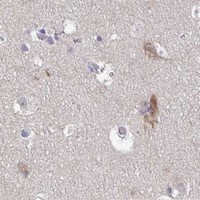


Comprehensive Human Protein Atlas IHC scoring per tissue (reliability: Uncertain — Low consistency between antibody staining and RNA expression data.). Rows are taken directly from the HPA tissue chart — click any row's HPA link to view the source.
| Tissue | Cell type | Level | Evidence | Source |
|---|---|---|---|---|
| Epididymis | Glandular cells | Medium | Protein (IHC) | HPA → |
| Lymph node | Non-germinal center cells | Medium | Protein (IHC) | HPA → |
| Testis | Leydig cells | Medium | Protein (IHC) | HPA → |
| Tonsil | Germinal center cells | Medium | Protein (IHC) | HPA → |
| Tissue | Cell type | Level | Evidence | Source |
|---|---|---|---|---|
| Adipose tissue | Adipocytes | Not detected | Protein (IHC) | HPA → |
| Breast | Adipocytes | Not detected | Protein (IHC) | HPA → |
| Bronchus | Respiratory epithelial cells | Not detected | Protein (IHC) | HPA → |
| Cerebellum | Cells in granular layer | Not detected | Protein (IHC) | HPA → |
| Cervix | Glandular cells | Not detected | Protein (IHC) | HPA → |
Troubleshoot CD1E staining in paraffin-section chromogenic IHC using its intracellular trafficking, processing, and tissue-expression evidence.
A06184 has an IHC image from human paraffin-embedded pancreas (catalog image caption); IF is a listed application, but no IF image is supplied (catalog applications; catalog image records).
A06184 will render with its human paraffin-embedded pancreas IHC image at 1:100 (catalog image caption). IF is listed for A06184, but no IF image is supplied (catalog applications; catalog image records).
Which to pick: Choose A06184 for human tissue IHC: its own image shows paraffin-embedded pancreas at 1:100; the fixative is unreported (catalog image caption). For IF, A06184 lists IF at 1:50; A06184-1 lists ICC and Human, Mouse, and Rat reactivity, but has no supplied IHC or IF image (catalog applications, dilutions, reactivity, and image records). Both list Rabbit as host, with no clone specified, so clonality cannot distinguish them; A06184-1 has no listed IHC application for cross-species tissue work (catalog host, clone, and applications).