CD247 / T-cell surface glycoprotein CD3 zeta chain · IHC design guide

Design Immunohistochemistry for CD247

Plan CD247 chromogenic IHC in paraffin sections using lymphoid immune cell staining as a reference (HPA tissue IHC). Interpret the expected membrane location alongside the tissue pattern, and account for CD247 in Fc receptor complexes (UniProt).

Evidence assembled Oct 2026 · For research use; verify linked source records and product datasheet before use
Immunohistochemistry protocol sheet for CD247 (IHC for CD247): expected localisation Cell membrane expected (UniProt); immune cells stain (HPA tissue IHC), antibody M02421, validated IHC image, and IHC protocol steps
Printable CD247 IHC protocol sheet — expected localisation Cell membrane expected (UniProt); immune cells stain (HPA tissue IHC), antibody M02421, controls and protocol steps. Open the full CD247 IHC guide →

CD247 Immunohistochemistry Experimental Design Guide

Expected localisation, validated protocols, controls and antibodies — the at-a-glance facts below, then the full design guide.

Must know before staining
Expected localisation Cell membrane expected (UniProt); immune cells stain (HPA tissue IHC)
Staining pattern Lymphoid immune cells stain (HPA tissue IHC); membrane expected (UniProt)
Antigen retrieval EDTA pH 8.0 HIER, heat-mediated (datasheet M02421)
Positive control ⓘ Appendix+4 more · see all
Negative control ⓘ Adipose tissue+4 more · see all
Important caveats
Reasons your staining may differ from the expected pattern.
Fixation Keep fixation conditions consistent across sections (standard IHC practice; not target-specific)
Caveat CD247 also occurs in Fc receptor complexes (UniProt)
Regulation TCR triggers ITAM phosphorylation (UniProt)
Isoform / epitope 3 isoforms; extracellular versus cytoplasmic epitope coverage is unknown (UniProt)
Section 1

Recommended CD247 IHC & IF Protocols

The catalog antibody protocol uses EDTA pH 8.0 heat retrieval (datasheet M02421). The published IHC protocols below cover ovarian cancer, kidney biopsies, skin lesions, and head and neck carcinoma (PMC6940041; PMC13018632; PMC12638995; PMC10683602).

Recommended immunohistochemistry (IHC-P) protocol parameters
SampleParaffin-embedded human prostate cancer tissue; fixative not specified (datasheet M02421)
FixationImage fixative and duration unreported (datasheet M02421); verify before use.
Sectioning4–5 µm sections on charged slides (standard)
DeparaffinisationXylene, graded ethanol series to water (standard)
Antigen retrievalHeat retrieval: EDTA pH 8.0 (datasheet M02421); 20 min, 95–100 °C (standard)
Peroxidase block3% H2O2, 10 min, room temperature (standard)
Blocking10% goat serum (datasheet M02421)
Primary antibodyRabbit monoclonal (clone IHI-3) anti-CD247, 1:50 (datasheet M02421)
Primary incubationOvernight at 4 °C (datasheet M02421)
DetectionHRP-conjugated secondary, DAB chromogen (datasheet M02421)
CounterstainHematoxylin, blue, dehydrate and mount (standard)
Expected resultCD247-positive staining in lymphoid tissue of appendix (HPA tissue IHC: High). HPA tissue profile: Selective expression in immune cells. No signal in the no-primary control.
💡Decision noteStart with EDTA pH 8.0 heat retrieval (datasheet M02421); a published skin protocol also uses EDTA pH 8.0 under high pressure (PMC12638995).
Section 2

What Is the Expected CD247 Staining Pattern?

CD247 is a membrane protein with a short extracellular segment and a long cytoplasmic tail (UniProt P20963 topology). In paraffin-section IHC, expect staining in immune-cell populations, especially lymphoid tissue and lymphocytes (HPA: selective expression in immune cells; UniProt P20963 tissue specificity). HPA rates its tissue IHC profile Enhanced, reflecting consistency between staining and RNA expression (HPA: Enhanced reliability).

What am I looking at on my slide?
Discrete staining outlines cells in lymphoid tissue, with little staining in surrounding non-immune cells.This fits a membrane-associated pattern (UniProt P20963: cell membrane) and the selective immune-cell profile (HPA: tissue IHC). Compare cell populations within the section; a tissue label alone does not identify every stained cell.
A predominantly nuclear or broad, uniform cytoplasmic pattern replaces the expected cell-edge signal.Treat this as a localisation mismatch, not proof of CD247 expression (UniProt P20963 topology). Review morphology and controls before scoring; chromogenic precipitate, background, or antibody behaviour can obscure a membrane pattern (general IHC practice).
Strong signal appears in adipocytes or respiratory epithelial cells.These are unexpected positive cell types: HPA reports CD247 as not detected in adipocytes of adipose tissue and respiratory epithelial cells of bronchus (HPA: tissue IHC). Consider cross-reactivity or detection-system activity; the slide alone cannot distinguish them.
Colour is spread across the section without clear cell boundaries or a difference between expected positive and negative areas.A diffuse pattern cannot be assigned confidently to CD247-positive cells (UniProt P20963: cell membrane; HPA: selective immune-cell expression). Reassess background with controls and inspect whether the signal follows tissue structures or reagent distribution (general IHC practice).
No convincing signal appears in appendix lymphoid tissue, tonsil non-germinal center cells, or another HPA-positive area.HPA reports high staining in those two annotated populations (HPA: tissue IHC). A blank result calls for checking section quality, the catalog antibody's IHC-P conditions, and detection controls before interpreting the specimen as CD247-negative (general IHC practice).
💡Expected CD247 appearanceCall a result positive when distinct immune cells show membrane-associated chromogenic staining, with high signal possible in HPA-listed lymphoid populations (UniProt P20963 topology; HPA: tissue IHC); isolated nuclear staining or uniform colour in HPA-negative non-immune cells is suspect (HPA: tissue IHC; general IHC practice).
How each factor affects the staining
Compartment and epitope orientationCD247 spans the membrane at residues 31–51, with residues 22–30 extracellular and 52–164 cytoplasmic (UniProt P20963 topology). These positions guide interpretation of antibody epitope information if available; the supplied record does not identify the catalog antibody's epitope.
Choice of comparison tissueAppendix lymphoid tissue, lymph-node germinal center cells, spleen red-pulp cells, and tonsil non-germinal center cells are listed as High; bone-marrow hematopoietic cells and endometrial lymphocytes are Medium (HPA: tissue IHC). Compare the annotated cells, since staining varies by population.
IHC-P retrieval and detection checksUse the catalog antibody's IHC-P instructions to choose retrieval and detection conditions (general IHC practice). HPA staining levels and UniProt topology do not establish a CD247-specific fixation effect or retrieval requirement; target-specific fixation sensitivity is unreported in the supplied sources.
IF/ICC: what localisation should be expected?A membrane-associated pattern is biologically consistent with CD247 (UniProt P20963: cell membrane; HPA: subcellular summary, Membrane). HPA supplies no ICC-IF image-bearing cell line or main-location assignment here, so that summary does not validate a particular IF staining appearance (HPA: subcellular record).
Why is my staining missing, weak or wrong?
SituationLikely causeNext action
Expected positive lymphoid cells show no staining.The assay may have failed, or the chosen section may lack an adequate annotated positive population (HPA: tissue IHC; general IHC practice).Check an HPA-listed High population on a control section, then review the catalog antibody's IHC-P instructions, reagent steps, and a detection control (HPA: tissue IHC; general IHC practice).
Only nuclei stain in otherwise suitable lymphoid tissue.Nuclear staining conflicts with the reported membrane location (UniProt P20963: cell membrane). Its origin cannot be assigned from appearance alone.Compare with a no-primary control and the expected cell-edge pattern; inspect whether nuclear colour persists independently of primary antibody (general IHC practice).
Adipocytes or bronchial epithelial cells stain strongly.Those populations are listed as Not detected (HPA: tissue IHC); cross-reactivity or endogenous detection activity is possible (general IHC practice).Inspect the same cell types in controls, including a no-primary control, before calling the signal CD247-specific (general IHC practice).
Red-pulp or bone-marrow sections show widespread chromogen.These specimens contain HPA-listed positive cells, while diffuse chromogen can also obscure cell-level interpretation (HPA: tissue IHC; general IHC practice).Score identifiable cells against adjacent unstained structures and check a no-primary control; for enzyme detection, review the blocking and wash steps (general IHC practice).
Signal is weak in endometrial lymphocytes or bone-marrow hematopoietic cells.HPA records Medium staining in those populations, versus High in several lymphoid populations (HPA: tissue IHC). Weakness alone does not show assay failure.Compare an HPA-listed High control in the same run before changing the assay or assigning a negative result (HPA: tissue IHC; general IHC practice).
Two IHC antibodies give different distributions.HPA lists both HPA008750 and CAB004651 as IHC Enhanced, but that status does not guarantee identical appearance in every section (HPA: antibody validation).Compare annotated cell types, compartment, and controls for each stain; resolve discordance before using either pattern as a scoring reference (HPA: tissue IHC; UniProt P20963 topology; general IHC practice).

Sample controls for CD247 IHC & IF

🧪Run appendix first: its lymphoid tissue should stain (HPA: High in appendix lymphoid tissue). Use adipose tissue as a negative comparator, assessing adipocytes (HPA: Not detected in adipocytes); on the appendix slide, nonlymphoid cells should show only background staining as an internal comparator (UniProt P20963: T-cell surface protein).
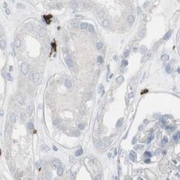
Positive control tissue: Appendix (Lymphoid tissue, HPA High)
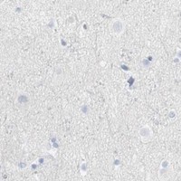
Negative control tissue: Adipose tissue (HPA Not detected)
ICC-IF cell lines (HPA subcellular resource): HPA carries no ICC-IF cell line for CD247; derive a cell-line control from the positive tissue's cell type (Lymphoid tissue) and confirm it by RNA or western blot first.
Technical controls: Include no-primary (secondary-only) and host- and immunoglobulin-class-matched rabbit isotype controls; the catalog caption identifies a rabbit primary but does not state its clonality (selected-SKU tissue-IHC caption: M02421). Use CD247-knockout tissue as a biological specificity control, and block endogenous peroxidase in the appendix section before HRP/DAB detection (selected-SKU tissue-IHC caption: HRP/DAB).
⚠️Feasibility: The selected-SKU paraffin-section caption reports heat-mediated EDTA retrieval at pH 8.0 and a 1:50 primary dilution overnight at 4°C, providing an IHC starting protocol; retrieval dependence has not been established by the supplied evidence (selected-SKU tissue-IHC caption: M02421). The fixative and any target-specific fixation window or fixation effect are unreported (selected-SKU tissue-IHC caption: fixative not stated). Whether frozen sections or IF are easier is unreported; appendix leukocytes may contribute endogenous peroxidase background during HRP/DAB detection (HPA: High in appendix lymphoid tissue; selected-SKU tissue-IHC caption: HRP/DAB).

HPA tissue IHC evidence for CD247

Comprehensive Human Protein Atlas IHC scoring per tissue (reliability: Enhanced — High consistency between antibody staining and RNA expression data. Antibody staining in cells/structures not annotated, view images.). Rows are taken directly from the HPA tissue chart — click any row's HPA link to view the source.

Positive expression · recommended positive controls

TissueCell typeLevelEvidenceSource
Appendix Lymphoid tissue High Protein (IHC) HPA →
Lymph node Germinal center cells High Protein (IHC) HPA →
Spleen Cells in red pulp High Protein (IHC) HPA →
Tonsil Non-germinal center cells High Protein (IHC) HPA →
Bone marrow Hematopoietic cells Medium Protein (IHC) HPA →

Undetected expression · recommended negative controls

TissueCell typeLevelEvidenceSource
Adipose tissue Adipocytes Not detected Protein (IHC) HPA →
Adrenal gland Glandular cells Not detected Protein (IHC) HPA →
Breast Adipocytes Not detected Protein (IHC) HPA →
Bronchus Respiratory epithelial cells Not detected Protein (IHC) HPA →
Caudate Glial cells Not detected Protein (IHC) HPA →
Section 3

Advanced CD247 IHC Tips

Use the catalog antibody’s paraffin section workflow as the IHC starting point, then assess staining by cell type, compartment and matched controls.

How should I retrieve CD247 in paraffin sections when staining is weak?
Start with heat mediated antigen retrieval in EDTA at pH 8.0 (datasheet M02421). The selected paraffin section example then used 10% goat serum blocking and a 1:50 primary antibody dilution overnight at 4°C (caption M02421). If staining remains weak, compare a shorter and longer heating interval on matched sections while keeping detection and counterstain constant (standard IHC practice). Score the change in lymphoid cells rather than overall tissue color, because CD247 expression is selective in immune cells and HPA reports high staining in appendix lymphoid tissue (HPA tissue IHC).
Can I attribute weak CD247 staining to the fixation method?
The selected paraffin section caption does not state its fixative, so target specific fixation sensitivity is unknown for this antibody (caption M02421: fixative not stated). Record the actual fixative and fixation duration for each specimen, and compare sections processed under the same conditions before attributing differences to CD247 abundance (standard IHC practice). If fixation varies, run a matched positive control through the EDTA pH 8.0 retrieval and 1:50 primary antibody conditions used in the selected example (caption M02421). Interpret loss of staining cautiously when tissue morphology or control staining also changes, since those changes can indicate a processing problem (standard IHC practice).
Where should CD247 staining appear, and how should diffuse cytoplasmic signal be judged?
CD247 is assigned to the cell membrane, with residues 22–30 extracellular, 31–51 transmembrane and 52–164 cytoplasmic (UniProt P20963 topology). Evaluate staining at the cell boundary alongside morphology, while allowing that a chromogenic section may not resolve a thin membrane distinctly around every small cell (standard IHC practice). Prioritize immune cell rich areas for assessment: HPA reports high staining in appendix lymphoid tissue and lymph node germinal center cells, and medium staining in endometrial lymphocytes (HPA tissue IHC). Diffuse staining across unrelated epithelial cells warrants a control review before being assigned to CD247 (HPA: selective expression in immune cells; standard IHC practice).
How do CD247 isoforms and epitope position affect a negative IHC result?
UniProt lists 3 CD247 isoforms and places most of the chain, residues 52–164, on the cytoplasmic side of the membrane (UniProt P20963). The record also identifies 3 cytoplasmic ITAMs and multiple modified residues, including phosphotyrosines at 64, 72, 83 and 111 (UniProt P20963). Without a mapped binding epitope for the catalog antibody, a negative stain cannot distinguish low expression from an inaccessible or absent epitope (standard IHC interpretation). Compare a negative specimen with a positive lymphoid control processed and retrieved together before making an isoform specific claim (HPA tissue IHC; standard IHC practice).
How should I adapt the CD247 question for a multiplex IF experiment?
Treat IF as a separate assay and first check whether the antibody has IF validation, since the supplied M02421 example demonstrates chromogenic staining of a paraffin section (caption M02421). Multiplex CD247 with a marker that identifies the expected immune cell population, then inspect signal within those cells rather than relying on colocalization across the whole field (HPA: selective expression in immune cells; standard IF practice). Choose a fluorophore channel after examining unstained tissue for autofluorescence, and include single stain controls to assess bleed through (standard IF practice). If the antibody binds the cytoplasmic portion at residues 52–164, test permeabilisation; an extracellular epitope at 22–30 calls for a separate access assessment (UniProt P20963 topology; standard IF practice).
What should I check when CD247 DAB staining appears widespread?
First inspect a no primary control for secondary reagent deposition and endogenous peroxidase signal, then compare it with the stained section under the same development conditions (standard IHC practice). A peroxidase blocking step belongs in an HRP and DAB workflow, but it is a general detection control rather than evidence about CD247 expression (standard IHC practice). The selected example used 10% goat serum blocking, a 1:50 rabbit primary antibody, an anti rabbit peroxidase secondary and DAB (caption M02421). Widespread color in cells unlike the immune cell distribution reported by HPA should prompt review of blocking, antibody dilution and DAB development time (HPA tissue IHC; standard IHC practice).
How should I quantify CD247 IHC across specimens with different immune cell content? ⚠ ANSWER MARKED FOR VERIFICATION
Define the tissue region and eligible cell population before scoring, then report the percentage of positive immune cells and staining intensity or an H score within that population (standard IHC practice). For spatial comparisons, count positive cells per mm² of viable tissue and also record total immune cell density in the same region (standard IHC practice). Normalize CD247 positive counts to the eligible immune cell count when the question concerns expression within infiltrates, since CD247 is selectively expressed in immune cells (HPA tissue IHC; standard IHC practice). Apply one threshold and counterstain rule across the batch, excluding folds, necrosis and tissue edges from both numerator and denominator (standard IHC practice).
How can I distinguish credible CD247 staining from an IHC artefact?
A credible pattern follows identifiable immune cells and is compatible with membrane associated CD247, while recognizing that chromogenic resolution can blur a narrow membrane outline (HPA: selective expression in immune cells; UniProt P20963 topology; standard IHC practice). Compare the test section with a lymphoid positive control, since HPA reports high staining in appendix lymphoid tissue and lymph node germinal center cells (HPA tissue IHC). Treat strong staining in an unexpected cell population, at tissue edges or in necrotic areas as a finding requiring further controls (HPA tissue IHC; standard IHC practice). If a no primary control retains DAB color, investigate endogenous enzyme activity or detection background before assigning the signal to CD247 (standard IHC practice).
Boster reagents

Best CD247 / T-cell surface glycoprotein CD3 zeta chain IHC Antibodies

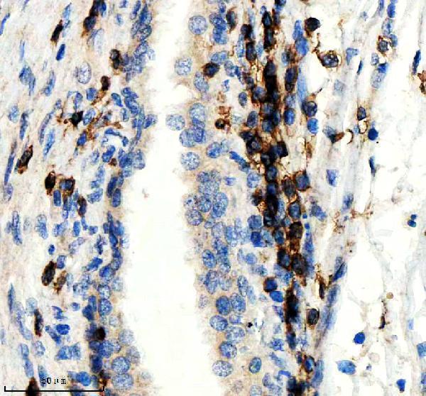
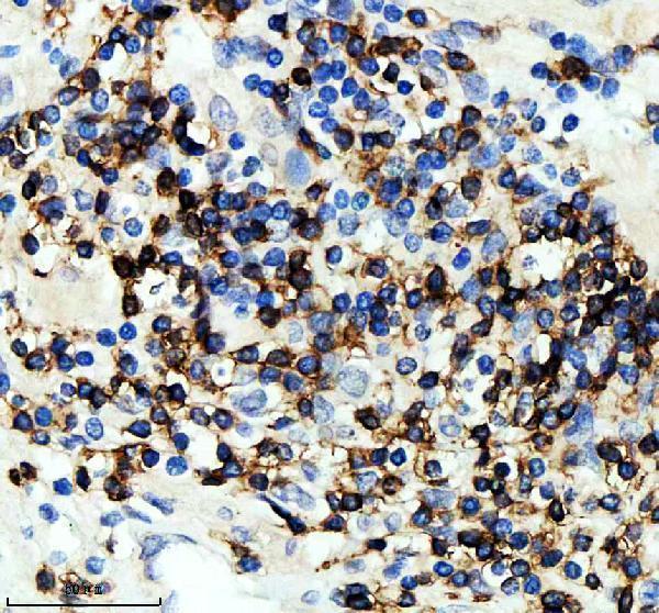
The catalog antibody has paraffin-section IHC images from human prostate cancer and lymphoma tissue (M02421 image captions); IF is listed for both SKUs, but neither has an IF figure (catalog payload).

Real IHC data IHC analysis of CD3Z using anti-CD3Z antibody (M02421). CD3Z was detected in a paraffin-embedded section of human prostate cancer tissue. Heat mediated antigen retrieval was performed in EDTA buffer (pH 8.0, epitope retrieval solution). The tissue section was blocked with 10% goat serum. The tissue section was then incubated with 1:50 rabbit anti-CD3Z Antibody (M02421) overnight at 4°C. Peroxidase Conjugated Goat Anti-rabbit IgG was used as secondary antibody and incubated for 30 minutes at 37°C. The tissue section was developed using HRP Conjugated Rabbit IgG Super Vision Assay Kit (Catalog # SV0002) with DAB as the chromogen.
Anti-CD3 zeta CD247 Rabbit Monoclonal Antibody
Cat # M02421

M02421 has IHC images from paraffin-embedded human prostate cancer and lymphoma sections (M02421 image captions); its listed reactivity is Human (M02421 catalog). P02421 lists IHC and IF/ICC applications and Human, Monkey, Mouse, and Rat reactivity, but has no IHC or IF image in the payload (P02421 catalog).

Which to pick: For human tissue IHC, choose monoclonal M02421: its own captions document paraffin-section staining at 1:50 with EDTA retrieval, while the fixative is unreported (M02421 image captions; M02421 catalog). For IF/ICC, both SKUs list those applications and an IF dilution of 1:50, but neither has an IF image here (M02421 and P02421 catalog). For work beyond human samples, consider polyclonal P02421 because it lists Monkey, Mouse, and Rat reactivity; its processing conditions are unreported because it has no IHC image caption (P02421 catalog).

Each figure is that product's own IHC / IF validation image from its datasheet.

References

  1. UniProt Consortium. UniProt entry P20963 (CD3Z_HUMAN, T-cell surface glycoprotein CD3 zeta chain).
  2. Human Protein Atlas. CD247 tissue IHC expression (reliability: Enhanced).
  3. Human Protein Atlas. CD247 subcellular location (ICC-IF): Membrane.
  4. Human Protein Atlas. CD247 antibody validation summary (2 antibodies).
  5. CD247 expression is associated with differentiation and classification in ovarian cancer. Medicine 2019 — PMC6940041.
  6. Integration of intragraft transcriptomics and urinary cytokines identifies CXCL10 and FasL signature in subclinical acute rejection. Scientific reports 2026 — PMC13018632.
  7. Immune genes CD247 STAT1 and LCK mediate host-pathogen interactions in sporotrichosis. Scientific reports 2025 — PMC12638995.
  8. Characterization of the immune cell function landscape in head and neck squamous carcinoma to assist in prognosis prediction and immunotherapy. Aging 2023 — PMC10683602.
  9. PubMed PMID:2974162 — UniProt-cited evidence.
  10. PubMed PMID:14702039 — UniProt-cited evidence.
  11. PubMed PMID:16710414 — UniProt-cited evidence.