CD38 / ADP-ribosyl cyclase/cyclic ADP-ribose hydrolase 1 · IHC design guide

Design Immunohistochemistry for CD38

Plan chromogenic CD38 IHC in paraffin sections using the observed immune cell staining pattern and the protein’s membrane topology (HPA tissue IHC; UniProt). Use lymph node or tonsil as a high staining reference and assess membranous and cytoplasmic signal by cell population (HPA tissue IHC).

Evidence assembled Oct 2026 · For research use; verify linked source records and product datasheet before use
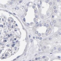
Immunohistochemistry protocol sheet for CD38 (IHC for CD38): expected localisation Membranous and cytoplasmic tissue staining (HPA tissue IHC), antibody A00193-3, validated IHC image, and IHC protocol steps
Printable CD38 IHC protocol sheet — expected localisation Membranous and cytoplasmic tissue staining (HPA tissue IHC), antibody A00193-3, controls and protocol steps. Open the full CD38 IHC guide →

CD38 Immunohistochemistry Experimental Design Guide

Expected localisation, validated protocols, controls and antibodies — the at-a-glance facts below, then the full design guide.

Must know before staining
Expected localisation Membranous and cytoplasmic tissue staining (HPA tissue IHC)
Staining pattern Membranous and cytoplasmic staining in immune cells (HPA tissue IHC)
Antigen retrieval EDTA pH 8.0 HIER, heat-mediated (datasheet A00193-3)
Positive control ⓘ Lymph node+4 more · see all
Negative control ⓘ Adipose tissue+4 more · see all
Important caveats
Reasons your staining may differ from the expected pattern.
Fixation Matched tissue-IHC evidence does not establish the fixation claim. Validate the specimen-specific method before use. (selected-SKU IHC image A00193-3)
Caveat Immune cells may complicate scoring in mixed tissues (HPA tissue IHC)
Regulation Induction not established in supplied evidence (UniProt)
Isoform / epitope 2 isoforms; epitope differences unspecified; check membrane side (UniProt)
Section 1

Recommended CD38 IHC & IF Protocols

The catalog antibody’s IHC-P protocol is followed by 4 published CD38 IHC protocols, covering liver, endometrium, breast tumors, and periapical lesions (PMC13357626; PMC10311816; PMC10607675; PMC6281009).

Recommended immunohistochemistry (IHC-P) protocol parameters
SampleParaffin-embedded human tonsil tissue; fixative not specified (datasheet A00193-3)
FixationImage fixative and duration unreported (datasheet A00193-3); verify before use.
Sectioning4–5 µm sections on charged slides (standard)
DeparaffinisationXylene, graded ethanol series to water (standard)
Antigen retrievalHeat retrieval: EDTA pH 8.0 (datasheet A00193-3); 20 min, 95–100 °C (standard)
Peroxidase block3% H2O2, 10 min, room temperature (standard)
Blocking10% goat serum (datasheet A00193-3)
Primary antibodyRabbit anti-CD38, 2μg/ml (datasheet A00193-3)
Primary incubationOvernight at 4 °C (datasheet A00193-3)
DetectionStreptavidin-biotin complex (SABC), DAB chromogen (datasheet A00193-3)
CounterstainHematoxylin, blue, dehydrate and mount (standard)
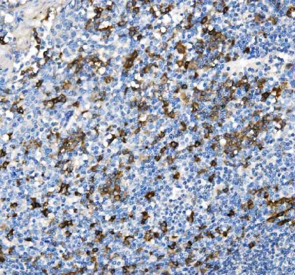
Expected resultCD38-positive staining in non-germinal center cells of lymph node (HPA tissue IHC: High). HPA tissue profile: Distinct cytoplasmic and membranous expression in hematopoietic and lymphoid tissue with immune cells in several tissues. No signal in the no-primary control.
💡Decision noteStart with heat-mediated EDTA retrieval at pH 8.0 for the catalog antibody (datasheet: A00193-3); the cited excerpts do not specify retrieval conditions.
Section 2

What Is the Expected CD38 Staining Pattern?

CD38 is a cell-surface protein with residues 43–300 extracellular and residues 1–21 cytoplasmic (UniProt P28907 topology). In paraffin-section IHC, expect membranous and sometimes cytoplasmic staining in hematopoietic and lymphoid cells, with high staining in lymph-node and tonsil non-germinal-center cells and in prostate and seminal-vesicle glandular cells (HPA tissue IHC). HPA rates the tissue pattern Enhanced, reflecting high agreement between staining and RNA expression (HPA tissue IHC).

What am I looking at on my slide?
Distinct membrane staining in lymph-node or tonsil non-germinal-center cells, with some cytoplasmic signal.This fits the cell-surface topology (UniProt P28907 topology) and the observed membranous and cytoplasmic lymphoid pattern (HPA tissue IHC). Judge signal by cell identity as well as compartment; adjacent cells need not stain equally.
High staining in prostate or seminal-vesicle glandular cells; medium staining in bone-marrow hematopoietic cells or splenic red-pulp cells.These are documented cell-specific patterns, not evidence that every cell in each organ is positive (HPA tissue IHC). Compare the named cell population with its expected level before calling a section unusually weak or strong.
Predominantly nuclear staining, with no convincing membrane or cell-associated cytoplasmic pattern.A nuclear-only pattern does not match the supported plasma-membrane location or the reported tissue IHC pattern (HPA subcellular ICC-IF; HPA tissue IHC). Treat it as suspect and check the matched negative control and detection background before scoring it as CD38.
Strong staining in adipocytes or bronchial respiratory epithelium, especially with little signal in expected positive cells.Those specified cell populations were not detected by HPA tissue IHC (HPA tissue IHC). Unexpected signal can reflect cross-reactivity or detection background; assess its location and controls. A negative call for those cells does not make the whole tissue a negative control.
Widespread haze across cells and surrounding tissue, or no signal in lymph-node non-germinal-center cells.Haze lacks the distinct cell-associated pattern reported for CD38, while absent signal in a documented high-staining population makes the run hard to interpret (HPA tissue IHC). Check controls and staining conditions before drawing a biological conclusion.
💡Expected CD38 appearanceCall a result positive when distinct membrane-associated, sometimes cytoplasmic staining marks the expected cells—high in lymph-node or tonsil non-germinal-center cells (HPA tissue IHC); nuclear-only signal or uniform haze is suspect against the reported pattern (HPA tissue IHC; HPA subcellular ICC-IF).
How each factor affects the staining
Topology and compartmentCD38 has one transmembrane segment at residues 22–42, an extracellular region at 43–300, and a cytoplasmic region at 1–21 (UniProt P28907 topology). The supported plasma-membrane location helps assess compartment; the record does not identify the catalog antibody's epitope (HPA subcellular ICC-IF).
Cell population and tissueHPA reports high signal in specified lymphoid and glandular populations, medium signal in appendix lymphoid tissue, bone-marrow hematopoietic cells, and splenic red-pulp cells, and no detected signal in specified adipocytes and other listed cells (HPA tissue IHC). Use the named cells, not an organ-wide label, for interpretation.
Independent pattern supportHPA rates the tissue IHC pattern Enhanced and lists four antibodies with Enhanced IHC status (HPA tissue IHC; HPA antibodies). This supports the reported pattern but does not establish that an unlisted antibody, dilution, or retrieval condition will reproduce it.
Protein variants and modificationUniProt lists two isoforms and four glycosylation sites at residues 100, 164, 209, and 219 (UniProt P28907). These annotations alone do not predict isoform-specific staining, epitope accessibility, or a fixation effect; interpret the observed cell pattern against HPA tissue IHC.
Why is my staining missing, weak or wrong?
SituationLikely causeNext action
No signal in lymph-node or tonsil non-germinal-center cellsA documented high-staining population should provide an informative positive control (HPA tissue IHC); the failed run alone cannot identify the cause.Check control tissue, antibody identity and working dilution, retrieval conditions, detection reagents, and counterstain visibility (general IHC practice).
Nuclear-only or sharply misplaced signalThe location conflicts with supported plasma-membrane localization and the reported tissue pattern (HPA subcellular ICC-IF; HPA tissue IHC).Review cell boundaries and compare a matched negative control; repeat with an IHC-validated antibody if the pattern persists (general IHC practice; HPA antibodies).
Uniform chromogenic haze or granular staining across unrelated cellsDiffuse signal obscures the distinct cell-associated pattern reported by HPA (HPA tissue IHC); background can arise during chromogenic detection (general IHC practice).Review blocking and washes; if using peroxidase detection, verify endogenous-peroxidase control and compare a no-primary control (general IHC practice).
Unexpected strong signal in adipocytes or bronchial respiratory epitheliumHPA did not detect CD38 in those specified cells (HPA tissue IHC); cross-reactivity or detection activity is possible, not proven.Confirm cell identity and compartment, examine the negative control, and compare an independently validated IHC antibody where available (general IHC practice; HPA antibodies).
A negative field is interpreted as a negative organHPA reports staining for particular cell populations: for example, adipocytes were not detected, while immune cells occur in several tissues (HPA tissue IHC).Score the named cell type and inspect a documented positive population on the same run before interpreting absence of signal (HPA tissue IHC; general IHC practice).
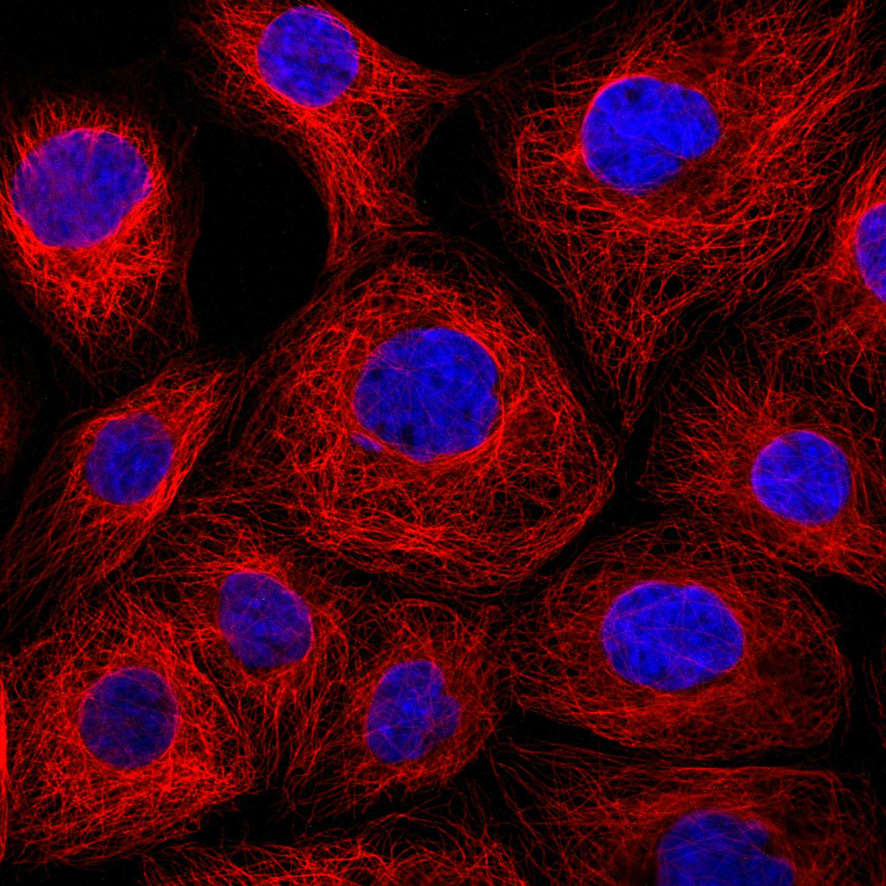
Can an ICC/IF image settle a disputed paraffin-section IHC result?HPA supports plasma-membrane localization in ICC-IF and lists A-431 and A-549 images, while its tissue IHC record describes membranous and cytoplasmic staining (HPA subcellular ICC-IF; HPA tissue IHC).Use ICC/IF as a compartment cross-check; judge the paraffin section against tissue IHC cell patterns and IHC controls (HPA tissue IHC; general IHC practice).

Sample controls for CD38 IHC & IF

🧪Run tonsil first and score its non-germinal center cells for CD38 staining (HPA: High in tonsil non-germinal center cells). Use bronchus respiratory epithelial cells as the negative tissue (HPA: Not detected in bronchus respiratory epithelial cells); on the tonsil slide, neighboring unstained cells should show counterstain without the specific membrane pattern, but their CD38 status should not be assumed from morphology alone (UniProt P28907: cell membrane localization).
Positive control tissue: Lymph node (Non-germinal center cells, HPA High)
Negative control tissue: Adipose tissue (HPA Not detected)
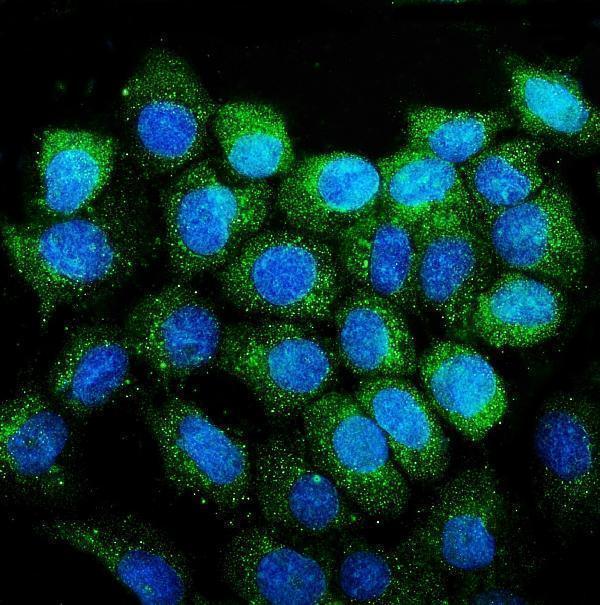
ICC-IF cell lines (HPA subcellular resource): HPA ICC-IF images show CD38 in A-431, A-549, with annotated localisation: Plasma membrane (supported) (HPA subcellular).
Technical controls: Include a no-primary, secondary-only control and a concentration-matched rabbit IgG isotype control (caption: rabbit anti-CD38 antibody; standard IHC practice). A matched CD38 knockout specimen can test biological specificity; in tonsil, quench endogenous peroxidase and check endogenous biotin background because the caption uses biotinylated secondary antibody, streptavidin–biotin detection, and DAB (caption: A00193-3; standard IHC practice).
⚠️Feasibility: The selected paraffin-section tonsil caption reports heat retrieval in EDTA at pH 8.0, but does not establish that retrieval is required (caption: A00193-3). Its fixative and any target-specific fixation window or fixation effect are unreported (caption: fixative not stated). The supplied evidence does not establish whether frozen sections or IF are easier; for this tonsil IHC setup, assess peroxidase and biotin background before interpreting staining (caption: biotinylated secondary antibody, streptavidin–biotin detection, DAB; standard IHC practice).

HPA tissue IHC evidence for CD38

Comprehensive Human Protein Atlas IHC scoring per tissue (reliability: Enhanced — High consistency between antibody staining and RNA expression data.). Rows are taken directly from the HPA tissue chart — click any row's HPA link to view the source.

Positive expression · recommended positive controls

TissueCell typeLevelEvidenceSource
Lymph node Non-germinal center cells High Protein (IHC) HPA →
Prostate Glandular cells High Protein (IHC) HPA →
Seminal vesicle Glandular cells High Protein (IHC) HPA →
Tonsil Non-germinal center cells High Protein (IHC) HPA →
Appendix Lymphoid tissue Medium Protein (IHC) HPA →

Undetected expression · recommended negative controls

TissueCell typeLevelEvidenceSource
Adipose tissue Adipocytes Not detected Protein (IHC) HPA →
Adrenal gland Glandular cells Not detected Protein (IHC) HPA →
Breast Adipocytes Not detected Protein (IHC) HPA →
Bronchus Respiratory epithelial cells Not detected Protein (IHC) HPA →
Caudate Glial cells Not detected Protein (IHC) HPA →
Section 3

Advanced CD38 IHC Tips

Use the paraffin-section tonsil image as the starting point for CD38 chromogenic IHC, then check cell identity, staining compartment, and controls (caption A00193-3; HPA tissue IHC).

How should I optimize retrieval when CD38 staining is weak in paraffin sections?
Start with heat-mediated retrieval in EDTA at pH 8.0 for paraffin sections (datasheet A00193-3). The selected tonsil image used that retrieval, a 10% goat-serum block, and 2 μg/mL primary antibody overnight at 4°C (caption A00193-3). If staining is weak, compare shorter and longer heating on adjacent sections while keeping the buffer, antibody concentration, and detection method constant (standard IHC practice). Check tissue morphology and a no-primary control, since excessive heating can damage sections or raise background (standard IHC practice). Assess non-germinal center cells in tonsil when comparing conditions; their reported staining level is high (HPA: Tonsil, non-germinal center cells High).
How should I troubleshoot variable CD38 staining between paraffin blocks?
CD38-specific fixation sensitivity is unknown from the supplied evidence; the selected paraffin tonsil caption does not report a fixative (caption A00193-3). Record each block’s fixative and processing history, then compare sections from the same block before attributing differences to fixation (standard IHC practice). Run the reported EDTA pH 8.0 retrieval and 2 μg/mL overnight primary condition consistently across the comparison (caption A00193-3). Include a tonsil control section and assess its non-germinal center cells, where staining is reported as high (HPA: Tonsil, non-germinal center cells High). If controls differ across runs, repeat the staining together before interpreting a biological difference (standard IHC practice).
Should I accept cytoplasmic CD38 staining in a tissue section?
CD38 is annotated at the cell surface and in membrane lipid domains, with one transmembrane segment at residues 22–42 (UniProt P28907 topology; UniProt P28907 subcellular). HPA reports both membranous and cytoplasmic staining in hematopoietic and lymphoid tissue, so record those compartments separately in chromogenic sections (HPA tissue IHC). First confirm that the stained cells match the tissue context: non-germinal center cells stain highly in tonsil and lymph node (HPA: Tonsil and Lymph node). Compare membrane-associated signal with adjacent unstained cells and a no-primary control before accepting diffuse cytoplasmic color (standard IHC practice). Document staining distribution and intensity rather than treating every brown deposit as equivalent CD38 expression (standard IHC practice).
What can I conclude if the antibody stains only some CD38-positive cells?
CD38 has 2 annotated isoforms, but the supplied catalog image does not identify the antibody’s epitope or establish isoform recognition (UniProt P28907 isoforms; caption A00193-3). The protein spans the membrane at residues 22–42, leaving residues 1–21 cytoplasmic and 43–300 extracellular (UniProt P28907 topology). Four glycosylation sites are annotated at residues 100, 164, 209, and 219; their effect on this antibody’s staining is unknown (UniProt P28907 glycosylation; caption A00193-3). Compare cell types and compartments in a consistent staining run before proposing an epitope-based explanation (standard IHC practice). An isoform-specific conclusion requires epitope or isoform-validation evidence beyond this image (standard IHC interpretation).
How can I investigate CD38 localisation with multiplex IF?
Treat multiplex IF as a separate assay requiring its own antibody and detection controls; the selected antibody image documents chromogenic paraffin-section IHC (caption A00193-3). Pair CD38 with a validated marker that identifies the lymphoid or hematopoietic cells under study, because those cells show CD38 staining in tissue (HPA tissue IHC). Choose a fluorophore channel after examining unstained tissue autofluorescence, and include single-stain controls to check spectral spillover (standard IF practice). CD38 residues 43–300 are extracellular, whereas residues 1–21 are cytoplasmic (UniProt P28907 topology). If the antibody epitope is unknown, compare permeabilized and nonpermeabilized preparations before interpreting differences in membrane or intracellular signal (standard IF practice).
How can I reduce widespread brown staining without losing CD38 signal?
Begin with a no-primary section to assess signal from the chromogenic detection system, and inspect whether brown color follows tissue edges or damaged areas (standard IHC practice). For peroxidase-based DAB detection, include an appropriate endogenous-peroxidase block and verify the secondary detection system independently (standard IHC practice). The selected tonsil procedure used 10% goat serum, 2 μg/mL primary antibody overnight at 4°C, and biotin-based DAB detection (caption A00193-3). If background persists, titrate primary antibody and shorten chromogen development while checking the tonsil control for retained cell-associated staining (standard IHC practice). Compare suspected background with the reported non-germinal center cell pattern, rather than judging color intensity alone (HPA: Tonsil, non-germinal center cells High).
How should I score CD38 across sections with different immune-cell content? ⚠ ANSWER MARKED FOR VERIFICATION
Define the cell population and tissue region before scoring, because CD38 staining differs across reported cell types and tissues (HPA tissue IHC). For a consistent compartment, report the percentage of positive target cells and staining intensity; an H-score can combine intensity categories 0–3 with their percentages (standard IHC scoring practice). If counting infiltrating cells, report CD38-positive cells per mm² of evaluable tissue and normalize to the number or density of the relevant cell population (standard IHC quantification practice). Exclude folds, edges, necrosis, and poorly preserved areas using the same rules for every section (standard IHC practice). Keep membranous and cytoplasmic scores separate, since both patterns are reported in lymphoid tissue (HPA tissue IHC).
How do I distinguish true CD38 staining from artefact in tonsil IHC?
Look for cell-associated staining in tonsil non-germinal center cells, where CD38 is reported at a high level (HPA: Tonsil, non-germinal center cells High). Membrane-associated staining fits CD38’s cell-surface annotation, while HPA also reports cytoplasmic staining in hematopoietic and lymphoid tissue (UniProt P28907 subcellular; HPA tissue IHC). Treat color limited to section edges, folds, necrotic areas, or unrelated tissue deposits cautiously, and compare those areas with a no-primary control (standard IHC practice). Check endogenous-peroxidase blocking when diffuse DAB color appears independently of plausible cell boundaries (standard IHC practice). Reconcile any positive call with cell identity, staining compartment, and control performance before assigning a score (standard IHC interpretation).
Boster reagents

Best CD38 / ADP-ribosyl cyclase/cyclic ADP-ribose hydrolase 1 IHC Antibodies

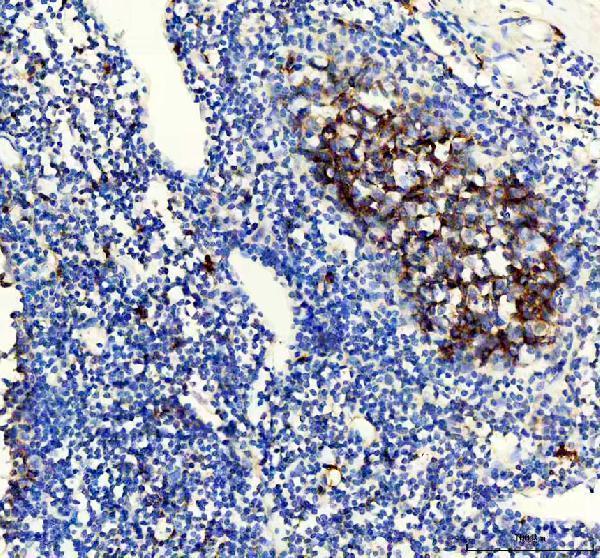
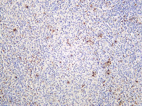
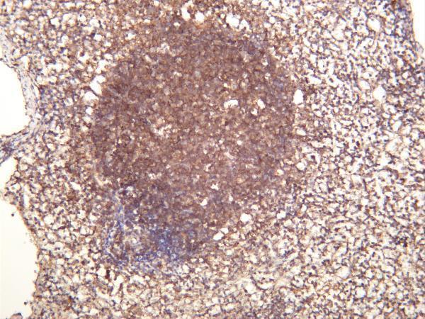
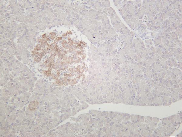
The catalog includes anti-CD38 IHC images from human tonsil, appendix and spleen, plus mouse and rat pancreas (IHC captions); an IF image shows A431 cells (A00193-3 IF caption).

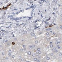
Real IHC data IHC analysis of CD38 using anti-CD38 antibody (A00193-3). CD38 was detected in paraffin-embedded section of human tonsil tissue. Heat mediated antigen retrieval was performed in EDTA buffer (pH8.0, epitope retrieval solution). The tissue section was blocked with 10% goat serum. The tissue section was then incubated with 2μg/ml rabbit anti-CD38 Antibody (A00193-3) overnight at 4°C. Biotinylated goat anti-rabbit IgG was used as secondary antibody and incubated for 30 minutes at 37°C. The tissue section was developed using Strepavidin-Biotin-Complex (SABC) (Catalog # SA1022) with DAB as the chromogen.
Anti-CD38 Antibody ®
Cat # A00193-3
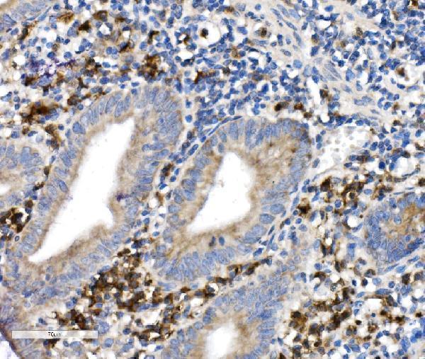
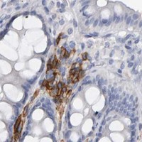
Real IHC data IHC analysis of CD38 using anti-CD38 antibody (M00193). CD38 was detected in a paraffin-embedded section of human appendix tissue. Heat mediated antigen retrieval was performed in EDTA buffer (pH 8.0, epitope retrieval solution). The tissue section was blocked with 10% goat serum. The tissue section was then incubated with 1:100 rabbit anti-CD38 Antibody (M00193) overnight at 4°C. Peroxidase Conjugated Goat Anti-rabbit IgG was used as secondary antibody and incubated for 30 minutes at 37°C. The tissue section was developed using HRP Conjugated Rabbit IgG Super Vision Assay Kit (Catalog # SV0002) with DAB as the chromogen.
Anti-CD38 Rabbit Monoclonal Antibody
Cat # M00193
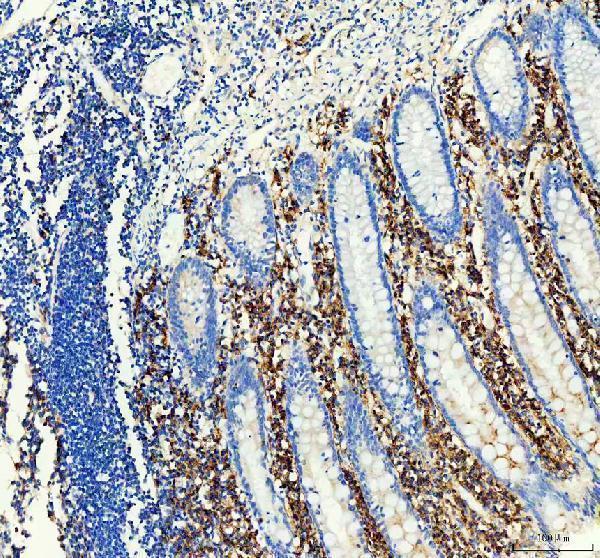
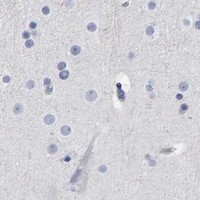
Real IHC data Human spleen was stained with anti-CD38 rabbit antibody
Anti-CD38 Rabbit Monoclonal Antibody
Cat # M00193-7
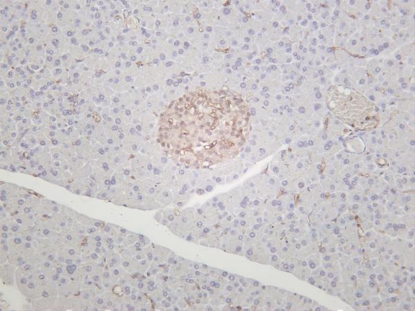
Real IHC data Rat pancreas was stained with anti-CD38 rabbit antibody
Anti-CD38 Rabbit Monoclonal Antibody
Cat # M00193-8

A00193-3 shows human tonsil IHC and A431 IF (A00193-3 image captions); M00193 shows human appendix IHC (M00193 IHC caption). M00193-7 shows human spleen and appendix IHC (M00193-7 IHC captions); M00193-8 shows rat and mouse pancreas IHC (M00193-8 IHC captions).

Which to pick: For human tissue IHC, M00193 is a rabbit monoclonal with an EDTA-retrieved paraffin appendix image; the fixative is unreported (catalog: M00193; M00193 IHC caption). For IF/ICC, A00193-3 lists both applications and has an A431 IF image; its tonsil IHC caption describes EDTA-retrieved paraffin sections but reports no fixative (catalog: A00193-3; A00193-3 image captions). For mouse or rat tissue IHC, M00193-8 is a rabbit monoclonal with both species listed and pancreas IHC images; those captions do not report processing or fixative (catalog: M00193-8; M00193-8 IHC captions).

Each figure is that product's own IHC / IF validation image from its datasheet.

References

  1. UniProt Consortium. UniProt entry P28907 (CD38_HUMAN, ADP-ribosyl cyclase/cyclic ADP-ribose hydrolase 1).
  2. Human Protein Atlas. CD38 tissue IHC expression (reliability: Enhanced).
  3. Human Protein Atlas. CD38 subcellular location (ICC-IF): Localized to the plasma membrane..
  4. Human Protein Atlas. CD38 antibody validation summary (4 antibodies).
  5. L-carnitine protects against methotrexate-induced hepatotoxicity via regulation of apoptosis and metabolic pathways. Naunyn-Schmiedeberg's archives of pharmacology 2026 — PMC13357626.
  6. The effect of chronic endometritis and treatment on patients with unexplained infertility. BMC women's health 2023 — PMC10311816.
  7. Prognostic Impact of CD38- and IgκC-Positive Tumor-Infiltrating Plasma Cells in Triple-Negative Breast Cancer. International journal of molecular sciences 2023 — PMC10607675.
  8. Inflammatory cell expression of Toll-like receptor-2 (TLR2) within refractory periapical granuloma. F1000Research 2018 — PMC6281009.
  9. PubMed PMID:2319135 — UniProt-cited evidence.
  10. PubMed PMID:9074508 — UniProt-cited evidence.
  11. PubMed PMID:15489334 — UniProt-cited evidence.