CD7 / T-cell antigen CD7 · IHC design guide

Design Immunohistochemistry for CD7

Plan CD7 paraffin IHC using the cytoplasmic immune-cell staining observed in tissue (HPA tissue IHC). Start with 0.5–1 μg/mL catalog antibody and use tonsil non-germinal center cells as a positive reference (datasheet A01974-2; HPA tissue IHC).

Evidence assembled Oct 2026 · For research use; verify linked source records and product datasheet before use
Immunohistochemistry protocol sheet for CD7 (IHC for CD7): expected localisation Cytoplasmic staining in an immune-cell subset (HPA tissue IHC), antibody A01974-2, validated IHC image, and IHC protocol steps
Printable CD7 IHC protocol sheet — expected localisation Cytoplasmic staining in an immune-cell subset (HPA tissue IHC), antibody A01974-2, controls and protocol steps. Open the full CD7 IHC guide →

CD7 Immunohistochemistry Experimental Design Guide

Expected localisation, validated protocols, controls and antibodies — the at-a-glance facts below, then the full design guide.

Must know before staining
Expected localisation Cytoplasmic staining in an immune-cell subset (HPA tissue IHC)
Staining pattern Subset of immune cells with cytoplasmic staining (HPA tissue IHC)
Antigen retrieval Citrate pH 6 HIER, heat-mediated (datasheet A01974-2)
Positive control ⓘ Appendix+2 more · see all
Negative control ⓘ Adipose tissue+4 more · see all
Important caveats
Reasons your staining may differ from the expected pattern.
Fixation Keep fixation consistent across paraffin sections. (standard IHC practice; not target-specific)
Caveat CD7 also occurs on NK cells; confirm cell identity (UniProt)
Regulation No abundance regulation specified (UniProt)
Isoform / epitope No listed isoforms; extracellular vs cytoplasmic epitope matters (UniProt)
Section 1

Recommended CD7 IHC & IF Protocols

The catalog antibody has a datasheet IHC-P protocol (datasheet: A01974-2); the four published CD7 protocols below provide tissue-specific examples (PMC12452609; PMC3961532; PMC9753314; PMC6299443).

Recommended immunohistochemistry (IHC-P) protocol parameters
SampleParaffin-embedded human tonsil tissue; fixative not specified (datasheet A01974-2)
FixationImage fixative and duration unreported (datasheet A01974-2); verify before use.
Sectioning4–5 µm sections on charged slides (standard)
DeparaffinisationXylene, graded ethanol series to water (standard)
Antigen retrievalHeat retrieval: Citrate pH 6, 20 min (datasheet A01974-2)
Peroxidase block3% H2O2, 10 min, room temperature (standard)
Blocking10% goat serum (datasheet A01974-2)
Primary antibodyRabbit anti-CD7, 0.5-1μg/ml (datasheet A01974-2)
Primary incubationOvernight at 4 °C (datasheet A01974-2)
DetectionStreptavidin-biotin complex (SABC), DAB chromogen (datasheet A01974-2)
CounterstainHematoxylin, blue, dehydrate and mount (standard)
Expected resultCD7-positive staining in non-germinal center cells of appendix (HPA tissue IHC: High). HPA tissue profile: Cytoplasmic expression in a subset of immune cells. No signal in the no-primary control.
💡Decision noteStart with heat-mediated citrate retrieval at pH 6 (datasheet: A01974-2); a published skin-biopsy protocol used pH 9 instead (PMC12452609).
Section 2

What Is the Expected CD7 Staining Pattern?

CD7 is a transmembrane protein expressed on T cells, natural killer (NK) cells and their precursors (UniProt P09564 tissue specificity and topology). In tissue IHC, HPA reports cytoplasmic staining in a subset of immune cells, with enhanced reliability based on agreement between staining and RNA data (HPA tissue IHC). Interpret the slide at both cell and compartment level: the tissue result and membrane topology describe different kinds of evidence.

What am I looking at on my slide?
A subset of non-germinal center immune cells stains in tonsil or appendix; nearby cells remain unstained.This matches HPA's high staining in those cell populations (HPA tissue IHC). CD7 expression in T and NK cells makes a restricted immune-cell pattern plausible (UniProt P09564 tissue specificity). The stain alone does not identify each positive cell as T or NK.
Positive immune cells appear cytoplasmic rather than as crisp membrane outlines.HPA explicitly describes cytoplasmic staining in a subset of immune cells (HPA tissue IHC). CD7 has an extracellular region, a transmembrane segment and a cytoplasmic tail (UniProt P09564 topology). These records do not establish why the reported IHC appearance differs from a membrane outline; judge it alongside cell distribution and controls.
Nuclei, broad extracellular areas or an entire epithelial population stain strongly.Those distributions are outside the supplied CD7 membrane topology and HPA immune-cell IHC profile (UniProt P09564 topology; HPA tissue IHC). Treat them as suspect and check controls before scoring them as CD7. Their appearance alone cannot identify the source of the unwanted signal (general IHC practice).
The negative control shows widespread color, or multiple unrelated cell types stain diffusely.Diffuse staining lacks the subset pattern reported by HPA (HPA tissue IHC). Background from the detection workflow or nonspecific antibody binding is possible (general IHC practice). A matched no-primary control helps distinguish detection background from primary-antibody-associated staining; it does not prove CD7 specificity by itself.
No cells stain in tonsil or appendix, including the expected non-germinal center population.An entirely blank result conflicts with HPA's high staining in those populations (HPA tissue IHC). Review tissue identity, antibody handling, retrieval and detection with the catalog IHC-P instructions (general IHC practice). A failed run cannot establish biological absence of CD7.
💡Expected CD7 appearanceCall the IHC result positive when a subset of immune cells stains, with high staining reported for non-germinal center cells in tonsil or appendix and cytoplasmic appearance reported by HPA; strong nuclear, extracellular or broad epithelial color is suspect (HPA tissue IHC; UniProt P09564 topology).
How each factor affects the staining
Cell distribution and control tissueHPA reports high staining in non-germinal center cells of tonsil and appendix, medium in splenic red-pulp cells, and low in lymph-node non-germinal center and marrow hematopoietic cells (HPA tissue IHC). Select the control and expected intensity with its named cell population in mind.
Topology versus observed IHC appearanceCD7 residues 26–180 are extracellular, 181–201 span the membrane and 202–240 are cytoplasmic (UniProt P09564 topology). HPA nevertheless reports cytoplasmic tissue IHC staining (HPA tissue IHC). The supplied records give no antibody epitope or mechanism that resolves the difference; avoid requiring a perfect membrane ring.
Antibody validationHPA lists two antibodies, HPA039079 and CAB002594, with IHC marked Enhanced (HPA antibodies). Its tissue profile also has enhanced reliability based on staining–RNA agreement (HPA tissue IHC). These summaries support confidence in the reported population pattern, but do not establish that every stained cell is CD7-positive.
Negative cell populationsHPA reports adipocytes in adipose tissue and respiratory epithelial cells in bronchus as not detected (HPA tissue IHC). These named populations can help flag broad off-target color. The label applies to those cells, not to every cell in their tissue sections (HPA tissue IHC).
Retrieval and fixation sensitivityThe supplied records give no CD7-specific fixation or retrieval response (UniProt P09564; HPA tissue IHC). Follow the IHC-validated antibody's stated retrieval conditions, then optimize against controls if needed (general IHC practice). Do not infer a CD7-specific fixation effect from topology or tissue staining level.
Why is my staining missing, weak or wrong?
SituationLikely causeNext action
Tonsil or appendix is completely unstained.The result conflicts with HPA's high staining in non-germinal center cells (HPA tissue IHC); a run failure is possible (general IHC practice).Confirm the expected cell area, then check the catalog antibody's IHC-P instructions, retrieval, reagent preparation and detection controls before interpreting absence (general IHC practice).
Color covers the whole section or appears in the no-primary control.This exceeds HPA's subset immune-cell pattern (HPA tissue IHC). Detection-system activity or inadequate background control is possible (general IHC practice).Check the matched no-primary control and the detection system; apply blocking steps appropriate to that system and re-evaluate only cell-restricted staining (general IHC practice).
Strong staining appears in adipocytes or bronchial respiratory epithelium.HPA lists these specific populations as not detected (HPA tissue IHC). Primary-antibody-associated cross-reactivity or detection background is possible (general IHC practice).Compare no-primary and positive-control sections, review staining across cell types, and avoid calling those cells CD7-positive from color alone (general IHC practice).
Only a cytoplasmic pattern is visible in otherwise expected immune cells.HPA reports cytoplasmic IHC staining, while UniProt places CD7 in the membrane (HPA tissue IHC; UniProt P09564 topology). The records do not explain the visual difference.Score the cell population and controls together; do not reject the result solely for lacking a sharp membrane outline (HPA tissue IHC; general IHC practice).
A weak result occurs in lymph node or bone marrow.HPA reports low staining in the specified non-germinal center or hematopoietic cell populations (HPA tissue IHC). Weak color there is less decisive than a blank high-staining control.Run tonsil or appendix alongside it, inspect the relevant cells, and compare background before changing the interpretation (HPA tissue IHC; general IHC practice).
IF/ICC: should the same visual pattern be expected?HPA summarizes the subcellular location as membrane but supplies no ICC-IF cell-line images or main-location assignment (HPA subcellular). Its cytoplasmic observation comes from tissue IHC (HPA tissue IHC).Use the separate IF/ICC guide for that application. Treat membrane localization as a starting expectation from UniProt topology and HPA summary, without presenting the tissue IHC appearance as an IF-validated result (UniProt P09564 topology; HPA subcellular).

Sample controls for CD7 IHC & IF

🧪Run tonsil first: non-germinal center cells should stain (HPA: High in tonsil non-germinal center cells). Use adipose tissue as a negative tissue, scoring adipocytes as unstained (HPA: Not detected in adipocytes); on the tonsil slide, morphologically identified non-lymphoid cells should provide an internal negative (UniProt P09564: expression on T cells and NK cells and their precursors).
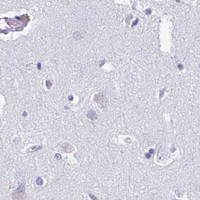
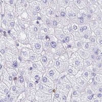
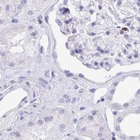
Positive control tissue: Appendix (Non-germinal center cells, HPA High)
Negative control tissue: Adipose tissue (HPA Not detected)
ICC-IF cell lines (HPA subcellular resource): HPA carries no ICC-IF cell line for CD7; derive a cell-line control from the positive tissue's cell type (Non-germinal center cells) and confirm it by RNA or western blot first.
Technical controls: Include no-primary (secondary-only) and host- and clonality-matched isotype controls (caption: rabbit primary antibody), plus CD7-knockout material processed alongside the positive tissue as a biological negative (standard IHC practice). Block endogenous peroxidase and check endogenous biotin background in tonsil when using the reported avidin–biotin/DAB detection method (caption: SABC with DAB; standard IHC practice).
⚠️Feasibility: A target-specific fixation window and fixation effect are unreported, and the exact selected-SKU paraffin-section caption does not state the fixative (caption: A01974-2). The demonstrated IHC workflow uses citrate retrieval at pH 6 for 20 minutes (caption: A01974-2); the evidence does not establish that retrieval is mandatory or that frozen sections or IF/ICC are easier. Dense positive lymphoid areas in tonsil require attention to background from endogenous peroxidase or biotin in the reported chromogenic workflow (HPA: High in tonsil non-germinal center cells; caption: SABC with DAB; standard IHC practice).

HPA tissue IHC evidence for CD7

Comprehensive Human Protein Atlas IHC scoring per tissue (reliability: Enhanced — High consistency between antibody staining and RNA expression data.). Rows are taken directly from the HPA tissue chart — click any row's HPA link to view the source.

Positive expression · recommended positive controls

TissueCell typeLevelEvidenceSource
Appendix Non-germinal center cells High Protein (IHC) HPA →
Tonsil Non-germinal center cells High Protein (IHC) HPA →
Spleen Cells in red pulp Medium Protein (IHC) HPA →

Undetected expression · recommended negative controls

TissueCell typeLevelEvidenceSource
Adipose tissue Adipocytes Not detected Protein (IHC) HPA →
Adrenal gland Glandular cells Not detected Protein (IHC) HPA →
Breast Adipocytes Not detected Protein (IHC) HPA →
Bronchus Respiratory epithelial cells Not detected Protein (IHC) HPA →
Caudate Glial cells Not detected Protein (IHC) HPA →
Section 3

Advanced CD7 IHC Tips

Troubleshoot CD7 staining in paraffin sections by checking retrieval, cell identity, and membrane localisation before interpreting chromogenic signal (UniProt P09564 topology; HPA tissue IHC).

How should I retrieve CD7 when paraffin sections stain weakly?
Start with heat-mediated retrieval in citrate buffer at pH 6 (datasheet A01974-2). The selected tonsil example used this buffer for 20 minutes, followed by 10% goat-serum blocking and 1 μg/ml primary antibody overnight at 4°C (caption A01974-2). If signal is weak, verify heating and cooling consistency across sections before changing primary concentration or detection conditions (standard IHC practice). Run a known positive tonsil section beside the test section so a failed retrieval run is easier to recognise (HPA tissue IHC: high staining in tonsil non-germinal center cells; standard IHC practice). Compare tissue preservation and background before accepting a stronger retrieval condition (standard IHC practice).
Could fixation explain weak or uneven CD7 staining?
CD7-specific sensitivity to fixation is unknown from the supplied evidence; the selected paraffin-section caption does not state a fixative (caption A01974-2). Record the fixative, fixation duration, processing history, and section age for each specimen before comparing staining intensity (standard IHC practice). Process a reference tonsil section alongside the test material using the same retrieval and detection run (HPA tissue IHC: high staining in tonsil non-germinal center cells; standard IHC practice). If only one specimen fails, inspect its morphology and processing record before adjusting the antibody concentration (standard IHC practice). Treat any apparent fixation effect as a working hypothesis until matched specimens support it (standard IHC practice).
Should CD7 staining appear on the membrane or in the cytoplasm?
Expect membrane-associated CD7 because its extracellular region spans residues 26–180, its transmembrane segment spans 181–201, and its cytoplasmic region spans 202–240 (UniProt P09564 topology). In chromogenic sections, assess whether staining outlines the expected immune cells while allowing for limited resolution of their thin membranes (UniProt P09564 tissue specificity; standard IHC practice). HPA also reports cytoplasmic expression in a subset of immune cells, so cytoplasmic staining alone is not automatically artefactual (HPA tissue IHC profile). Compare the pattern with adjacent negative cell populations and a positive tonsil control (HPA tissue IHC: tonsil non-germinal center cells high; standard IHC practice). Diffuse staining across unrelated cell types warrants a background check (standard IHC practice).
How do epitope location and glycosylation affect CD7 staining?
CD7 has no listed isoforms, so the supplied record does not support an isoform-specific staining explanation (UniProt P09564 isoforms). Its extracellular region spans residues 26–180, with an Ig-like domain at 26–130 and glycosylation sites at 45 and 96 (UniProt P09564 topology, domains, glycosylation). Check the antibody's documented epitope, if available, before interpreting differences between surface-associated and cytoplasmic signal (standard IHC practice). The supplied caption establishes a retrieval method but does not identify the catalog antibody's epitope (caption A01974-2). Compare candidate conditions on serial sections with the same positive control and score both signal and background (standard IHC practice).
How can I assess CD7 by multiplex immunofluorescence?
Use a T-cell or NK-cell marker in a separate channel to assess whether CD7 signal belongs to an expected cell population (UniProt P09564 tissue specificity; standard IF practice). Choose spectrally separated fluorophores after inspecting unstained tissue for autofluorescence, and include single-stain controls for channel spillover (standard IF practice). If the antibody recognises an extracellular epitope, test staining without permeabilisation; a cytoplasmic-side epitope requires access across the membrane (UniProt P09564 topology; standard IF practice). The supplied IHC caption does not establish an IF fixation or permeabilisation method for this antibody (caption A01974-2). Optimise those steps using IF controls and compare localisation with the expected membrane pattern (UniProt P09564 subcellular location; standard IF practice).
What should I check when CD7 DAB staining looks widespread?
First compare the widespread signal with a section processed without primary antibody and with an expected negative cell population (standard IHC practice). CD7 is reported on T cells, NK cells, and their precursors, while HPA reports no detection in adipocytes from adipose tissue (UniProt P09564 tissue specificity; HPA tissue IHC). Check the peroxidase block, secondary detection, and DAB development time before interpreting diffuse brown deposit as CD7 (standard chromogenic IHC practice). The selected tonsil example used biotinylated secondary antibody, streptavidin-biotin detection, and DAB, so examine detection-related background in that workflow (caption A01974-2; standard IHC practice). Also inspect section edges and damaged areas separately from intact tissue (standard IHC practice).
How should I quantify CD7 across differently cellular sections? ⚠ ANSWER MARKED FOR VERIFICATION
Define the tissue region and eligible immune-cell population before scoring, using the same inclusion rules for every section (standard IHC practice). Report the percentage of positive eligible cells and, when intensity is reproducible, an H-score based on intensity categories (standard IHC practice). For spatial comparisons, report positive-cell density per mm² of analysed tissue and the number of eligible cells assessed (standard IHC practice). Normalise the positive fraction to the eligible cell count, or the density to analysed tissue area, rather than raw brown-pixel area (standard IHC practice). Exclude folds, necrosis, and section edges by predefined rules, and keep retrieval and DAB development consistent across the comparison (standard IHC practice).
When is a CD7-positive cell convincing rather than artefact?
A convincing result places CD7 signal in an expected immune-cell population with membrane-associated staining, while recognising that HPA describes cytoplasmic expression in a subset of immune cells (UniProt P09564 tissue specificity and topology; HPA tissue IHC profile). Use tonsil non-germinal center cells as a positive reference because HPA reports high staining there (HPA tissue IHC). Question signal concentrated at section edges or necrotic areas, or staining that persists in a no-primary control (standard IHC practice). Diffuse staining in unrelated cell populations calls for review of endogenous enzyme activity, blocking, and detection background (standard chromogenic IHC practice). Interpret weak cells only after comparing morphology, localisation, and controls from the same run (standard IHC practice).
Boster reagents

Best CD7 / T-cell antigen CD7 IHC Antibodies

The catalog lists human-reactive anti-CD7 antibodies for IHC and IF/ICC, with a tissue IHC image supplied for A01974-2 (catalog applications and reactivity; A01974-2 image caption).

Real IHC data IHC analysis of CD7 using anti-CD7 antibody (A01974-2). CD7 was detected in paraffin-embedded section of human tonsil tissue . Heat mediated antigen retrieval was performed in citrate buffer (pH6, epitope retrieval solution) for 20 mins. The tissue section was blocked with 10% goat serum. The tissue section was then incubated with 1μg/ml rabbit anti-CD7 Antibody (A01974-2) overnight at 4°C. Biotinylated goat anti-rabbit IgG was used as secondary antibody and incubated for 30 minutes at 37°C. The tissue section was developed using Strepavidin-Biotin-Complex (SABC)(Catalog # SA1022) with DAB as the chromogen.
Anti-CD7 Antibody ®
Cat # A01974-2

A01974-2 lists human IHC and ICC applications and shows staining in a paraffin-embedded human tonsil section (catalog applications and reactivity; A01974-2 image caption). M01974 lists human IHC, ICC, and IF applications, but has no supplied IHC or IF image (catalog applications and reactivity; catalog image alts).

Which to pick: For tissue IHC, choose A01974-2 because its own image documents a human paraffin section; the fixative is unreported (A01974-2 image caption). For IF/ICC, consider monoclonal M01974 because both applications are listed, while recognizing that no IF image is supplied (catalog clone, applications, and image alts). Neither SKU has documented cross-species reactivity; both list human only (catalog reactivity).

Each figure is that product's own IHC / IF validation image from its datasheet.

References

  1. UniProt Consortium. UniProt entry P09564 (CD7_HUMAN, T-cell antigen CD7).
  2. Human Protein Atlas. CD7 tissue IHC expression (reliability: Enhanced).
  3. Human Protein Atlas. CD7 subcellular location (ICC-IF): Membrane.
  4. Human Protein Atlas. CD7 antibody validation summary (2 antibodies).
  5. Prognostic Value of Immunohistochemical T-Cell Marker Loss in Early-Stage Mycosis Fungoides: A Single-Center Cohort Study. Dermatopathology (Basel, Switzerland) 2025 — PMC12452609.
  6. SECTM1 produced by tumor cells attracts human monocytes via CD7-mediated activation of the PI3K pathway. The Journal of investigative dermatology 2014 — PMC3961532.
  7. Naturally selected CD7 CAR-T therapy without genetic editing demonstrates significant antitumour efficacy against relapsed and refractory acute myeloid leukaemia (R/R-AML). Journal of translational medicine 2022 — PMC9753314.
  8. T-cell functionality testing is highly relevant to developing novel immuno-tracers monitoring T cells in the context of immunotherapies and revealed CD7 as an attractive target. Theranostics 2018 — PMC6299443.
  9. PubMed PMID:3501369 — UniProt-cited evidence.
  10. PubMed PMID:1703303 — UniProt-cited evidence.
  11. PubMed PMID:15489334 — UniProt-cited evidence.