This website uses cookies to ensure you get the best experience on our website.


- Table of Contents
Plan CD83 IHC on paraffin sections using tissue staining examples and controls. Expect cytoplasmic staining in subsets of immune cells (HPA tissue IHC); CD83 is a membrane protein with an extracellular domain (UniProt).

Expected localisation, validated protocols, controls and antibodies — the at-a-glance facts below, then the full design guide.
| Expected localisation | Cytoplasmic staining in immune-cell subsets (HPA tissue IHC) | |
| Staining pattern | Cytoplasmic staining in subsets of immune cells (HPA tissue IHC) | |
| Antigen retrieval | Citrate pH 6.0 HIER, 95–98 °C, 20 min (rule: cytoplasmic / membrane antigen) | |
| Positive control | Cerebellum+4 more · see all | |
| Negative control | Adipose tissue+4 more · see all |
| Fixation | Keep fixation conditions consistent across sections. (standard IHC practice; not target-specific) | |
| Caveat | Presumed off-target staining was observed and disregarded (HPA tissue IHC) | |
| Regulation | Expressed by activated lymphocytes and dendritic cells (UniProt) | |
| Isoform / epitope | No isoforms; epitope side matters: residues 20–144 vs 167–205 (UniProt) |
The catalog antibody protocol is accompanied by published CD83 IHC methods for periapical, breast, cervical and thyroid specimens (PMC6281009; PMC10868929; PMC11554423; PMC4433839).
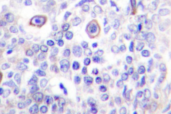
| Sample | Paraffin-embedded human lung carcinoma tissue; fixative not specified (datasheet A01777) |
| Fixation | Image fixative and duration unreported (datasheet A01777); verify before use. |
| Sectioning | 4–5 µm sections on charged slides (standard) |
| Deparaffinisation | Xylene, graded ethanol series to water (standard) |
| Antigen retrieval | Heat-induced epitope retrieval in citrate buffer, pH 6.0, 20 min at 95–98 °C (standard rule: cytoplasmic / membrane antigen) |
| Peroxidase block | 3% H2O2, 10 min, room temperature (standard) |
| Blocking | 10% normal serum of the secondary host, 30 min, room temperature (standard) |
| Primary antibody | Rabbit anti-CD83, 1:50-1:200 (datasheet A01777) |
| Primary incubation | Overnight at 4 °C (standard) |
| Detection | HRP-polymer secondary, DAB chromogen 5–10 min (standard) |
| Counterstain | Hematoxylin, blue, dehydrate and mount (standard) |
| Expected result | CD83-positive staining in purkinje cells - cytoplasm/membrane of cerebellum (HPA tissue IHC: High). HPA tissue profile: Cytoplasmic expression in a subsets of immune cells in several tissues, including lymphoid organs. Additional expression in brain. No signal in the no-primary control. |
CD83 is a surface glycoprotein with an extracellular region at residues 20–144 and a transmembrane segment at 145–166 (UniProt Q01151 topology). In paraffin-section IHC, expect staining in subsets of immune cells, often cytoplasmic by HPA observation (HPA tissue IHC). Interpret positives cautiously: HPA rates the tissue profile Enhanced but reports medium agreement with RNA data and disregarded presumed off-target staining (HPA tissue IHC).
| Discrete membrane-associated or cytoplasmic staining in scattered immune cells within lymphoid tissue. | This fits CD83 surface localization and HPA's observed cytoplasmic staining in immune-cell subsets (UniProt Q01151 topology; HPA tissue IHC). HPA reports medium staining in appendix and tonsil germinal-center cells, lymph-node non-germinal-center cells, and splenic red-pulp cells (HPA tissue IHC). Score the stained cells and compartment, rather than calling the entire tissue positive. |
| Strong, widespread nuclear staining dominates a paraffin section. | Reassess specificity and detection background before accepting this as CD83 IHC: UniProt places CD83 at the membrane, while HPA's nuclear ICC-IF locations are marked uncertain (UniProt Q01151 subcellular location; HPA subcellular ICC-IF). HPA does report cytoplasmic tissue staining, so cytoplasm alone is not a reason to reject a result (HPA tissue IHC). |
| Chromogen appears in cells expected to lack CD83, such as adipocytes. | HPA did not detect staining in adipocytes in adipose tissue or breast (HPA tissue IHC). Check whether the signal follows cell boundaries and whether negative-reagent controls also stain; cross-reactivity or endogenous detection activity are possible explanations (general IHC practice). Do not treat every cell in an otherwise positive organ as a CD83-positive cell. |
| A diffuse haze covers cells, stroma, or the whole section without a discernible cellular pattern. | This cannot be scored as the discrete immune-cell staining HPA describes (HPA tissue IHC). Review blocking, antibody concentration, washing, and chromogen development using matched controls (general IHC practice). HPA notes presumed off-target binding in its tissue assessment, so diffuse staining needs independent scrutiny (HPA tissue IHC). |
| A run shows no cellular staining in an expected-positive control section. | Check the section and IHC run before interpreting study samples as CD83-negative (general IHC practice). HPA reports medium staining in tonsil germinal-center cells and bone-marrow hematopoietic cells; neither designation guarantees staining in every specimen or cell (HPA tissue IHC). Verify that the chosen control contains the relevant cell population. |
| Cell population and tissue context | CD83 is expressed by activated lymphocytes, Langerhans cells, and activated dendritic cells (UniProt Q01151 tissue specificity). HPA observes immune-cell subsets across several tissues, including lymphoid organs, and also reports staining in Purkinje and Leydig cells (HPA tissue IHC). Identify the stained population before comparing sections. |
| Epitope position and mature protein | The signal peptide spans residues 1–19; the annotated mature chain spans 20–205, with extracellular residues 20–144 (UniProt Q01151 processing and topology). An antibody's epitope would determine which region it recognizes (general antibody practice). No epitope or CD83-specific retrieval condition is supplied here, so do not infer one. |
| Glycosylated extracellular region | UniProt lists glycosylation sites at residues 79, 96, and 117 within the extracellular region (UniProt Q01151 glycosylation and topology). This describes the antigen, but does not establish how fixation or retrieval affects the IHC signal for a particular antibody. |
| Strength of tissue evidence | HPA labels the tissue IHC profile Enhanced and lists two antibodies with Enhanced IHC validation, HPA041454 and CAB002509 (HPA tissue IHC; HPA antibodies). HPA also reports medium consistency with RNA data and presumed off-target staining that it disregarded; assess unexpected cells independently (HPA tissue IHC). |
| IF/ICC interpretation | For the separate IF/ICC application, HPA approves plasma-membrane and Golgi localization but marks nucleoplasm and nucleoli uncertain (HPA subcellular ICC-IF). Those cell-image observations do not establish a paraffin-section IHC pattern or an IF/ICC protocol. |
| Situation | Likely cause | Next action |
|---|---|---|
| Expected immune-cell staining is absent in the positive-control section. | The relevant cells may be missing from the section, or an IHC run step may have failed (general IHC practice). HPA's medium tissue level is an observation, not a guaranteed signal in every section (HPA tissue IHC). | Confirm the control contains the annotated cell population, then review reagent omission controls, antibody dilution, detection, and chromogen development according to the validated IHC workflow (general IHC practice). |
| Nuclear staining is stronger than membrane or cytoplasmic staining. | Dominant nuclear staining has weak support for tissue IHC: CD83 is membrane-associated, and HPA marks its nuclear ICC-IF locations uncertain (UniProt Q01151 subcellular location; HPA subcellular ICC-IF). | Compare cellular morphology and negative-reagent controls, and repeat with an independently IHC-validated antibody if available (general IHC practice; HPA antibodies). Keep the uncertain IF nuclear observation separate from IHC scoring. |
| Adipocytes or another HPA-negative cell population stains. | The finding conflicts with HPA's absence of adipocyte staining; cross-reactivity or endogenous detection activity may contribute (HPA tissue IHC; general IHC practice). Nearby immune cells require separate assessment. | Inspect cell identity, run appropriate negative-reagent and endogenous-activity controls, and compare staining with the expected immune-cell distribution (general IHC practice; HPA tissue IHC). |
| A broad brown haze obscures individual positive cells. | Nonspecific reagent binding, inadequate washing, or excess chromogen development can produce diffuse background (general IHC practice). HPA reports disregarded presumed off-target staining in its tissue assessment (HPA tissue IHC). | Review blocking, dilution, washes, and development against control sections; score only a resolvable cellular pattern (general IHC practice). |
| A lymphoid section appears uniformly positive. | HPA describes staining in subsets, with particular compartments reported in appendix, tonsil, lymph node, and spleen (HPA tissue IHC). Uniform color may reflect background or an overbroad cell call (general IHC practice). | Score the named cell compartment and staining location at higher magnification, then compare with negative-reagent controls (HPA tissue IHC; general IHC practice). |
| Does nucleoplasmic IF/ICC staining confirm a positive CD83 IHC section? | HPA marks nucleoplasm uncertain in ICC-IF, while plasma membrane is approved; the applications provide different observations (HPA subcellular ICC-IF). | Use the IF/ICC finding only with its stated uncertainty. Judge paraffin-section IHC against its own cellular pattern and controls (HPA tissue IHC; general IHC practice). |


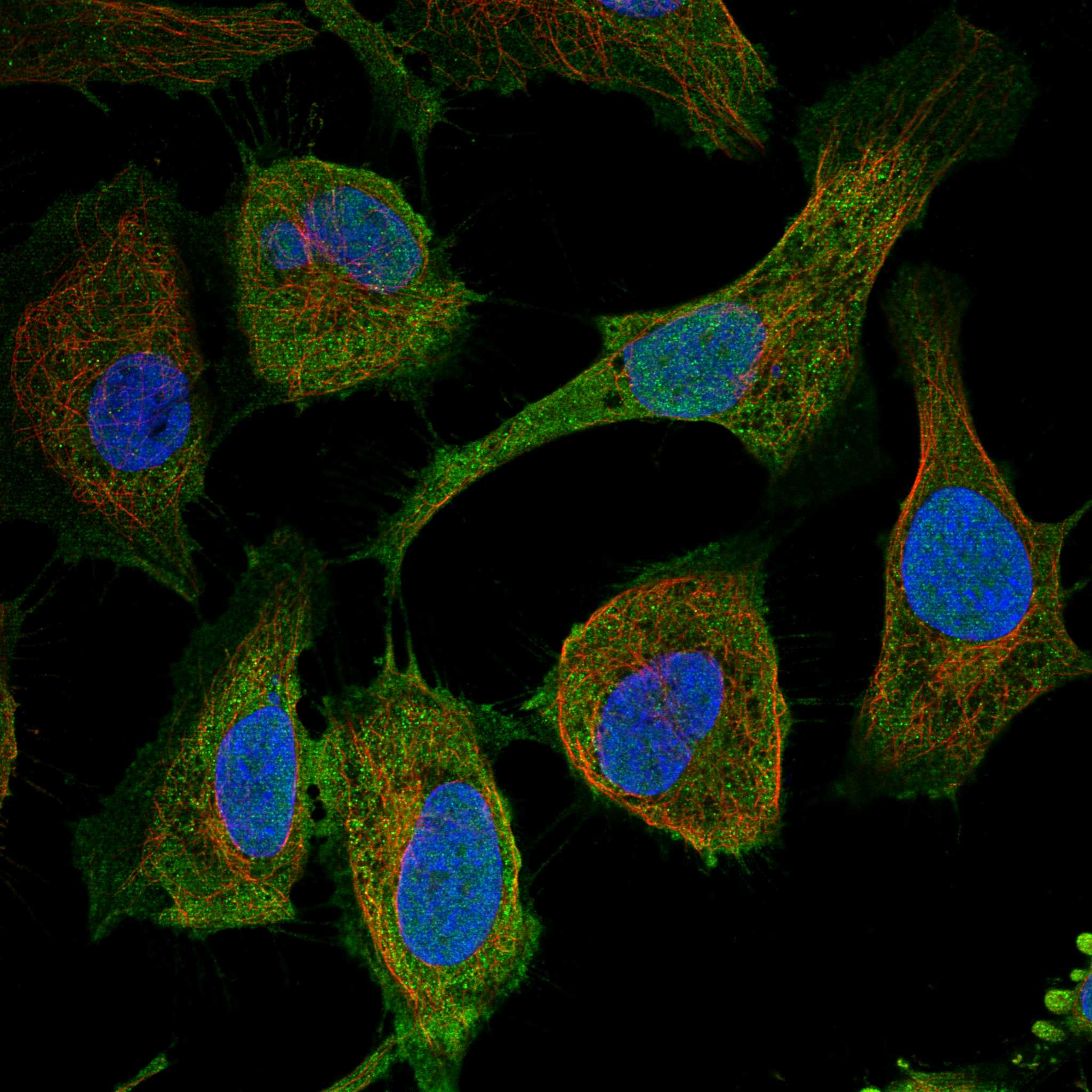
Comprehensive Human Protein Atlas IHC scoring per tissue (reliability: Enhanced — Medium consistency between antibody staining and RNA expression data. Presumed off target binding observed and disregarded. Antibody staining in cells/structures not annotated, view images.). Rows are taken directly from the HPA tissue chart — click any row's HPA link to view the source.
| Tissue | Cell type | Level | Evidence | Source |
|---|---|---|---|---|
| Cerebellum | Purkinje cells - cytoplasm/membrane | High | Protein (IHC) | HPA → |
| Appendix | Germinal center cells | Medium | Protein (IHC) | HPA → |
| Bone marrow | Hematopoietic cells | Medium | Protein (IHC) | HPA → |
| Lung | Macrophages | Medium | Protein (IHC) | HPA → |
| Lymph node | Non-germinal center cells | Medium | Protein (IHC) | HPA → |
| Tissue | Cell type | Level | Evidence | Source |
|---|---|---|---|---|
| Adipose tissue | Adipocytes | Not detected | Protein (IHC) | HPA → |
| Adrenal gland | Glandular cells | Not detected | Protein (IHC) | HPA → |
| Breast | Adipocytes | Not detected | Protein (IHC) | HPA → |
| Bronchus | Respiratory epithelial cells | Not detected | Protein (IHC) | HPA → |
| Cervix | Glandular cells | Not detected | Protein (IHC) | HPA → |
Troubleshoot chromogenic CD83 staining in paraffin sections by checking retrieval, compartment, cell identity and controls before comparing staining scores.
A01777 has an IHC image from paraffin-embedded human lung carcinoma tissue (catalog image caption) and lists human, mouse and rat reactivity (catalog: reactivity).
A01777 is listed for IHC in human, mouse and rat (catalog: applications; reactivity). Its IHC figure shows paraffin-embedded human lung carcinoma tissue (catalog image caption); no IF figure is supplied (catalog: IF image alts).
Which to pick: For tissue IHC, choose A01777; its own image shows paraffin-embedded human lung carcinoma tissue, but the fixative is unreported (catalog image caption). No listed SKU is validated for IF/ICC (catalog: applications; IF image alts). For cross-species IHC planning, A01777 lists human, mouse and rat reactivity, although its IHC image shows human tissue only (catalog: reactivity; image caption).