This website uses cookies to ensure you get the best experience on our website.


- Table of Contents
Plan chromogenic IHC on paraffin sections around nuclear and cytoplasmic staining, with lymph node germinal center cells as a high-staining reference (HPA tissue IHC). The catalog antibody has an IHC-P starting range of 2–5 μg/mL (datasheet PB9488).

Expected localisation, validated protocols, controls and antibodies — the at-a-glance facts below, then the full design guide.
| Expected localisation | Nuclear and cytoplasmic tissue staining (HPA tissue IHC) | |
| Staining pattern | Nuclear/cytoplasmic; high in germinal center cells (HPA tissue IHC) | |
| Antigen retrieval | EDTA pH 8.0 HIER, heat-mediated (datasheet PB9488) | |
| Positive control | Lymph node+4 more · see all | |
| Negative control | Adrenal gland+4 more · see all |
| Fixation | Matched tissue-IHC evidence does not establish the fixation claim. Validate the specimen-specific method before use. (selected-SKU IHC image PB9488) | |
| Caveat | Antibody staining and RNA show medium consistency (HPA tissue IHC) | |
| Regulation | Expression regulation unreported (UniProt) | |
| Isoform / epitope | 4 isoforms; antibody epitope coverage is unspecified (UniProt; datasheet PB9488) |
Compare the catalog antibody’s EDTA pH 8.0 IHC protocol (datasheet: PB9488) with four published CDC25B chromogenic IHC methods (PMC8435635; PMC2887779; PMC11555179; PMC8981514).
| Sample | Paraffin-embedded human stomach cancer tissue; fixative not specified (datasheet PB9488) |
| Fixation | Image fixative and duration unreported (datasheet PB9488); verify before use. |
| Sectioning | 4–5 µm sections on charged slides (standard) |
| Deparaffinisation | Xylene, graded ethanol series to water (standard) |
| Antigen retrieval | Heat retrieval: EDTA pH 8.0 (datasheet PB9488); 20 min, 95–100 °C (standard) |
| Peroxidase block | 3% H2O2, 10 min, room temperature (standard) |
| Blocking | 10% goat serum (datasheet PB9488) |
| Primary antibody | Rabbit anti-CDC25B, 2-5μg/ml (datasheet PB9488) |
| Primary incubation | Overnight at 4 °C (datasheet PB9488) |
| Detection | HRP-conjugated secondary, DAB chromogen (datasheet PB9488) |
| Counterstain | Hematoxylin, blue, dehydrate and mount (standard) |
| Expected result | CDC25B-positive staining in germinal center cells of lymph node (HPA tissue IHC: High). HPA tissue profile: Nuclear and cytoplasmic expression in several tissue types, most abundant in the gastrointestinal tract and lymphoid tissues. No signal in the no-primary control. |
In paraffin-section IHC, expect CDC25B staining in nuclei and cytoplasm across several tissues, with high staining in lymph-node germinal center cells and medium staining in rectal glandular cells (HPA tissue IHC: Enhanced). Cytoplasmic staining is consistent with reported centrosome and spindle-pole localization (UniProt P30305). CDC25B has no transmembrane segment, so a dominant cell-surface pattern needs scrutiny (UniProt P30305 topology).
| Germinal center cells stain strongly, with nuclear and cytoplasmic signal; rectal glandular cells stain less intensely. | This matches high germinal center and medium rectal glandular staining (HPA tissue IHC). Compare compartments within the identified cells; the HPA profile describes both nuclear and cytoplasmic expression (HPA tissue IHC). |
| A crisp cell-surface rim dominates while nuclei and cytoplasm show little signal. | A surface-dominant result is discordant with the nuclear and cytoplasmic IHC profile (HPA tissue IHC) and lack of a transmembrane segment (UniProt P30305 topology). Review morphology and antibody controls before calling it CDC25B. |
| Strong signal appears in bronchial respiratory epithelial cells or adrenal glandular cells. | Those specific cell populations were not detected in the HPA tissue survey (HPA tissue IHC). Consider cross-reactivity or endogenous chromogen-generating activity (general IHC practice); staining elsewhere in either organ requires separate assessment. |
| Brown color spreads across stroma, lumina, or most cells without clear cellular boundaries. | This is less interpretable than the cell-resolved nuclear and cytoplasmic profile (HPA tissue IHC). Background from detection reagents, endogenous activity, or incomplete blocking is possible (general IHC practice); check reagent controls. |
| Germinal center cells remain unstained in a section expected to be positive. | High staining is reported for these cells (HPA tissue IHC), so an absent result warrants an assay check. It does not alone establish that the specimen lacks CDC25B; verify cell identity and review assay controls (general IHC practice). |
| Tissue and cell selection | Use identified lymph-node germinal center cells as the high-staining comparator; breast myoepithelial, rectal glandular, and several other listed cell populations are medium (HPA tissue IHC). An organ name alone is not a cell-level score. |
| Compartment and cell-cycle context | HPA describes nuclear and cytoplasmic tissue staining (HPA tissue IHC). UniProt places CDC25B at centrosomes and spindle poles and links it to G2/M progression (UniProt P30305); these annotations do not require a visible spindle in every paraffin section. |
| Strength of tissue evidence | The tissue-IHC reliability is Enhanced, with medium consistency between antibody staining and RNA data (HPA tissue IHC). HPA lists two antibodies with Enhanced IHC validation; interpret a new antibody's pattern against tissue and reagent controls, not that label alone. |
| Isoforms and modified residues | Four isoforms and several phosphorylated residues are annotated (UniProt P30305). Their effect on staining cannot be predicted without the antibody's epitope and validation details; avoid assigning a compartment or intensity difference to one isoform from this record. |
| Processing and topology | UniProt lists one chain spanning residues 1–580, with no signal peptide, propeptide, or transmembrane segment (UniProt P30305). There is no supplied basis to interpret surface staining as expected processing or shedding. |
| Situation | Likely cause | Next action |
|---|---|---|
| No stain in the high-staining comparator | Germinal center cells are reported as High (HPA tissue IHC); absent staining could reflect specimen selection or an assay problem (general IHC practice). | Confirm germinal center morphology, then check the IHC-validated antibody's stated IHC-P procedure, reagent controls, retrieval, and detection steps (general IHC practice). |
| Signal is dominated by a membrane-like rim | That pattern conflicts with the reported nuclear and cytoplasmic IHC profile (HPA tissue IHC) and no-transmembrane topology (UniProt P30305). | Inspect the same cell types at higher magnification and compare reagent controls; withhold a CDC25B-positive call until the cellular pattern is supported (general IHC practice). |
| Unexpected cells stain strongly | Bronchial respiratory epithelial and adrenal glandular cells were not detected in the HPA survey (HPA tissue IHC); cross-reactivity or endogenous detection activity is possible (general IHC practice). | Check the precise cell identity, use appropriate detection controls, and compare with a known-positive section (general IHC practice). |
| Diffuse chromogen obscures compartments | Cell boundaries cannot be assessed against the nuclear and cytoplasmic reference profile (HPA tissue IHC); background may arise during blocking, washing, or detection (general IHC practice). | Review reagent-only controls, blocking and wash steps, and development time before scoring individual cells (general IHC practice). |
| Only one compartment appears positive | HPA reports both nuclear and cytoplasmic tissue expression, but its summary does not require both compartments in every cell (HPA tissue IHC). | Record nuclear and cytoplasmic staining separately, compare identified positive cells, and avoid treating one compartment alone as proof of artefact (HPA tissue IHC; general IHC practice). |
| Can the tissue-IHC pattern be used as an IF/ICC protocol? | HPA's ICC-IF summary places CDC25B mainly in vesicles and additionally on the mitotic spindle, with both locations marked uncertain (HPA subcellular ICC-IF). | Treat that localization as a tentative IF/ICC comparison, not an IHC-P staining requirement or protocol; use the separate IF/ICC guide for assay setup (HPA subcellular ICC-IF; HPA tissue IHC). |


Comprehensive Human Protein Atlas IHC scoring per tissue (reliability: Enhanced — Medium consistency between antibody staining and RNA expression data.). Rows are taken directly from the HPA tissue chart — click any row's HPA link to view the source.
| Tissue | Cell type | Level | Evidence | Source |
|---|---|---|---|---|
| Lymph node | Germinal center cells | High | Protein (IHC) | HPA → |
| Breast | Myoepithelial cells | Medium | Protein (IHC) | HPA → |
| Cerebral cortex | Endothelial cells | Medium | Protein (IHC) | HPA → |
| Colon | Endothelial cells | Medium | Protein (IHC) | HPA → |
| Kidney | Cells in glomeruli | Medium | Protein (IHC) | HPA → |
| Tissue | Cell type | Level | Evidence | Source |
|---|---|---|---|---|
| Adrenal gland | Glandular cells | Not detected | Protein (IHC) | HPA → |
| Bronchus | Respiratory epithelial cells | Not detected | Protein (IHC) | HPA → |
| Caudate | Glial cells | Not detected | Protein (IHC) | HPA → |
| Cerebellum | Cells in granular layer | Not detected | Protein (IHC) | HPA → |
| Cervix | Glandular cells | Not detected | Protein (IHC) | HPA → |
Use the catalog antibody’s paraffin-section example as a starting point, and interpret CDC25B staining alongside cell identity and tissue architecture (datasheet PB9488; HPA tissue IHC).
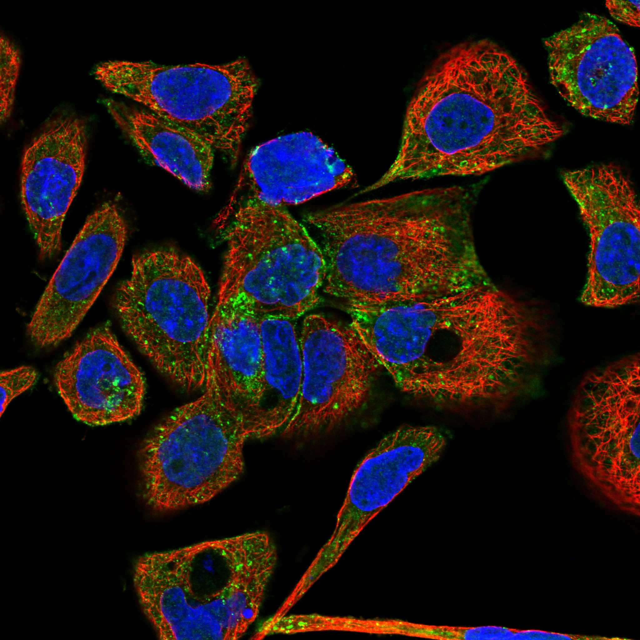
PB9488 has IHC data from paraffin-embedded human stomach cancer tissue (PB9488 IHC caption) and IF/ICC data from U2OS cells (PB9488 IF caption). Catalog reactivity lists human, mouse, and rat (PB9488 catalog).
PB9488 will render with an IHC figure from a paraffin-embedded human stomach cancer section (PB9488 IHC caption). Its IF/ICC figure shows U2OS cells, while the catalog lists human, mouse, and rat reactivity (PB9488 IF caption; PB9488 catalog).
Which to pick: Choose PB9488 for paraffin-section IHC: its own caption documents staining at 2 μg/ml, heat retrieval in EDTA at pH 8.0, and DAB detection; the fixative is unreported (PB9488 IHC caption). Choose the same SKU for IF/ICC because its own caption documents staining in U2OS cells at 5 μg/ml (PB9488 IF caption). For other species, PB9488 lists mouse and rat reactivity, but the supplied IHC and IF figures show human samples only; the catalog identifies a rabbit host and does not specify clonality (PB9488 catalog; PB9488 IHC and IF captions).