This website uses cookies to ensure you get the best experience on our website.


- Table of Contents
For paraffin CDC27 IHC, start with 2–5 μg/mL A03905-3 (datasheet: IHC-P). Expect widespread nuclear staining, with high signal in bone marrow hematopoietic cells and no detected signal in cardiomyocytes (HPA tissue IHC).

Expected localisation, validated protocols, controls and antibodies — the at-a-glance facts below, then the full design guide.
| Expected localisation | Predominantly nuclear tissue staining (HPA tissue IHC) | |
| Staining pattern | Ubiquitous nuclear staining across tissue cell types (HPA tissue IHC) | |
| Antigen retrieval | EDTA pH 8.0 HIER, heat-mediated (datasheet A03905-3) | |
| Positive control | Bone marrow+4 more · see all | |
| Negative control | Heart muscle |
| Fixation | Keep fixation consistent across sections. (standard IHC practice; not target-specific) | |
| Caveat | Cardiomyocytes may lack detectable staining (HPA tissue IHC) | |
| Regulation | Expression regulation not specified (UniProt) | |
| Isoform / epitope | 2 isoforms; effect on IHC epitope recognition unknown (UniProt) |
The catalog antibody’s IHC-P protocol (datasheet: A03905-3) is accompanied by four published CDC27 IHC workflows (PMC9185143; PMC4955705; PMC5351633; PMC8886130).
| Sample | Paraffin-embedded human bladder cancer tissue; fixative not specified (datasheet A03905-3) |
| Fixation | Image fixative and duration unreported (datasheet A03905-3); verify before use. |
| Sectioning | 4–5 µm sections on charged slides (standard) |
| Deparaffinisation | Xylene, graded ethanol series to water (standard) |
| Antigen retrieval | Heat retrieval: EDTA pH 8.0 (datasheet A03905-3); 20 min, 95–100 °C (standard) |
| Peroxidase block | 3% H2O2, 10 min, room temperature (standard) |
| Blocking | 10% goat serum (datasheet A03905-3) |
| Primary antibody | Rabbit anti-CDC27, 2-5μg/ml (datasheet A03905-3) |
| Primary incubation | Overnight at 4 °C (datasheet A03905-3) |
| Detection | Streptavidin-biotin complex (SABC), DAB chromogen (datasheet A03905-3) |
| Counterstain | Hematoxylin, blue, dehydrate and mount (standard) |
| Expected result | CDC27-positive staining in hematopoietic cells of bone marrow (HPA tissue IHC: High). HPA tissue profile: Ubiquitous nuclear expression. No signal in the no-primary control. |
Expect CDC27 staining chiefly in nuclei across many tissue cell types: HPA describes ubiquitous nuclear expression and rates its tissue IHC profile Supported because staining agrees with RNA data (HPA: tissue IHC). High staining is reported in selected hematopoietic, epithelial, glandular, endothelial, and tubular cells (HPA: tissue IHC). UniProt also places CDC27 in the cytoplasm and spindle and reports no transmembrane segment (UniProt P30260: subcellular location and topology).
| Clear nuclear chromogen in kidney tubular cells or bone marrow hematopoietic cells, with cell boundaries still discernible. | This matches the expected compartment and two reported high-staining populations (HPA: ubiquitous nuclear expression; High in kidney tubular and bone marrow hematopoietic cells). Compare the intended cells with adjacent structures when reading a section; a positive tissue control supports interpretation of the run (general IHC practice). |
| Predominantly cytoplasmic staining across a section, with little nuclear signal. | This departs from the reported tissue IHC profile (HPA: ubiquitous nuclear expression). Check the staining controls and scoring before calling it CDC27. UniProt also lists cytoplasmic and spindle locations, so a localized nonnuclear signal alone cannot establish an artefact; the supplied evidence does not define its expected frequency in tissue sections (UniProt P30260: subcellular location). |
| Strong signal in heart muscle cardiomyocytes, while the expected nuclear pattern is absent. | HPA reports CDC27 as not detected in cardiomyocytes; assess possible cross-reactivity or endogenous detection activity with appropriate controls (HPA: heart muscle cardiomyocytes, Not detected; general IHC practice). That observation concerns cardiomyocytes specifically and should not be extended to every cell in a heart section (HPA: tissue IHC). |
| Chromogen appears broadly across nuclei, cytoplasm, and tissue spaces, obscuring individual cells. | This cannot be scored confidently as the HPA nuclear pattern (HPA: ubiquitous nuclear expression). Assess background with a control omitting the primary antibody, then review blocking, washing, and detection conditions; these are general IHC checks, not established CDC27-specific effects (general IHC practice). |
| No nuclear signal in kidney tubular cells or bone marrow hematopoietic cells. | These populations are reported High and are useful positive tissue checks (HPA: kidney tubular and bone marrow hematopoietic cells, High). A failed signal warrants review of the antibody, retrieval, and detection run before inferring absence of CDC27; the supplied sources do not establish a CDC27-specific retrieval requirement (general IHC practice). |
| Cell population chosen for interpretation (HPA: tissue IHC). | HPA reports High staining in bronchial basal cells, colon endothelial cells, and several other listed populations; hepatocytes, prostate glandular cells, and ovarian follicle cells are Low, while cardiomyocytes are Not detected (HPA: tissue IHC). Score the named population rather than treating a whole organ as uniformly positive or negative (general IHC practice). |
| Antibody validation (HPA: antibody records). | HPA rates tissue IHC for HPA028129 and CAB016315 as Supported; its ICC ratings are Enhanced and Supported, respectively (HPA: antibody records). These are application-specific validation summaries, so an ICC rating does not establish how an antibody will stain a paraffin section (HPA: antibody records; general IHC practice). |
| Epitope and CDC27 variants (UniProt P30260: isoforms and modified residues). | UniProt lists 2 isoforms and 15 modified residues, including phosphorylation sites (UniProt P30260: isoforms and modified residues). No antibody epitope or variant-specific staining result is supplied, so these features cannot predict a particular IHC intensity or establish why a tissue is negative. |
| Processing and cellular topology (UniProt P30260: processing and topology). | UniProt lists a single chain spanning residues 1–824, with no signal peptide, propeptide, or transmembrane segment (UniProt P30260: processing and topology). Those annotations support interpreting CDC27 as an intracellular target; they do not establish an antigen-retrieval setting or a tissue-specific staining level. |
| IF/ICC Q&A: Where should signal appear? | In the supplied ICC-IF profile, HPA identifies the nucleoplasm as the enhanced main location; UniProt additionally lists cytoplasm and spindle (HPA: subcellular ICC-IF; UniProt P30260: subcellular location). This is an IF/ICC localisation answer, not a paraffin IHC protocol or a reason to expect every compartment in every cell. |
| Situation | Likely cause | Next action |
|---|---|---|
| A known high-staining population is blank. | The run may have failed, or the chosen antibody and conditions may not produce detectable signal in that section; these are possibilities, not CDC27-specific findings (general IHC practice). | Check a positive tissue control and the detection reagents; review the antibody instructions and antigen-retrieval conditions used for paraffin IHC (general IHC practice). Kidney tubular cells are reported High, but no CDC27-specific retrieval condition is supplied (HPA: kidney tubular cells, High). |
| Most cells show cytoplasmic staining without convincing nuclei. | The pattern conflicts with HPA's ubiquitous nuclear tissue profile, although UniProt also records cytoplasmic and spindle localisation (HPA: tissue IHC; UniProt P30260: subcellular location). | Review the positive control and compare nuclear detail with the counterstain; assess background and antibody specificity before assigning the signal to CDC27 (general IHC practice). |
| Cardiomyocytes stain strongly. | That result disagrees with HPA's Not detected call for cardiomyocytes; cross-reactivity or endogenous detection activity is possible, rather than established by this observation alone (HPA: heart muscle cardiomyocytes, Not detected; general IHC practice). | Identify the stained cell type carefully and run an appropriate control omitting the primary antibody; inspect detection-related background before scoring (general IHC practice). |
| Staining is diffuse and cell boundaries are hard to read. | Background may be obscuring the nuclear pattern reported by HPA (HPA: ubiquitous nuclear expression; general IHC practice). | Compare a control omitting the primary antibody; review blocking, washing, and detection conditions, then score only signal whose cellular location can be resolved (general IHC practice). |
| A low-staining tissue appears weaker than the positive control. | This can agree with HPA: hepatocytes, prostate glandular cells, and ovarian follicle cells are Low, whereas kidney tubular cells are High (HPA: tissue IHC). | Compare the named cell populations under the same run conditions and report intensity with its cell type; weak staining alone does not prove technical failure (HPA: tissue IHC; general IHC practice). |
| An IF/ICC image shows nucleoplasmic signal, but the IHC section looks different. | HPA's enhanced nucleoplasmic location comes from ICC-IF, while its tissue IHC summary reports ubiquitous nuclear expression; the records describe different applications (HPA: subcellular ICC-IF and tissue IHC). | Interpret the paraffin section against the tissue IHC profile and its controls. Use the ICC-IF location as supporting context, without treating it as an IHC protocol or an exact intensity standard (HPA: tissue IHC and subcellular ICC-IF; general IHC practice). |




Comprehensive Human Protein Atlas IHC scoring per tissue (reliability: Supported — High consistency between antibody staining and RNA expression data.). Rows are taken directly from the HPA tissue chart — click any row's HPA link to view the source.
| Tissue | Cell type | Level | Evidence | Source |
|---|---|---|---|---|
| Bone marrow | Hematopoietic cells | High | Protein (IHC) | HPA → |
| Bronchus | Basal cells | High | Protein (IHC) | HPA → |
| Cervix | Glandular cells | High | Protein (IHC) | HPA → |
| Colon | Endothelial cells | High | Protein (IHC) | HPA → |
| Duodenum | Glandular cells | High | Protein (IHC) | HPA → |
| Tissue | Cell type | Level | Evidence | Source |
|---|---|---|---|---|
| Heart muscle | Cardiomyocytes | Not detected | Protein (IHC) | HPA → |
Troubleshoot CDC27 staining in paraffin sections by checking retrieval, compartment, controls and scoring before interpreting chromogenic signal.
The catalog shows CDC27 IHC data from human paraffin sections and rat liver, plus IF/ICC data from A431 cells (catalog image captions).
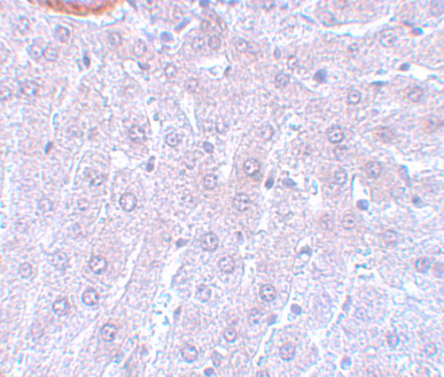
A03905 has a rat liver IHC image at 5 μg/mL (A03905 IHC image caption). A03905-3 has human bladder cancer paraffin-section IHC data and A431-cell IF/ICC data (A03905-3 image captions).
Which to pick: For human tissue IHC, choose A03905-3: its image shows a paraffin-embedded bladder cancer section, with the fixative unreported (A03905-3 IHC image caption). For rat tissue IHC, A03905 has a rat liver image; for IF/ICC, choose A03905-3, which lists those applications and has an A431-cell IF image (A03905 IHC image caption; A03905-3 application list and IF image caption). For cross-species planning, both list Human, Mouse, and Rat reactivity, while the shown IHC images document human tissue for A03905-3 and rat tissue for A03905; both are rabbit antibodies with no clone reported (catalog reactivity, host and clone fields; IHC image captions).