This website uses cookies to ensure you get the best experience on our website.


- Table of Contents
Plan CDC45 staining in paraffin sections around the nuclear pattern seen in most tissues (HPA tissue IHC). Use testicular pachytene spermatocytes as a strong positive reference and compare nuclear staining across cell types (HPA tissue IHC).

Expected localisation, validated protocols, controls and antibodies — the at-a-glance facts below, then the full design guide.
| Expected localisation | Nuclear staining in most tissues (HPA tissue IHC) | |
| Staining pattern | Nuclear; strongest in testicular pachytene spermatocytes (HPA tissue IHC) | |
| Antigen retrieval | EDTA pH 8.0 HIER, heat-mediated (datasheet M01367-2) | |
| Positive control | Testis+3 more · see all | |
| Negative control | Adrenal gland+4 more · see all |
| Fixation | Keep fixation consistent across paraffin sections. (standard IHC practice; not target-specific) | |
| Caveat | Staining and RNA show only medium consistency (HPA tissue IHC) | |
| Regulation | Highest in adult testis and thymus (UniProt) | |
| Isoform / epitope | 3 isoforms; epitope coverage needs checking (UniProt) |
The catalog antibody protocol uses heat-mediated EDTA retrieval at pH 8.0 (datasheet: M01367-2). The published IHC methods below provide three tissue-based examples (PMC4714687; PMC7919231; PMC10956518).
| Sample | Paraffin-embedded human ovarian cancer tissue; fixative not specified (datasheet M01367-2) |
| Fixation | Image fixative and duration unreported (datasheet M01367-2); verify before use. |
| Sectioning | 4–5 µm sections on charged slides (standard) |
| Deparaffinisation | Xylene, graded ethanol series to water (standard) |
| Antigen retrieval | Heat retrieval: EDTA pH 8.0 (datasheet M01367-2); 20 min, 95–100 °C (standard) |
| Peroxidase block | 3% H2O2, 10 min, room temperature (standard) |
| Blocking | 10% goat serum (datasheet M01367-2) |
| Primary antibody | Mouse monoclonal (clone 6H6) anti-CDC45, 2μg/ml (datasheet M01367-2) |
| Primary incubation | Overnight at 4 °C (datasheet M01367-2) |
| Detection | Streptavidin-biotin complex (SABC), DAB chromogen (datasheet M01367-2) |
| Counterstain | Hematoxylin, blue, dehydrate and mount (standard) |
| Expected result | CDC45-positive staining in pachytene spermatocytes of testis (HPA tissue IHC: High). HPA tissue profile: Nuclear expression in most tissues, most abundant in testicular seminiferous duct. No signal in the no-primary control. |
CDC45 is a chromatin-associated nuclear protein with no transmembrane segment (UniProt O75419: subcellular location and topology). In paraffin-section IHC, expect nuclear staining, especially in testicular pachytene spermatocytes; lymphoid cells may stain at lower levels (HPA tissue IHC: High in pachytene spermatocytes; Medium in sampled lymphoid populations). HPA rates the tissue profile Enhanced, while reporting medium consistency between antibody staining and RNA expression (HPA tissue IHC: reliability).
| Distinct nuclear stain in pachytene spermatocytes, with weaker stain in sampled lymphoid populations (HPA tissue IHC). | This matches the strongest listed IHC signal in testis and Medium staining in appendix, lymph node, and tonsil populations (HPA tissue IHC). |
| Predominantly cytoplasmic or membranous chromogen with little nuclear staining. | Review as a possible IHC artefact because CDC45 associates with chromatin and the HPA tissue profile is nuclear (UniProt O75419; HPA tissue IHC). |
| Strong stain in colon glandular cells or adrenal glandular cells. | Those sampled cells were Not detected by HPA; assess cross-reactivity or endogenous detection activity before calling them positive (HPA tissue IHC; standard IHC practice). |
| Hazy chromogen covers nuclei, cytoplasm, and surrounding tissue. | Diffuse background prevents a nuclear call; check blocking, antibody concentration, and wash conditions using standard IHC practice. HPA's CDC45 profile describes nuclear expression (HPA tissue IHC). |
| No discernible nuclear signal in testicular pachytene spermatocytes. | Treat the run as inconclusive until controls and the IHC workflow are checked; these cells are High in the HPA tissue profile (HPA tissue IHC; standard IHC practice). |
| Cell compartment | Score the nuclear compartment in IHC: UniProt places CDC45 in the nucleus and on chromatin, and HPA reports nuclear expression across most tissues (UniProt O75419; HPA tissue IHC). |
| Choice of comparison cells | Testicular pachytene spermatocytes provide the strongest listed positive example; sampled colon and adrenal glandular cells are Not detected (HPA tissue IHC). Compare the specified cells, not an entire section. |
| Antibody evidence | Two listed antibodies, HPA000614 and CAB009459, have Enhanced IHC status; the tissue profile still has medium staining–RNA consistency (HPA antibodies; HPA tissue IHC). Interpret unexpected cells cautiously. |
| Isoform coverage | UniProt lists three CDC45 isoforms, but the payload gives no epitope or isoform recognition data for either antibody (UniProt O75419; HPA antibodies). Do not assign a staining difference to an isoform. |
| IF/ICC Q&A: where should signal appear? | Mainly in the nucleoplasm, with additional cytosol and centrosome localization reported in ICC-IF (HPA subcellular). This IF observation does not establish a cytoplasmic IHC pattern. |
| Situation | Likely cause | Next action |
|---|---|---|
| The testis positive control has no nuclear stain. | A failed staining run is possible; pachytene spermatocytes are High in the reference profile (HPA tissue IHC). | Check control integrity, retrieval execution, primary-antibody application, and detection reagents using standard IHC practice; do not score samples from this run. |
| Chromogen appears mainly outside nuclei. | The pattern conflicts with nuclear, chromatin-associated CDC45 and HPA's nuclear tissue profile (UniProt O75419; HPA tissue IHC). | Inspect morphology and nuclear counterstain, then check blocking, washes, and detection controls using standard IHC practice. |
| Colon or adrenal glandular cells stain strongly. | These sampled cell types are Not detected in HPA tissue IHC; cross-reactivity or endogenous detection activity is possible (HPA tissue IHC; standard IHC practice). | Run a no-primary control and compare the stain with cell boundaries and the expected nuclear compartment (standard IHC practice; UniProt O75419). |
| Most of the section has diffuse color. | Background may obscure cell-specific nuclear stain; HPA describes nuclear expression (HPA tissue IHC; standard IHC practice). | Review blocking, wash stringency, and primary-antibody concentration; repeat alongside an appropriate control using standard IHC practice. |
| A low-staining tissue has no visible positive cells. | HPA reports Low staining in several sampled populations, including lung alveolar cells and stomach glandular cells (HPA tissue IHC). | Check a testis positive control and score only the relevant cell type; absence of a readily visible signal in a Low category alone does not establish reagent failure (HPA tissue IHC). |
| Two antibodies give different cell-level patterns. | Both listed antibodies have Enhanced IHC status, but the HPA tissue profile has medium staining–RNA consistency (HPA antibodies; HPA tissue IHC). | Compare matched sections and controls, then report the discrepancy by cell type and compartment rather than assigning an unsupported isoform explanation (UniProt O75419: three isoforms; standard IHC practice). |
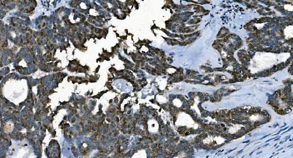
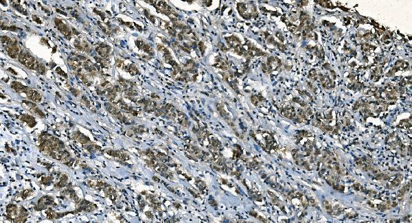
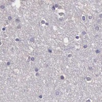
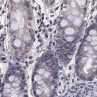
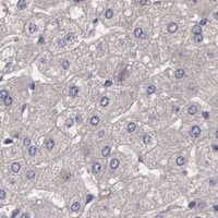


Comprehensive Human Protein Atlas IHC scoring per tissue (reliability: Enhanced — Medium consistency between antibody staining and RNA expression data.). Rows are taken directly from the HPA tissue chart — click any row's HPA link to view the source.
| Tissue | Cell type | Level | Evidence | Source |
|---|---|---|---|---|
| Testis | Pachytene spermatocytes | High | Protein (IHC) | HPA → |
| Appendix | Lymphoid tissue | Medium | Protein (IHC) | HPA → |
| Lymph node | Germinal center cells | Medium | Protein (IHC) | HPA → |
| Tonsil | Non-germinal center cells | Medium | Protein (IHC) | HPA → |
| Tissue | Cell type | Level | Evidence | Source |
|---|---|---|---|---|
| Adrenal gland | Glandular cells | Not detected | Protein (IHC) | HPA → |
| Caudate | Glial cells | Not detected | Protein (IHC) | HPA → |
| Cerebellum | Cells in granular layer | Not detected | Protein (IHC) | HPA → |
| Cerebral cortex | Glial cells | Not detected | Protein (IHC) | HPA → |
| Cervix | Glandular cells | Not detected | Protein (IHC) | HPA → |
Troubleshoot CDC45 staining in paraffin-section chromogenic IHC by checking retrieval, nuclear localisation, controls and scoring against the reported tissue pattern.
The catalog antibody has real IHC data from human ovarian cancer paraffin sections (image caption: M01367-2); the catalog lists human, mouse and rat reactivity (catalog: M01367-2).
M01367-2 is the sole SKU with a rendered card; its IHC image shows a human ovarian cancer paraffin section (image caption: M01367-2). The catalog lists IHC and human, mouse and rat reactivity for M01367-2 (catalog: M01367-2); no IF image is supplied for it (catalog: M01367-2).
Which to pick: For tissue IHC, choose M01367-2: its mouse monoclonal clone 6H6 has a human ovarian cancer paraffin-section image (catalog and image caption: M01367-2); the caption does not report the fixative (image caption: M01367-2). For IF/ICC, choose rabbit monoclonal clone AD-3, M01367, because IF and ICC are listed applications, although no IF image is supplied (catalog: M01367). For mouse or rat samples, both SKUs list reactivity, but the supplied IHC image for M01367-2 documents human tissue only (catalog: M01367 and M01367-2; image caption: M01367-2).