CDH13 / Cadherin-13 · IHC design guide

Design Immunohistochemistry for CDH13

Plan chromogenic IHC on paraffin sections with the IHC-validated antibody at 1–2 μg/mL (datasheet A01986-1). Use glomerular cells as a high-staining reference and score membranous and cytoplasmic signal by cell type (HPA tissue IHC).

Evidence assembled Oct 2026 · For research use; verify linked source records and product datasheet before use
Immunohistochemistry protocol sheet for CDH13 (IHC for CDH13): expected localisation Membranous and cytoplasmic tissue staining (HPA tissue IHC), antibody A01986-1, validated IHC image, and IHC protocol steps
Printable CDH13 IHC protocol sheet — expected localisation Membranous and cytoplasmic tissue staining (HPA tissue IHC), antibody A01986-1, controls and protocol steps. Open the full CDH13 IHC guide →

CDH13 Immunohistochemistry Experimental Design Guide

Expected localisation, validated protocols, controls and antibodies — the at-a-glance facts below, then the full design guide.

Must know before staining
Expected localisation Membranous and cytoplasmic tissue staining (HPA tissue IHC)
Staining pattern Muscle, glomeruli, endothelium, basal cells: membrane/cytoplasm (HPA tissue IHC)
Antigen retrieval EDTA pH 8.0 HIER, heat-mediated (datasheet A01986-1)
Positive control ⓘ Kidney+4 more · see all
Negative control ⓘ Adipose tissue+4 more · see all
Important caveats
Reasons your staining may differ from the expected pattern.
Fixation Keep fixation consistent across sections (standard IHC practice; not target-specific)
Caveat Appendix glandular cells may lack signal in a positive section (HPA tissue IHC; datasheet A01986-1)
Regulation Expression varies by tissue and cell type (HPA tissue IHC)
Isoform / epitope 5 isoforms; map epitopes to mature aa 139–693, which lacks a cytoplasmic tail (UniProt)
Section 1

Recommended CDH13 IHC & IF Protocols

Compare the catalog antibody’s IHC-P protocol (datasheet A01986-1) with published CDH13 staining in lung tumors, mouse brain, aorta, and hepatocellular carcinoma (PMC13114730; PMC4930129; PMC9339483; PMC12817644).

Recommended immunohistochemistry (IHC-P) protocol parameters
SampleParaffin-embedded human appendicitis tissue; fixative not specified (datasheet A01986-1)
FixationImage fixative and duration unreported (datasheet A01986-1); verify before use.
Sectioning4–5 µm sections on charged slides (standard)
DeparaffinisationXylene, graded ethanol series to water (standard)
Antigen retrievalHeat retrieval: EDTA pH 8.0 (datasheet A01986-1); 20 min, 95–100 °C (standard)
Peroxidase block3% H2O2, 10 min, room temperature (standard)
Blocking10% goat serum (datasheet A01986-1)
Primary antibodyRabbit anti-CDH13, 1-2μg/ml (datasheet A01986-1)
Primary incubationOvernight at 4 °C (datasheet A01986-1)
DetectionStreptavidin-biotin complex (SABC), DAB chromogen (datasheet A01986-1)
CounterstainHematoxylin, blue, dehydrate and mount (standard)
Expected resultCDH13-positive staining in cells in glomeruli of kidney (HPA tissue IHC: High). HPA tissue profile: Distinct membranous and cytoplasmic expression in muscle tissues, renal glomeruli, cerebral cortex, in endothelial cells and in basal cells of squamous epithelia. No signal in the no-primary control.
💡Decision noteStart with heat-mediated EDTA pH 8.0 retrieval for the catalog antibody (datasheet A01986-1); use each article’s stated conditions when reproducing its protocol.
Section 2

What Is the Expected CDH13 Staining Pattern?

CDH13 should show membranous staining, with cytoplasmic staining also reported in tissue IHC (HPA: tissue IHC profile). Look for signal in renal glomerular cells, cardiomyocytes, myocytes, smooth muscle cells, and basal cells of squamous epithelia (HPA: tissue IHC). The tissue profile has Enhanced reliability, reflecting high consistency with RNA expression (HPA: reliability). CDH13 has a GPI anchor and no transmembrane segment (UniProt P55290 topology and keywords).

What am I looking at on my slide?
Glomerular cells stain strongly; cardiomyocytes, skeletal myocytes, or smooth muscle cells stain less intensely.This matches HPA's High glomerular and Medium muscle-cell staining. Judge intensity within the identified cell type, rather than treating every cell in a positive tissue as positive (HPA: tissue IHC).
Membranous staining is accompanied by cytoplasmic staining in expected cells.Both compartments occur in HPA tissue IHC, while HPA ICC-IF supports plasma-membrane localization. Cytoplasmic tissue staining alone does not establish mislocalization; assess its cell-type distribution and the control slide (HPA: tissue IHC and subcellular ICC-IF).
Predominantly nuclear staining appears without the expected cell-boundary pattern.Nuclear localization is absent from the supplied HPA and UniProt localization records. Treat this as suspect staining and review morphology, controls, and detection background before assigning it to CDH13 (HPA: localization; UniProt P55290 subcellular location; general IHC practice).
Strong signal appears in adipocytes or cerebellar granular-layer cells.HPA reports CDH13 as not detected in those cell types. Check whether the signal belongs to neighboring structures; if it persists in the specified cells, investigate nonspecific antibody binding or endogenous detection activity (HPA: tissue IHC; general IHC practice).
No signal appears in renal glomerular cells on the test section.HPA reports High staining in glomerular cells, so an absent signal warrants a run-control review. It does not by itself prove that the specimen lacks CDH13 (HPA: kidney IHC; general IHC practice).
💡Expected CDH13 appearanceCall a positive result when cell-associated membranous staining, possibly with cytoplasmic staining, appears in the expected cells: High in renal glomerular cells or Medium in listed muscle cells; isolated nuclear or broadly diffuse staining is suspect (HPA: tissue IHC; general IHC practice).
How each factor affects the staining
IHC validation and tissue contextHPA rates tissue IHC reliability Enhanced and lists two antibodies with Enhanced IHC validation, HPA001380 and CAB025863. This supports the reported pattern, while cell identity remains essential when reading a mixed tissue section (HPA: reliability and antibodies; general IHC practice).
Topology and compartmentCDH13 is GPI-anchored and lacks a transmembrane segment (UniProt P55290 topology and keywords). HPA reports membranous and cytoplasmic tissue staining, so membrane signal is expected without requiring exclusively membrane staining in IHC (HPA: tissue IHC).
Processing, isoforms, and epitopeUniProt lists a mature chain at residues 139–693, processed terminal regions, and 5 isoforms. The supplied records give no epitope for the test antibody; do not infer which forms it detects from staining alone (UniProt P55290 processing and isoforms).
IF/ICC Q&A: What localization should an IF image show?HPA supports plasma-membrane localization in ICC-IF and lists HaCaT and SiHa image sets. This is an interpretation reference for IF/ICC; compare tissue IHC against its separate HPA tissue profile (HPA: subcellular ICC-IF and tissue IHC).
Why is my staining missing, weak or wrong?
SituationLikely causeNext action
Glomerular cells lack signal while the section has interpretable tissue morphology.A failed staining or detection step is possible; HPA reports High glomerular staining but provides no CDH13-specific retrieval or fixation-effect evidence (HPA: kidney IHC; general IHC practice).Review a known-positive control, primary-antibody application, detection reagents, and counterstain; then optimize retrieval under the antibody's IHC-P instructions without assuming a CDH13-specific retrieval requirement (general IHC practice).
Staining is mainly nuclear or appears detached from recognizable cell structures.The pattern conflicts with the supplied membrane and cytoplasmic localizations; nonspecific staining or detection artefact is possible (HPA: localization; UniProt P55290 subcellular location; general IHC practice).Recheck focus and cell boundaries, compare the known-positive control, and repeat with appropriate negative detection controls before interpreting the compartment (general IHC practice).
Adipocytes stain as strongly as glomerular cells.HPA reports adipocytes as not detected and glomerular cells as High; signal assigned to adipocytes may reflect misidentified neighboring structures or nonspecific staining (HPA: tissue IHC; general IHC practice).Confirm cell identity on the counterstained section and inspect a negative control for background; score staining by cell type rather than by whole-tissue brightness (general IHC practice).
Color is diffuse across the section, obscuring cell boundaries.Diffuse background can arise from inadequate blocking, washing, or detection controls in chromogenic IHC; this appearance is not the distinct cellular pattern reported for CDH13 (general IHC practice; HPA: tissue IHC profile).Inspect negative controls, review blocking and wash steps, and adjust detection conditions according to the assay instructions before scoring CDH13 localization (general IHC practice).
Cytoplasmic staining is present alongside membrane staining in expected cells.HPA describes both membranous and cytoplasmic tissue IHC, although its ICC-IF localization is supported at the plasma membrane (HPA: tissue IHC and subcellular ICC-IF).Record both compartments and their cell-type distribution. Use the tissue IHC profile when interpreting the paraffin section; do not reject an otherwise matching result solely for cytoplasmic signal (HPA: tissue IHC; general IHC practice).
A muscle section has weaker staining than a glomerular control.That intensity difference can fit HPA's Medium staining in listed muscle cells versus High staining in glomerular cells (HPA: tissue IHC).Compare the correct cell types and compartment patterns before calling the muscle section negative; investigate the run if its positive control also fails (HPA: tissue IHC; general IHC practice).

Sample controls for CDH13 IHC & IF

🧪Run kidney first: cells in glomeruli should stain (HPA: High in cells in glomeruli). Use adrenal gland glandular cells as the negative tissue (HPA: Not detected in adrenal gland glandular cells); on the kidney slide, assess non-glomerular cells that lack specific staining as a background reference, without assuming all non-glomerular cells are CDH13-negative.
Positive control tissue: Kidney (Cells in glomeruli, HPA High)
Negative control tissue: Adipose tissue (HPA Not detected)
ICC-IF cell lines (HPA subcellular resource): HPA ICC-IF images show CDH13 in HaCaT, SiHa, with annotated localisation: Plasma membrane (supported) (HPA subcellular).
Technical controls: Include a no-primary, secondary-only control, a concentration-matched nonimmune rabbit IgG isotype control appropriate to the primary antibody’s clonality, and CDH13-knockout tissue or cells as a biological negative (selected-SKU caption: rabbit primary; standard IHC practice). In kidney sections, block endogenous peroxidase and check endogenous biotin if using the caption’s biotin–streptavidin detection system (selected-SKU caption: biotinylated secondary and streptavidin–biotin complex; standard IHC practice).
⚠️Feasibility: No target-specific fixation window or fixation effect is reported, and the selected-SKU paraffin-section caption does not state the fixative (selected-SKU caption: fixative unreported). The caption reports heat-mediated retrieval in EDTA at pH 8.0, but does not establish that retrieval is required under all conditions (selected-SKU caption: EDTA retrieval). The supplied evidence does not establish whether frozen sections or IF/ICC are easier; for kidney chromogenic IHC, endogenous biotin can complicate the caption’s detection method (selected-SKU caption: biotin–streptavidin detection; standard IHC practice).

HPA tissue IHC evidence for CDH13

Comprehensive Human Protein Atlas IHC scoring per tissue (reliability: Enhanced — High consistency between antibody staining and RNA expression data.). Rows are taken directly from the HPA tissue chart — click any row's HPA link to view the source.

Positive expression · recommended positive controls

TissueCell typeLevelEvidenceSource
Kidney Cells in glomeruli High Protein (IHC) HPA →
Heart muscle Cardiomyocytes Medium Protein (IHC) HPA →
Skeletal muscle Myocytes Medium Protein (IHC) HPA →
Skin Cells in basal layer Medium Protein (IHC) HPA →
Smooth muscle Smooth muscle cells Medium Protein (IHC) HPA →

Undetected expression · recommended negative controls

TissueCell typeLevelEvidenceSource
Adipose tissue Adipocytes Not detected Protein (IHC) HPA →
Adrenal gland Glandular cells Not detected Protein (IHC) HPA →
Appendix Glandular cells Not detected Protein (IHC) HPA →
Bone marrow Hematopoietic cells Not detected Protein (IHC) HPA →
Breast Adipocytes Not detected Protein (IHC) HPA →
Section 3

Advanced CDH13 IHC Tips

Troubleshoot CDH13 staining by checking retrieval, cell type and compartment, then compare each run with appropriate positive and negative controls.

What retrieval should I use when CDH13 staining is weak in paraffin sections?
Start with heat-mediated retrieval in EDTA pH 8.0 for paraffin sections (datasheet A01986-1). The selected image used this retrieval before overnight primary-antibody incubation at 4°C and chromogenic detection (caption A01986-1). If signal is weak, first compare retrieval time and heating consistency across sections, then titrate the primary around the caption's 2 µg/mL condition while holding detection constant (caption A01986-1). Check a tissue compartment with expected staining, such as renal glomerular cells, before treating a negative sample as technical failure (HPA: high in kidney glomerular cells).
Could fixation explain weak CDH13 staining in my paraffin sections?
The selected paraffin-section caption does not state a fixative, so CDH13 sensitivity to a particular fixative or fixation duration is unknown (caption A01986-1). Record the fixative, fixation interval and processing schedule for each specimen, and compare matched sections processed together before attributing a difference to biology (standard IHC practice). Keep retrieval at EDTA pH 8.0 and the primary incubation consistent while evaluating the fixation variable (datasheet A01986-1). If both an expected positive compartment and the study region stain weakly, test fixation and retrieval conditions on adjacent sections with matched detection settings (HPA: high in kidney glomerular cells; standard IHC practice).
Should CDH13 staining appear on membranes or in cytoplasm?
Expect prominent cell-boundary staining because CDH13 is reported at the plasma membrane, with cytoplasmic expression also described in tissue IHC (HPA subcellular: supported plasma membrane; HPA tissue: membranous and cytoplasmic expression). Its membrane association is consistent with a GPI anchor and no transmembrane segment, so a diffuse cytoplasmic signal alone needs careful validation (UniProt P55290 topology and keywords). Compare adjacent cells within the same anatomical compartment and score membrane and cytoplasmic patterns separately (standard IHC practice). If a section shows only uniform cytoplasmic haze, review the negative control and detection background before assigning that pattern to CDH13 (standard IHC practice).
How do isoforms and processing affect interpretation of CDH13 staining?
CDH13 has five listed isoforms, but the supplied antibody evidence does not map its epitope to any one of them (UniProt P55290 isoforms; caption A01986-1). The precursor includes a signal peptide and propeptide regions, while the reported cadherin chain spans residues 139–693 (UniProt P55290 processing). Multiple predicted glycosylation sites and cadherin domains make epitope accessibility a reasonable variable to test during retrieval optimisation, without assigning a specific effect to this antibody (UniProt P55290 glycosylation and domains; standard IHC practice). Confirm the catalog antibody's stated epitope before claiming isoform-specific staining, and interpret a negative section against a matched positive control (standard IHC practice).
How should I adapt CDH13 localisation checks for multiplex immunofluorescence?
For a separate IF experiment, pair CDH13 with a marker that identifies the expected cell population, such as endothelial cells, and inspect whether signal follows cell boundaries (HPA tissue: endothelial expression; HPA subcellular: supported plasma membrane). Choose fluorophores and imaging channels after checking tissue autofluorescence, and include single-label controls to assess bleed-through (standard IF practice). Use minimal permeabilisation when testing a surface-accessible epitope; if the antibody's epitope faces the cytoplasm, optimise permeabilisation on matched samples instead (UniProt P55290: GPI-anchor keyword and no transmembrane segment; standard IF practice). HPA lists ICC/IF images in HaCaT and SiHa, which provide localisation context without establishing a tissue-IF protocol for this antibody (HPA subcellular).
How can I reduce diffuse or patchy chromogenic background?
The selected paraffin-section example used 10% goat serum blocking, a biotinylated secondary and an avidin-biotin detection complex with DAB (caption A01986-1). If staining is widespread, compare a no-primary section and inspect endogenous peroxidase or biotin contributions before changing the primary concentration (standard IHC practice). Include a peroxidase block in the chromogenic workflow and titrate blocking, washing and DAB development against an expected positive compartment (standard IHC practice; HPA: high in kidney glomerular cells). Persistent signal in the no-primary control indicates detection background; a clean control with diffuse primary-dependent staining calls for antibody titration and localisation review (standard IHC practice).
What is a defensible way to quantify CDH13 IHC across sections? ⚠ ANSWER MARKED FOR VERIFICATION
Define the anatomical compartment and scoring rule before reviewing outcomes, then report either an H-score, percentage of positive cells or positive-cell density per mm² (standard IHC practice). Score membrane and cytoplasmic staining separately because both patterns are reported for CDH13 in tissues (HPA tissue: membranous and cytoplasmic expression). Normalise cell counts to the number of eligible cells or measured compartment area, and keep exposure, DAB development and thresholding consistent within a comparison (standard IHC practice). Exclude folds, torn edges and necrotic regions using the same prespecified rule for every section, and retain an expected positive control in each staining run (standard IHC practice).
How do I distinguish true CDH13 staining from artefact in an inflamed appendix?
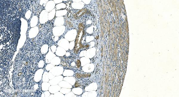
The selected image shows CDH13 staining in paraffin-embedded human appendicitis tissue, but its caption does not identify the positive cell type (caption A01986-1). HPA reports appendix glandular cells as not detected, while endothelial cells and several other compartments show CDH13 expression, so identify stained cells before interpreting the image as glandular positivity (HPA tissue). Favour cell-boundary staining in an expected compartment, while assessing cytoplasmic staining against morphology and controls (HPA subcellular: supported plasma membrane; HPA tissue: membranous and cytoplasmic expression). Treat edge staining, necrotic deposits and signal reproduced in a no-primary or peroxidase-control section as possible artefacts (standard IHC practice).
Boster reagents

Best CDH13 / Cadherin-13 IHC Antibodies

These anti-CDH13 antibodies have IHC images from human and mouse tissues and IF images from cultured cells (catalog image captions); listed species reactivity spans human, mouse, and rat (catalog reactivity).

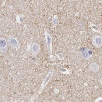
Real IHC data IHC analysis of H Cadherin/CDH13 using anti-H Cadherin/CDH13 antibody (A01986-1). H Cadherin/CDH13 was detected in paraffin-embedded section of human appendicitis tissue. Heat mediated antigen retrieval was performed in EDTA buffer (pH8.0, epitope retrieval solution). The tissue section was blocked with 10% goat serum. The tissue section was then incubated with 2μg/ml rabbit anti-H Cadherin/CDH13 Antibody (A01986-1) overnight at 4°C. Biotinylated goat anti-rabbit IgG was used as secondary antibody and incubated for 30 minutes at 37°C. The tissue section was developed using Strepavidin-Biotin-Complex (SABC) (Catalog # SA1022) with DAB as the chromogen.
Anti-H Cadherin/CDH13 Antibody ®
Cat # A01986-1
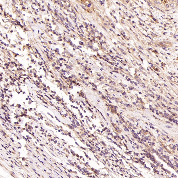
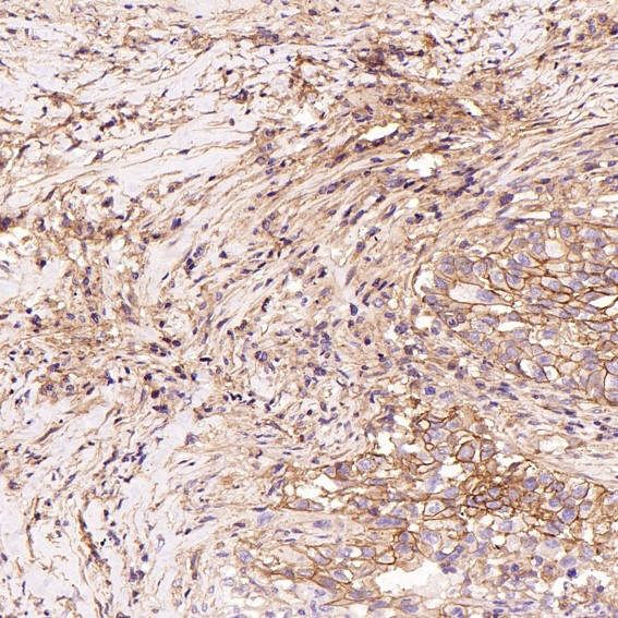
Real IHC data Immunohistochemical analysis of paraffin-embedded Human cervical cancer, using the Antibody at 1:100 dilution.
Anti-H Cadherin Rabbit Monoclonal Antibody
Cat # M01986-1
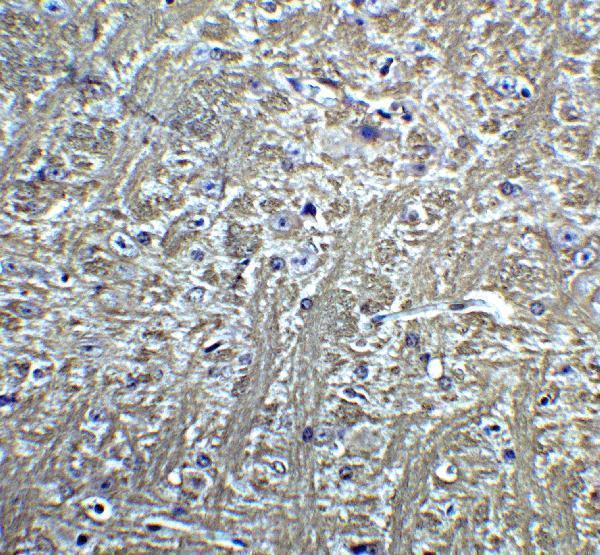
Real IHC data Immunohistochemistry of T cadherin in mouse brain tissue with T cadherin Antibody at 5 μg/mL.
Anti-T-cadherin CDH13 Antibody
Cat # A10606

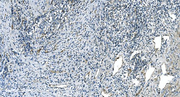
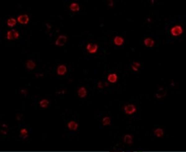
A01986-1 has IHC images of human appendicitis, gastric cancer, renal carcinoma, and melanoma sections, plus IF in SIHA cells (A01986-1 image captions). M01986-1 has IHC images of human cervical cancer, human squamous cell carcinoma, and mouse skeletal muscle; A10606 has IHC in mouse brain and IF in K562 and 3T3 cells (M01986-1 and A10606 image captions).

Which to pick: For paraffin-section IHC, choose A01986-1 when its documented EDTA retrieval at pH 8.0 and 2 μg/mL staining conditions fit the experiment; M01986-1 also has paraffin-section images, while the A10606 mouse-brain caption does not specify section processing (respective IHC image captions). The paraffin-section captions do not report a fixative (A01986-1 and M01986-1 IHC image captions). For IF/ICC, A01986-1 has a SIHA-cell IF image and lists both applications; for broader listed species reactivity in tissue IHC, M01986-1 is a rabbit monoclonal reactive with human, mouse, and rat, with IHC images from human and mouse tissue (catalog applications, host, clone, reactivity, and image captions).

Each figure is that product's own IHC / IF validation image from its datasheet.

References

  1. UniProt Consortium. UniProt entry P55290 (CAD13_HUMAN, Cadherin-13).
  2. Human Protein Atlas. CDH13 tissue IHC expression (reliability: Enhanced).
  3. Human Protein Atlas. CDH13 subcellular location (ICC-IF): Localized to the plasma membrane..
  4. Human Protein Atlas. CDH13 antibody validation summary (2 antibodies).
  5. CDKN2A/p16 Exon 2 Hypermethylation in Lung Squamous Cell Carcinoma Associated with Interstitial and Emphysematous Lung Diseases: A Comparative Analysis of Tumor, Adjacent and Distant Lung Tissues. Current oncology (Toronto, Ont.) 2026 — PMC13114730.
  6. Cadherin-13, a risk gene for ADHD and comorbid disorders, impacts GABAergic function in hippocampus and cognition. Translational psychiatry 2015 — PMC4930129.
  7. Aging Alters the Aortic Proteome in Health and Thoracic Aortic Aneurysm. Arteriosclerosis, thrombosis, and vascular biology 2022 — PMC9339483.
  8. Multimodal analysis reveals potential association of CDH13 with endothelial cells and its overexpression in hepatocellular carcinoma. European journal of medical research 2025 — PMC12817644.
  9. PubMed PMID:7982033 — UniProt-cited evidence.
  10. PubMed PMID:8673923 — UniProt-cited evidence.
  11. PubMed PMID:9737784 — UniProt-cited evidence.