This website uses cookies to ensure you get the best experience on our website.


- Table of Contents
Plan chromogenic CDH17 IHC on paraffin sections with the IHC-validated antibody at 1–2 μg/ml (datasheet A06389-1). Assess membranous and cytoplasmic staining in gastrointestinal glands, accounting for differences between intestinal endocrine cells and stomach glandular cells (HPA tissue IHC).

Expected localisation, validated protocols, controls and antibodies — the at-a-glance facts below, then the full design guide.
| Expected localisation | Membranous and cytoplasmic staining in GI glands (HPA tissue IHC) | |
| Staining pattern | GI glands show membrane and cytoplasmic signal; endocrine cells stain strongly (HPA tissue IHC) | |
| Antigen retrieval | EDTA pH 8.0 HIER, heat-mediated (datasheet A06389-1) | |
| Positive control | Appendix+4 more · see all | |
| Negative control | Adipose tissue+4 more · see all |
| Fixation | Keep paraffin-section fixation consistent across samples. (standard IHC practice; not target-specific) | |
| Caveat | Stomach glands stain less strongly than intestinal endocrine cells (HPA tissue IHC) | |
| Regulation | Intestine-enriched expression (HPA tissue IHC) | |
| Isoform / epitope | No isoforms annotated; map the epitope to the extracellular or cytoplasmic side (UniProt) |
The catalog antibody uses EDTA pH 8.0 retrieval (datasheet: A06389-1). The published CDH17 IHC protocols below describe colorectal, gastric, renal and pancreatic tumor staining (PMC12294824; PMC6970965; PMC4360917; PMC4302003).
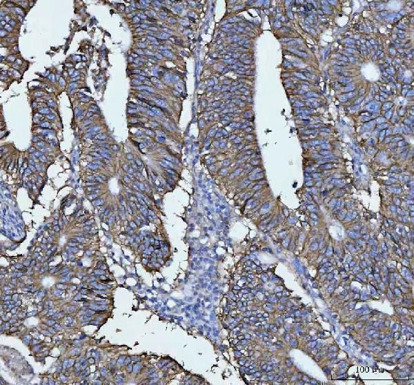
| Sample | Paraffin-embedded human rectal cancer tissue; fixative not specified (datasheet A06389-1) |
| Fixation | Image fixative and duration unreported (datasheet A06389-1); verify before use. |
| Sectioning | 4–5 µm sections on charged slides (standard) |
| Deparaffinisation | Xylene, graded ethanol series to water (standard) |
| Antigen retrieval | Heat retrieval: EDTA pH 8.0 (datasheet A06389-1); 20 min, 95–100 °C (standard) |
| Peroxidase block | 3% H2O2, 10 min, room temperature (standard) |
| Blocking | 10% goat serum (datasheet A06389-1) |
| Primary antibody | Rabbit anti-CDH17, 1-2 μg/ml (datasheet A06389-1) |
| Primary incubation | Overnight at 4 °C (datasheet A06389-1) |
| Detection | Streptavidin-biotin complex (SABC), DAB chromogen (datasheet A06389-1) |
| Counterstain | Hematoxylin, blue, dehydrate and mount (standard) |
| Expected result | CDH17-positive staining in endocrine cells of appendix (HPA tissue IHC: High). HPA tissue profile: Distinct membranous and cytoplasmic expression in gastrointestinal glands. No signal in the no-primary control. |
CDH17 is a cell-membrane protein with an extracellular region at residues 23–787 and a transmembrane segment at 788–808 (UniProt Q12864 topology). In paraffin-section IHC, expect membranous and cytoplasmic staining in gastrointestinal glands, especially the specified endocrine cells of appendix, colon, duodenum, rectum and small intestine (HPA tissue IHC). HPA rates the tissue pattern Enhanced, citing consistency between antibody staining and RNA expression (HPA tissue IHC).
| Distinct cell-border staining, with some cytoplasmic signal, in gastrointestinal glands (HPA tissue IHC). | This matches the reported tissue pattern; the cell-border component agrees with CDH17 membrane localization (HPA tissue IHC; UniProt Q12864 subcellular location). Judge the signal by its compartment and identified cell type together, since cytoplasmic staining also appears in HPA's IHC description (HPA tissue IHC). |
| Predominantly nuclear staining in a paraffin section, with little convincing membrane signal. | This departs from the expected IHC pattern and warrants an artefact check (HPA tissue IHC; UniProt Q12864 subcellular location). HPA reports an uncertain additional nucleoplasmic location in ICC-IF; that observation does not establish a nuclear IHC pattern (HPA ICC-IF). |
| Strong staining in adipocytes or adrenal glandular cells, rather than the identified gastrointestinal positive cells. | These cells are reported as not detected in the listed HPA tissue observations (HPA tissue IHC). Consider antibody cross-reactivity or endogenous detection activity, and compare with a control lacking primary antibody (standard IHC practice). |
| Diffuse color across tissue, including cells outside the expected pattern. | Treat this as background until a distinct cell-associated pattern is resolved (standard IHC practice). Excess detection signal or inadequate blocking can contribute to diffuse staining; HPA's described pattern is distinct, with membranous and cytoplasmic expression in gastrointestinal glands (standard IHC practice; HPA tissue IHC). |
| No staining in a correctly identified gastrointestinal positive control. | An absent result in a listed High positive cell population calls the run into question; it cannot establish that the specimen lacks CDH17 (HPA tissue IHC; standard IHC practice). Check the control section and detection workflow before interpreting other negative fields (standard IHC practice). |
| Membrane topology (UniProt Q12864 topology) | CDH17 has an extracellular region at 23–787, one membrane span at 788–808, and a short cytoplasmic region at 809–832 (UniProt Q12864 topology). These positions support a membrane expectation, but do not identify the catalog antibody's epitope or dictate a retrieval method. |
| Cell and tissue selection (HPA tissue IHC) | HPA reports High staining in endocrine cells of appendix, colon, duodenum, rectum and small intestine; stomach glandular cells are Medium, and gallbladder glandular cells are listed as low (HPA tissue IHC). Match the scored cell type when choosing controls. |
| IHC antibody validation (HPA antibodies) | The four listed antibodies have Enhanced IHC status: HPA023614, HPA023616, HPA026556 and CAB025143 (HPA antibodies). That supports the reported pattern, but validation status does not establish the performance of an unlisted antibody or resolve an unexpected compartment. |
| Retrieval and detection conditions (standard IHC practice) | Antigen retrieval and chromogenic detection can be optimized against positive and negative controls (standard IHC practice). The supplied UniProt and HPA records give no CDH17-specific fixation sensitivity, retrieval condition or dilution; do not infer one from topology or staining intensity. |
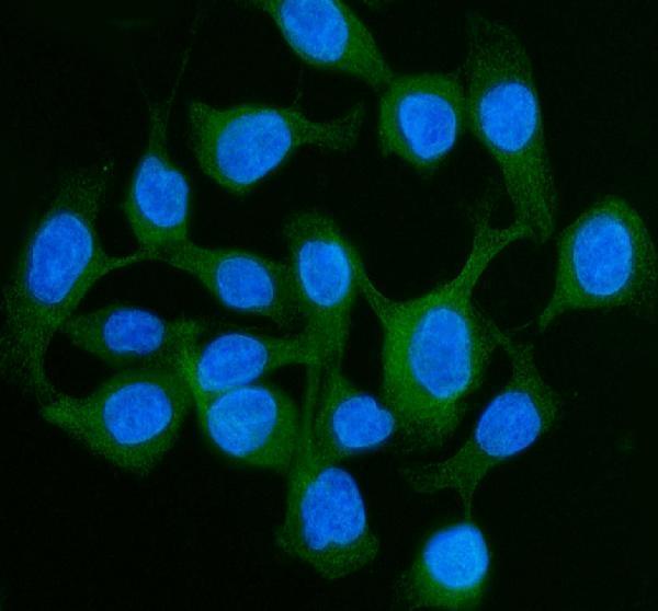
| IF/ICC Q&A: Is nucleoplasmic signal expected? | HPA's ICC-IF summary places CDH17 mainly at cell junctions and lists additional nucleoplasmic localization as uncertain (HPA ICC-IF). Treat nucleoplasmic IF signal cautiously; this separate observation does not change the expected paraffin-section IHC pattern (HPA ICC-IF; HPA tissue IHC). |
| Situation | Likely cause | Next action |
|---|---|---|
| The positive intestinal control is blank. | A blank control may reflect a failed staining run; HPA lists High signal in specified intestinal endocrine cells (standard IHC practice; HPA tissue IHC). | Confirm that the expected cells are present, then check primary antibody, retrieval and detection steps with a positive control (standard IHC practice). |
| Nuclei dominate the chromogenic signal. | Nuclear dominance conflicts with the reported membranous and cytoplasmic IHC pattern (HPA tissue IHC). | Inspect a control lacking primary antibody and compare the cell borders with a positive control before scoring (standard IHC practice). |
| Adrenal glandular cells or adipocytes stain strongly. | HPA reports CDH17 as not detected in those cell types; cross-reactivity or endogenous detection activity is possible (HPA tissue IHC; standard IHC practice). | Use a control lacking primary antibody to assess the detection system, then reassess antibody specificity (standard IHC practice). |
| Color is widespread and cell borders cannot be resolved. | Diffuse background can obscure the distinct gastrointestinal pattern (standard IHC practice; HPA tissue IHC). | Check blocking, antibody concentration, washes and chromogen development against the negative control (standard IHC practice). |
| Stomach looks weaker than an intestinal positive control. | That difference can agree with HPA's Medium stomach glandular staining and High staining in the listed intestinal endocrine cells (HPA tissue IHC). | Identify and score the specified cell types before treating the intensity difference as a run failure (HPA tissue IHC; standard IHC practice). |
| Cytoplasmic signal accompanies clear membrane staining. | HPA describes both membranous and cytoplasmic expression in gastrointestinal glands (HPA tissue IHC). | Keep the cell type and distinct membrane component in view when scoring; investigate only if diffuse color prevents localization (HPA tissue IHC; standard IHC practice). |


Comprehensive Human Protein Atlas IHC scoring per tissue (reliability: Enhanced — High consistency between antibody staining and RNA expression data.). Rows are taken directly from the HPA tissue chart — click any row's HPA link to view the source.
| Tissue | Cell type | Level | Evidence | Source |
|---|---|---|---|---|
| Appendix | Endocrine cells | High | Protein (IHC) | HPA → |
| Colon | Endocrine cells | High | Protein (IHC) | HPA → |
| Duodenum | Endocrine cells | High | Protein (IHC) | HPA → |
| Rectum | Endocrine cells | High | Protein (IHC) | HPA → |
| Small intestine | Endocrine cells | High | Protein (IHC) | HPA → |
| Tissue | Cell type | Level | Evidence | Source |
|---|---|---|---|---|
| Adipose tissue | Adipocytes | Not detected | Protein (IHC) | HPA → |
| Adrenal gland | Glandular cells | Not detected | Protein (IHC) | HPA → |
| Bone marrow | Hematopoietic cells | Not detected | Protein (IHC) | HPA → |
| Breast | Adipocytes | Not detected | Protein (IHC) | HPA → |
| Bronchus | Respiratory epithelial cells | Not detected | Protein (IHC) | HPA → |
Troubleshoot CDH17 staining in paraffin sections by checking retrieval, compartment, tissue context and controls before comparing staining intensity (UniProt Q12864; HPA tissue IHC).
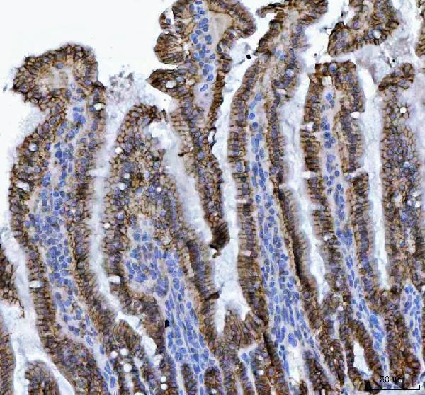
A06389-1 has IHC images from human rectal cancer and mouse and rat colon paraffin sections, plus IF images from human samples (catalog image captions).
A06389-1 has IHC images from human rectal cancer and mouse and rat colon paraffin sections, and IF images from CACO-2 cells and human intestinal cancer paraffin sections (catalog image captions). M06389 lists human reactivity and IHC, with no IHC or IF image in the supplied catalog payload (catalog: applications, reactivity, image captions).
Which to pick: For tissue IHC, choose A06389-1 when an illustrated paraffin-section procedure is useful: its caption reports EDTA retrieval at pH 8.0 and 2 μg/ml primary antibody; the fixative is unreported (A06389-1 IHC image caption). For IF/ICC, choose A06389-1 because both applications and human IF images are supplied; M06389 lists IHC but no IF/ICC application, and its rabbit monoclonal clone is AFFC-3 (catalog: applications, IF image captions, clone). For human, mouse, or rat IHC, A06389-1 has species-matched paraffin-section images, while M06389 lists human reactivity only and has no supplied IHC image (catalog: reactivity and IHC image captions).