This website uses cookies to ensure you get the best experience on our website.


- Table of Contents
Plan CDK13 staining in paraffin sections using the catalog antibody’s IHC protocol. Compare nuclear speckle expectations (UniProt) with tissue staining cautiously because HPA rates its IHC evidence uncertain (HPA tissue IHC).

Expected localisation, validated protocols, controls and antibodies — the at-a-glance facts below, then the full design guide.
| Expected localisation | Nuclear speckles expected; tissue compartment unverified (UniProt) | |
| Staining pattern | High in duodenal glandular and kidney tubular cells (HPA tissue IHC) | |
| Antigen retrieval | EDTA pH 8.0 HIER, heat-mediated (datasheet A05292-1) | |
| Positive control | Duodenum+4 more · see all | |
| Negative control | Adipose tissue+4 more · see all |
| Fixation | Keep fixation consistent across samples. (standard IHC practice; not target-specific) | |
| Caveat | Staining has low concordance with RNA expression (HPA tissue IHC) | |
| Regulation | No expression regulator specified (UniProt) | |
| Isoform / epitope | 2 isoforms; check epitope coverage (UniProt) |
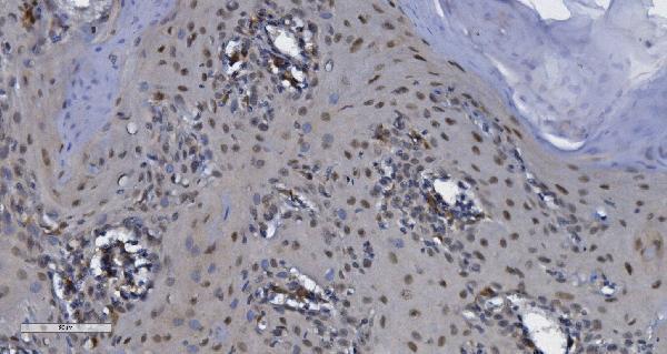
The catalog antibody uses EDTA pH 8.0 retrieval (datasheet: A05292-1). The published CDK13 IHC protocols below cover gastric cancer and glioblastoma tissue (PMC8427521; PMC13179391).
| Sample | Paraffin-embedded human esophageal squamous carcinoma tissue; fixative not specified (datasheet A05292-1) |
| Fixation | Image fixative and duration unreported (datasheet A05292-1); verify before use. |
| Sectioning | 4–5 µm sections on charged slides (standard) |
| Deparaffinisation | Xylene, graded ethanol series to water (standard) |
| Antigen retrieval | Heat retrieval: EDTA pH 8.0 (datasheet A05292-1); 20 min, 95–100 °C (standard) |
| Peroxidase block | 3% H2O2, 10 min, room temperature (standard) |
| Blocking | 10% goat serum (datasheet A05292-1) |
| Primary antibody | Rabbit anti-CDK13, 2-5μg/ml (datasheet A05292-1) |
| Primary incubation | Overnight at 4 °C (datasheet A05292-1) |
| Detection | Streptavidin-biotin complex (SABC), DAB chromogen (datasheet A05292-1) |
| Counterstain | Hematoxylin, blue, dehydrate and mount (standard) |
| Expected result | CDK13-positive staining in glandular cells of duodenum (HPA tissue IHC: High). HPA tissue profile: Expression in several tissues. No signal in the no-primary control. |
CDK13 is expected mainly in nuclear speckles (UniProt Q14004; HPA: nuclear speckles supported). In paraffin IHC, look for predominantly nuclear staining in duodenal glandular cells and kidney tubular cells, both reported as high (HPA: tissue IHC). Interpret intensity cautiously: HPA rates the tissue IHC pattern uncertain because antibody staining has low consistency with RNA expression (HPA: tissue IHC reliability). CDK13 has no transmembrane segment (UniProt Q14004 topology).
| Predominantly nuclear staining in duodenal glandular cells or kidney tubular cells, possibly speckled where detail is resolved. | This fits the reported high staining in those cell types (HPA: tissue IHC) and the nuclear speckle location (UniProt Q14004; HPA: ICC-IF supported). Chromogenic IHC may show a nuclear signal without resolving individual speckles (general IHC practice). Score the relevant cells and compartment rather than treating all staining across a section as equivalent. |
| Strong, broadly cytoplasmic staining with little nuclear staining. | Treat this as a questionable CDK13 pattern and check controls, retrieval and detection background (general IHC practice). Nuclear speckles are the supported location; Golgi apparatus and cytosol are additional uncertain locations in ICC-IF (HPA: subcellular). A cytoplasmic signal alone therefore does not establish specific CDK13 staining, but it is not proof of an artefact. |
| Strong staining in an unexpected cell population, especially alongside weak staining in the chosen positive cells. | Compare the exact cell type before assigning a result: HPA reports high staining in kidney tubular cells but no detection in bone marrow hematopoietic cells (HPA: tissue IHC). An unexpected pattern may reflect cross-reactivity or endogenous detection activity (general IHC practice); it cannot be resolved from tissue identity alone because HPA rates CDK13 tissue IHC uncertain (HPA: reliability). |
| Diffuse color across nuclei, cytoplasm and extracellular areas, including areas without expected positive cells. | This limits compartment and cell-type scoring and suggests background from the staining workflow (general IHC practice). Review the reagent-omission control, blocking, washes and detection chemistry (general IHC practice). The reported CDK13 pattern is mainly nuclear speckles (UniProt Q14004; HPA: subcellular), while the tissue IHC evidence remains uncertain (HPA: reliability). |
| No convincing signal in duodenal glandular cells or kidney tubular cells. | These are useful positive-reference cell populations because HPA reports high IHC staining in each (HPA: tissue IHC). First check whether the relevant cells are present and whether the run's positive control stained; then review retrieval, antibody dilution and detection steps (general IHC practice). Absence in one specimen alone does not settle CDK13 expression, given the uncertain HPA tissue IHC reliability (HPA: reliability). |
| Subcellular location | Nuclear speckles are supported by ICC-IF and listed by UniProt (HPA: subcellular; UniProt Q14004). Golgi apparatus and cytosol are uncertain additional ICC-IF locations (HPA: subcellular). Use nuclear enrichment as the principal localization check in IHC; do not require individually visible speckles in a chromogenic section (general IHC practice). |
| Tissue pattern and evidence strength | HPA reports high staining in duodenal glandular, kidney tubular, small-intestinal glandular and seminiferous-duct cells (HPA: tissue IHC). Its overall tissue IHC reliability is uncertain because staining and RNA data have low consistency (HPA: reliability). Treat these as observed reference patterns rather than definitive proof of specificity in every specimen. |
| Protein architecture and isoforms | CDK13 is a 1,512-residue protein with no signal peptide, propeptide or transmembrane segment; UniProt lists two isoforms and a kinase domain at residues 705–998 (UniProt Q14004). These facts support a nonmembrane localization expectation, but they do not identify the catalog antibody's epitope or predict retrieval performance (UniProt Q14004). |
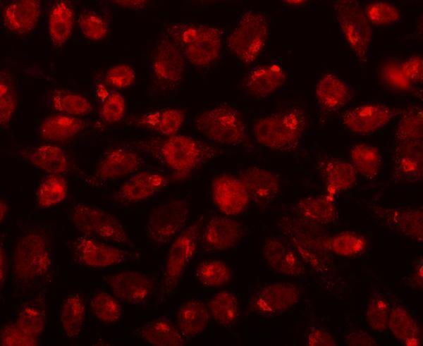
| IF/ICC: what localization should be expected? | Mainly nuclear speckles, supported in HPA ICC-IF and consistent with UniProt; Golgi apparatus and cytosol are additional uncertain locations (HPA: subcellular; UniProt Q14004). HPA lists HeLa, SiHa, U2OS and KOLF2.1J among cell lines with ICC-IF images (HPA: subcellular). This localization evidence informs interpretation here; IF/ICC methods belong on the separate guide page. |
| Situation | Likely cause | Next action |
|---|---|---|
| Positive-reference tissue stains weakly or not at all. | The selected section may lack the relevant cells, or the IHC run may have a workflow failure (general IHC practice). HPA reports high staining in duodenal glandular and kidney tubular cells, with uncertain overall reliability (HPA: tissue IHC). | Confirm the reference cells are present, review the run's positive control, then check retrieval, catalog antibody dilution and detection according to the validated IHC procedure (general IHC practice). Avoid declaring a biological negative from one failed section. |
| Staining is predominantly cytoplasmic. | The signal conflicts with the supported nuclear-speckle localization, although cytosol is an uncertain additional ICC-IF location (HPA: subcellular). Background or nonspecific binding is possible (general IHC practice). | Compare nuclear and cytoplasmic signal in the expected positive cells, examine reagent-omission controls, and repeat with the validated IHC conditions if controls are unsatisfactory (general IHC practice; HPA: tissue IHC high in duodenal glandular and kidney tubular cells). |
| Strong signal appears in cells reported as not detected. | Cross-reactivity or endogenous detection activity is possible (general IHC practice); HPA's tissue IHC reliability is uncertain, so a discrepancy alone does not identify the cause (HPA: reliability). | Verify the cell identity and compare an expected positive cell population in the same run; inspect reagent-omission controls and detection blocking (general IHC practice). For example, HPA reports bone marrow hematopoietic cells as not detected (HPA: tissue IHC). |
| Diffuse chromogen obscures nuclei and tissue boundaries. | Excess background can prevent compartment scoring (general IHC practice), while the expected main CDK13 location is nuclear speckles (UniProt Q14004; HPA: subcellular). | Check reagent-omission controls, blocking, wash steps, detection exposure and antibody dilution against the validated IHC procedure (general IHC practice). Reassess only where individual cells and nuclei remain interpretable. |
| Different tissues give inconsistent intensity. | HPA reports several high, medium, low and not-detected cell-type patterns, and rates the overall tissue IHC evidence uncertain because staining and RNA data have low consistency (HPA: tissue IHC reliability). | Record the specific cell type, compartment and intensity for each specimen; compare expected positive and negative-reference cells in the same run before inferring a biological difference (general IHC practice; HPA: tissue IHC). |
| A nuclear signal is visible, but distinct speckles cannot be resolved. | Chromogenic paraffin IHC may lack the spatial detail of ICC-IF for fine intranuclear structures (general IHC practice). Nuclear speckles are the supported ICC-IF location (HPA: subcellular). | Judge whether staining is predominantly nuclear in the expected cells and whether controls are acceptable (HPA: tissue IHC high in duodenal glandular and kidney tubular cells; general IHC practice). Use the separate IF/ICC guide if speckle resolution is required. |


Comprehensive Human Protein Atlas IHC scoring per tissue (reliability: Uncertain — Low consistency between antibody staining and RNA expression data. Pending external verification.). Rows are taken directly from the HPA tissue chart — click any row's HPA link to view the source.
| Tissue | Cell type | Level | Evidence | Source |
|---|---|---|---|---|
| Duodenum | Glandular cells | High | Protein (IHC) | HPA → |
| Kidney | Cells in tubules | High | Protein (IHC) | HPA → |
| Small intestine | Glandular cells | High | Protein (IHC) | HPA → |
| Testis | Cells in seminiferous ducts | High | Protein (IHC) | HPA → |
| Fallopian tube | Glandular cells | Medium | Protein (IHC) | HPA → |
| Tissue | Cell type | Level | Evidence | Source |
|---|---|---|---|---|
| Adipose tissue | Adipocytes | Not detected | Protein (IHC) | HPA → |
| Adrenal gland | Glandular cells | Not detected | Protein (IHC) | HPA → |
| Bone marrow | Hematopoietic cells | Not detected | Protein (IHC) | HPA → |
| Breast | Adipocytes | Not detected | Protein (IHC) | HPA → |
| Caudate | Glial cells | Not detected | Protein (IHC) | HPA → |
Troubleshoot chromogenic CDK13 IHC in paraffin sections by checking retrieval, nuclear localisation, tissue controls and staining quality before scoring.
A05292-1 has human paraffin-section IHC data from esophageal squamous carcinoma and IF/ICC data from HELA cells (catalog image captions; catalog applications/reactivity).
A05292-1 was demonstrated by chromogenic IHC on a paraffin-embedded human esophageal squamous carcinoma section (catalog IHC image caption). The same SKU was demonstrated by IF/ICC on HELA cells (catalog IF image caption).
Which to pick: Choose A05292-1 for human paraffin-section IHC; its IHC image used 2 μg/ml, EDTA retrieval at pH 8.0, and DAB detection (catalog IHC image caption). Choose A05292-1 for IF/ICC on HELA cells; its IF image used 5 μg/ml and a fluorescent secondary antibody (catalog IF image caption). Cross-species reactivity is unreported (catalog reactivity: Human only), and the paraffin-section IHC caption does not report the fixative (catalog IHC image caption).