CDK5RAP3 / CDK5 regulatory subunit-associated protein 3 · IHC design guide

Design Immunohistochemistry for CDK5RAP3

Plan chromogenic CDK5RAP3 IHC in paraffin sections with the catalog antibody at 1:100–1:300 (datasheet: A05770-1). Assess cytoplasmic staining against the high signal reported in respiratory epithelial cells (HPA tissue IHC).

Evidence assembled Oct 2026 · For research use; verify linked source records and product datasheet before use
Immunohistochemistry protocol sheet for CDK5RAP3 (IHC for CDK5RAP3): expected localisation Cytoplasm in tissue (HPA tissue IHC); nucleus possible (UniProt), antibody A05770-1, validated IHC image, and IHC protocol steps
Printable CDK5RAP3 IHC protocol sheet — expected localisation Cytoplasm in tissue (HPA tissue IHC); nucleus possible (UniProt), antibody A05770-1, controls and protocol steps. Open the full CDK5RAP3 IHC guide →

CDK5RAP3 Immunohistochemistry Experimental Design Guide

Expected localisation, validated protocols, controls and antibodies — the at-a-glance facts below, then the full design guide.

Must know before staining
Expected localisation Cytoplasm in tissue (HPA tissue IHC); nucleus possible (UniProt)
Staining pattern Cytoplasmic staining in respiratory epithelial cells (HPA tissue IHC)
Antigen retrieval Tris-EDTA pH 8.0 HIER, heat-mediated (datasheet A05770-1)
Positive control ⓘ Bronchus+4 more · see all
Negative control ⓘ Adipose tissue+2 more · see all
Important caveats
Reasons your staining may differ from the expected pattern.
Fixation Keep fixation consistent across sections. (standard IHC practice; not target-specific)
Caveat Staining varies by cell type within tissues (HPA tissue IHC)
Regulation Ubiquitously expressed (UniProt)
Isoform / epitope 4 isoforms; epitope coverage is unresolved (UniProt)
Section 1

Recommended CDK5RAP3 IHC & IF Protocols

The catalog antibody has a datasheet IHC-P protocol (datasheet A05770-1). Three published CDK5RAP3 IHC protocols provide additional paraffin-section conditions (PMC6141336; PMC6839262; PMC6466961).

Recommended immunohistochemistry (IHC-P) protocol parameters
SampleParaffin-embedded Human brain tissue; fixative not specified (datasheet A05770-1)
FixationImage fixative and duration unreported (datasheet A05770-1); verify before use.
Sectioning4–5 µm sections on charged slides (standard)
DeparaffinisationXylene, graded ethanol series to water (standard)
Antigen retrievalHeat retrieval: Tris-EDTA pH 8.0 (datasheet A05770-1); 20 min, 95–100 °C (standard)
Peroxidase block3% H2O2, 10 min, room temperature (standard)
Blocking10% normal serum of the secondary host, 30 min, room temperature (standard)
Primary antibodyRabbit anti-CDK5RAP3, 1:100-1:300 (datasheet A05770-1)
Primary incubationOvernight at 4 °C (standard)
DetectionHRP-polymer secondary, DAB chromogen 5–10 min (standard)
CounterstainHematoxylin, blue, dehydrate and mount (standard)
Expected resultCDK5RAP3-positive staining in respiratory epithelial cells of bronchus (HPA tissue IHC: High). HPA tissue profile: General cytoplasmic expression. No signal in the no-primary control.
💡Decision noteStart with heat-mediated Tris-EDTA retrieval at pH 8.0 (datasheet A05770-1); if optimizing, compare citrate retrieval at pH 6.0 (PMC6141336; PMC6839262).
Section 2

What Is the Expected CDK5RAP3 Staining Pattern?

CDK5RAP3 should appear mainly cytoplasmic in tissue IHC (HPA: general cytoplasmic expression), consistent with its cytoplasmic and endoplasmic reticulum associations (UniProt Q96JB5: subcellular location). Expect prominent staining in selected epithelial, endocrine, glandular and immune cell populations (HPA: High in the listed populations). HPA rates the tissue pattern Supported, with medium consistency between antibody staining and RNA data (HPA: tissue IHC reliability). CDK5RAP3 has no transmembrane segment (UniProt Q96JB5: topology).

What am I looking at on my slide?
Cytoplasmic chromogen is strong in bronchial respiratory epithelium or lung alveolar type II cells (HPA: High in both populations).This matches the reported tissue pattern and is a useful positive result (HPA: general cytoplasmic expression; High in those cells). Judge signal within the named cells: HPA also reports High endocrine cells in colon and duodenum, glandular cells in epididymis and gallbladder, and non-germinal center cells in lymph node (HPA: tissue IHC).
A sharp cell-surface rim or exclusively nuclear chromogen dominates the section.Reassess compartment assignment and controls before calling it CDK5RAP3: HPA describes general cytoplasmic tissue staining, and UniProt lists no transmembrane segment (HPA: tissue IHC; UniProt Q96JB5: topology). Some nuclear localization is biologically plausible, however (UniProt Q96JB5: nucleus); compartment alone cannot prove artefact.
Adipocytes, skeletal myocytes or vaginal squamous epithelial cells stain as strongly as the selected positive cells.That conflicts with HPA's Not detected calls for those cell populations (HPA: adipocytes, myocytes and vaginal squamous epithelium). Check whether the stain follows tissue structures or endogenous detection activity; cross-reactivity and endogenous activity are possible explanations, not diagnoses from one section (general IHC practice).
Weak, diffuse color covers cells and surrounding tissue without a readable cytoplasmic boundary.Treat the pattern as background until controls separate specific signal from diffuse deposition (general IHC practice). CDK5RAP3's expected tissue profile is cellular and generally cytoplasmic (HPA: tissue IHC); broad expression alone does not make uniform extracellular color a convincing positive result (UniProt Q96JB5: ubiquitous expression; general IHC practice).
No cytoplasmic signal appears in a selected HPA High population, including bronchial respiratory epithelial cells.First verify that the named cells are present and that the positive control worked (general IHC practice; HPA: High in bronchial respiratory epithelial cells). A blank section leaves assay performance unresolved; HPA's Supported designation has only medium consistency with RNA data, so it does not guarantee every specimen will stain (HPA: tissue IHC reliability).
💡Expected CDK5RAP3 appearanceCall a result positive when named HPA High cells show clear, predominantly cytoplasmic chromogen (HPA: tissue IHC); a dominant cell-surface rim or diffuse tissue-wide color needs control review (UniProt Q96JB5: topology; general IHC practice).
How each factor affects the staining
Compartment and membrane associationCDK5RAP3 is cytoplasmic and tethered to the endoplasmic reticulum through the UREL complex (UniProt Q96JB5: subcellular location). An intracellular cytoplasmic pattern fits; a cell-surface outline should be questioned because no transmembrane segment is annotated (UniProt Q96JB5: topology).
Cell population chosen for comparisonHPA reports High signal in specific populations, Low in hepatocytes and Not detected in skeletal myocytes (HPA: tissue IHC). Score the identified cell population rather than assigning one expected intensity to every cell in an organ (HPA: tissue IHC; general IHC practice).
Isoforms and antibody recognitionFour isoforms are listed, and isoform 3 is reported in kidney, liver, skeletal muscle and placenta (UniProt Q96JB5: isoforms; tissue specificity). The supplied sources do not identify the catalog antibody's epitope, so isoform-specific staining cannot be inferred from these records.
Processing and topologyUniProt annotates one chain spanning residues 1–506, with no signal peptide, propeptide or transmembrane segment (UniProt Q96JB5: processing; topology). The record provides no basis for predicting a shed extracellular staining pattern or a processed fragment with a different location.
IF/ICC: what localization is expected?Mainly cytosolic signal, with additional nucleolar and vesicular localization, is reported in ICC-IF (HPA: subcellular, cytosol Supported; nucleoli and vesicles Approved). Use that as a compartment cross-check; it is an ICC-IF observation, not an IHC-P protocol or a promise of visible nucleoli in chromogenic sections.
Why is my staining missing, weak or wrong?
SituationLikely causeNext action
The positive-control section is blank.Detection or tissue preparation may have failed (general IHC practice); a blank control cannot establish CDK5RAP3 absence.Confirm the expected HPA High cells are present, then review retrieval, primary antibody incubation and detection with the assay's controls (HPA: tissue IHC; general IHC practice). No CDK5RAP3-specific retrieval condition is supplied.
Low-staining hepatocytes are used as the only positive check.HPA rates hepatocytes Low, so a weak result there is difficult to interpret (HPA: liver hepatocytes).Include a population reported High, such as bronchial respiratory epithelial cells, for the positive check (HPA: High in bronchus). Compare cells within the same named population (general IHC practice).
Color persists in a no-primary control.Primary antibody-independent signal, including endogenous detection activity, is possible (general IHC practice).Review the chromogenic detection controls and any blocking appropriate to the detection chemistry before interpreting the tissue pattern (general IHC practice).
Only nuclei or plasma-membrane rims stain.Compartment assignment or nonspecific staining may be involved (general IHC practice); HPA's tissue profile is generally cytoplasmic (HPA: tissue IHC).Compare with a positive tissue and control section. Allow for possible nuclear CDK5RAP3, but investigate a dominant surface rim (UniProt Q96JB5: nucleus; no transmembrane segment).
The whole section has diffuse brown haze.Background from detection or insufficient washing may obscure cellular localization (general IHC practice).Check the no-primary control, washing and detection conditions; score only interpretable cellular staining (general IHC practice; HPA: general cytoplasmic expression).
A nominally negative population shows a few stained cells.Cell mixtures and identification errors can complicate a tissue-level comparison (general IHC practice); HPA calls apply to named cell populations (HPA: tissue IHC).Identify the stained cell type with morphology and counterstain before assigning a discrepancy. Treat HPA Not detected calls as observed assay results, not proof of absolute protein absence (HPA: tissue IHC; general IHC practice).

Sample controls for CDK5RAP3 IHC & IF

🧪Start with bronchus: respiratory epithelial cells should stain (HPA: High in respiratory epithelial cells). Run adipose tissue as a negative comparator (HPA: Not detected in adipocytes); on the bronchus slide, use any cells without specific staining as a background reference, without assuming a particular cell type is negative (standard IHC practice).
Positive control tissue: Bronchus (Respiratory epithelial cells, HPA High)
Negative control tissue: Adipose tissue (HPA Not detected)
ICC-IF cell lines (HPA subcellular resource): HPA ICC-IF images show CDK5RAP3 in A-431, U-251MG, U2OS, A-549, with annotated localisation: Cytosol (supported) (HPA subcellular).
Technical controls: Include no-primary (secondary-only) and host-species- and clonality-matched isotype controls, plus a CDK5RAP3 knockout or immunogen-peptide-blocked antibody control (standard IHC practice; selected-SKU caption: peptide-blocked control). Quench endogenous peroxidase and check background in the bronchus section before interpreting chromogenic staining (standard IHC practice).
⚠️Feasibility: A target-specific fixation window or fixation effect is unreported in the supplied evidence, and the selected-SKU paraffin-section caption does not state a fixative (selected-SKU caption: fixative unreported). The caption uses high-pressure, high-temperature Tris-EDTA retrieval at pH 8.0, but does not establish that retrieval is required under other conditions (selected-SKU caption: Tris-EDTA, pH 8.0). The supplied evidence does not establish whether frozen sections or IF are easier, or identify a bronchus-specific artefact; assess local background with the controls above (standard IHC practice).

HPA tissue IHC evidence for CDK5RAP3

Comprehensive Human Protein Atlas IHC scoring per tissue (reliability: Supported — Medium consistency between antibody staining and RNA expression data.). Rows are taken directly from the HPA tissue chart — click any row's HPA link to view the source.

Positive expression · recommended positive controls

TissueCell typeLevelEvidenceSource
Bronchus Respiratory epithelial cells High Protein (IHC) HPA →
Colon Endocrine cells High Protein (IHC) HPA →
Duodenum Endocrine cells High Protein (IHC) HPA →
Epididymis Glandular cells High Protein (IHC) HPA →
Gallbladder Glandular cells High Protein (IHC) HPA →

Undetected expression · recommended negative controls

TissueCell typeLevelEvidenceSource
Adipose tissue Adipocytes Not detected Protein (IHC) HPA →
Skeletal muscle Myocytes Not detected Protein (IHC) HPA →
Vagina Squamous epithelial cells Not detected Protein (IHC) HPA →
Section 3

Advanced CDK5RAP3 IHC Tips

Troubleshoot CDK5RAP3 staining in paraffin sections by checking retrieval, controls, cellular distribution and scoring before interpreting biological differences.

How should I retrieve CDK5RAP3 when paraffin sections stain weakly?
Start with heat-mediated retrieval in Tris-EDTA, pH 8.0 (datasheet A05770-1). The selected brain-section image used high-pressure, high-temperature retrieval with that buffer, followed by the catalog antibody at 1:100 overnight at 4°C (caption A05770-1). If staining remains weak, compare a longer heating interval on matched sections while monitoring tissue damage; treat any alternative buffer or pH as a fallback requiring its own validation (standard IHC practice). Keep section thickness, cooling and detection constant across that comparison, and include a known positive section plus a no-primary control so a retrieval change is distinguishable from background (standard IHC practice).
Could fixation explain weak or uneven CDK5RAP3 staining?
Target-specific fixation sensitivity is unknown: the selected paraffin-section caption does not report a fixative (caption A05770-1). Record the fixative, fixation duration and time before processing for every specimen, then compare staining only among sections with comparable handling (standard IHC practice). If a weak sample differs in handling, test matched material using the same Tris-EDTA, pH 8.0 retrieval and detection conditions before assigning the difference to CDK5RAP3 expression (datasheet A05770-1; standard IHC practice). Inspect tissue preservation and staining uniformity within each section, and document any systematic variation before using the sample for quantitative comparisons (standard IHC practice).
Which staining compartments are credible for CDK5RAP3 in tissue?
Prioritise cytoplasmic staining when assessing paraffin sections: the tissue profile reports general cytoplasmic expression (HPA tissue IHC). Cytosolic staining is the main supported subcellular location, with nucleolar and vesicular signal additionally reported in cell imaging (HPA subcellular). The protein is also annotated in the nucleus, cytoskeleton, centrosome and at the endoplasmic reticulum membrane as part of the UREL complex (UniProt Q96JB5 localisation). Score cytoplasmic and nuclear signal separately, and verify an unusual compartment against the no-primary and peptide-absorbed controls before calling it biological redistribution (standard IHC practice; caption A05770-1).
Can this stain distinguish CDK5RAP3 isoforms or an ER-facing epitope?
Do not assign a stained cell to a particular isoform without an epitope map and isoform-specific validation: 4 isoforms are listed, but the supplied antibody caption does not define its epitope (UniProt Q96JB5 isoforms; caption A05770-1). The annotated protein has no transmembrane segment and can be tethered to the endoplasmic reticulum membrane within the UREL complex (UniProt Q96JB5 topology and localisation). Therefore, membrane-associated staining alone cannot establish which molecular surface the antibody recognizes (UniProt Q96JB5 topology; standard IHC interpretation). Request the immunogen sequence, check its presence in each isoform, and validate any isoform claim with a suitable independent assay (standard IHC practice).
How can IF help investigate ambiguous CDK5RAP3 IHC staining?
Use IF as a complementary localisation check when chromogenic boundaries are ambiguous, while interpreting the paraffin-section result on its own terms (standard IHC/IF practice). Multiplex CDK5RAP3 with a validated marker for the cell type being scored; for example, alveolar type II cells show high tissue-IHC staining, so confirm cell identity before comparing signals (HPA tissue IHC; standard IF practice). Choose a fluorophore channel after measuring tissue autofluorescence, and include single-channel controls to assess bleed-through (standard IF practice). Because CDK5RAP3 lacks a transmembrane segment yet associates with the endoplasmic reticulum, select permeabilisation based on the mapped epitope and verify access with controls (UniProt Q96JB5 topology and localisation; standard IF practice).
How do I reduce diffuse or granular background in CDK5RAP3 IHC?
Compare the stained section with a no-primary control and, where available, the caption's immunogen-peptide-absorbed control before adjusting the antibody concentration (caption A05770-1; standard IHC practice). The caption used 1:100 overnight at 4°C; treat that as an evidence-based starting condition, then titrate on matched sections if nonspecific colour persists (caption A05770-1; standard IHC practice). Check blocking, washes, endogenous peroxidase quenching and DAB development time, since each can affect chromogenic background (standard IHC practice). Interpret granular cytoplasmic signal carefully because vesicular localisation is reported, but require cell-shaped staining and reproducibility before accepting it as specific (HPA subcellular; standard IHC practice).
How should I score CDK5RAP3 across paraffin sections? ⚠ ANSWER MARKED FOR VERIFICATION
Define the target compartment and cell population before scoring, then apply the same rule to every section (standard IHC practice). For per-cell chromogenic staining, record the percentage of positive cells and intensity grades 0–3; an H-score can summarize these as 0–300 (standard IHC practice). Normalize cell counts to the number of eligible cells, or report positive-cell density per mm² of evaluable tissue when cell counts are impractical (standard IHC practice). Keep exposure to stain, counterstain, thresholds and excluded damaged regions consistent, and report cytoplasmic and nuclear results separately because both localisations are annotated (standard IHC practice; UniProt Q96JB5 localisation).
When is CDK5RAP3 staining convincing rather than artefactual?
Seek reproducible, cell-shaped cytoplasmic staining, which agrees with the general tissue-IHC profile and supported cytosolic location (HPA tissue IHC; HPA subcellular). Check whether the stained cells fit the sampled tissue: alveolar type II cells are reported high, whereas adipocytes and skeletal myocytes are reported not detected in the tissue-IHC profile (HPA tissue IHC). Treat isolated section-edge colour, necrotic deposits or staining reproduced by the no-primary control as possible artefacts, including endogenous enzyme activity in chromogenic detection (standard IHC practice). Nuclear signal is biologically plausible, but confirm its cell boundaries and control behaviour before interpreting it as redistribution (UniProt Q96JB5 localisation; standard IHC practice).
Boster reagents

Best CDK5RAP3 / CDK5 regulatory subunit-associated protein 3 IHC Antibodies

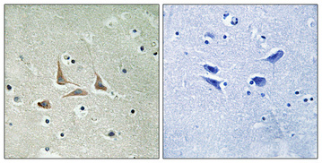
The catalog antibody A05770-1 lists IHC and IF applications and Human, Mouse, and Rat reactivity (catalog: applications and reactivity); its figure shows paraffin-embedded Human brain IHC (IHC image caption).

Real IHC data Immunohistochemical analysis of paraffin-embedded Human brain. Antibody was diluted at 1:100 (4° overnight). High-pressure and temperature Tris-EDTA, pH8.0 was used for antigen retrieval. Negetive contrl (right) obtaned from antibody was pre-absorbed by immunogen peptide.
Anti-HSF-27 CDK5RAP3 Antibody
Cat # A05770-1

A05770-1 is the sole SKU shown (catalog: SKU and applications). Its IHC figure uses paraffin-embedded Human brain, while IF is listed as an application without an IF figure (IHC image caption; catalog: applications and IF image alts).

Which to pick: Choose A05770-1 for paraffin-section IHC: its figure shows Human brain at 1:100 with Tris-EDTA retrieval at pH 8.0 (IHC image caption). For IF, A05770-1 lists a 1:50 dilution, but no IF figure is supplied; ICC is not listed (catalog: IF dilution, IF image alts, applications). The antibody is rabbit polyclonal and lists Human, Mouse, and Rat reactivity, while the pictured IHC evidence is Human only; the caption does not report the fixative (catalog: host, clonality, reactivity; IHC image caption).

Each figure is that product's own IHC / IF validation image from its datasheet.

References

  1. UniProt Consortium. UniProt entry Q96JB5 (CK5P3_HUMAN, CDK5 regulatory subunit-associated protein 3).
  2. Human Protein Atlas. CDK5RAP3 tissue IHC expression (reliability: Supported).
  3. Human Protein Atlas. CDK5RAP3 subcellular location (ICC-IF): Mainly localized to the cytosol. In addition localized to the nucleoli and vesicles..
  4. Human Protein Atlas. CDK5RAP3 antibody validation summary (3 antibodies).
  5. Low expression of CDK5RAP3 and DDRGK1 indicates a poor prognosis in patients with gastric cancer. World journal of gastroenterology 2018 — PMC6141336.
  6. CDK5RAP3 inhibits angiogenesis in gastric neuroendocrine carcinoma by modulating AKT/HIF-1α/VEGFA signaling. Cancer cell international 2019 — PMC6839262.
  7. CDK5RAP3 Participates in Autophagy Regulation and Is Downregulated in Renal Cancer. Disease markers 2019 — PMC6466961.
  8. Endoplasmic reticulum stress-related genes as prognostic and immunogenic biomarkers in prostate cancer. European journal of medical research 2024 — PMC11031923.
  9. PubMed PMID:12054757 — UniProt-cited evidence.
  10. PubMed PMID:12737517 — UniProt-cited evidence.
  11. PubMed PMID:14702039 — UniProt-cited evidence.