This website uses cookies to ensure you get the best experience on our website.


- Table of Contents
Plan CDNF staining in paraffin sections using the reported cytoplasmic and nuclear tissue pattern (HPA tissue IHC). Compare controls and interpret staining cautiously because the tissue evidence has uncertain reliability (HPA tissue IHC).

Expected localisation, validated protocols, controls and antibodies — the at-a-glance facts below, then the full design guide.
| Expected localisation | Cytoplasmic and nuclear tissue staining (HPA tissue IHC) | |
| Staining pattern | Most tissues: cytoplasmic and nuclear cell staining (HPA tissue IHC) | |
| Antigen retrieval | Citrate pH 6.0 HIER, 95–98 °C, 20 min (rule: cytoplasmic / membrane antigen) | |
| Positive control | Adrenal gland+4 more · see all | |
| Negative control | Adipose tissue+4 more · see all |
| Fixation | Matched tissue-IHC evidence does not establish the fixation claim. Validate the specimen-specific method before use. (selected-SKU IHC image A10069-1) | |
| Caveat | Secreted CDNF may appear away from its site of synthesis (UniProt; HPA tissue IHC) | |
| Regulation | No specific expression regulator documented (UniProt) | |
| Isoform / epitope | 2 isoforms; signal peptide 1–24; epitope coverage unknown (UniProt) |
Compare the catalog antibody’s IHC-P protocol with four published CDNF chromogenic IHC protocols: two paraffin-section methods and two free-floating or cryostat-section methods (PMC9941578; PMC6580362; PMC7645584; PMC10593617).
| Sample | Tissue sections; selected-image fixative not specified (standard IHC workflow) |
| Fixation | Image fixative and duration unreported (datasheet A10069-1); verify before use. |
| Sectioning | 4–5 µm sections on charged slides (standard) |
| Deparaffinisation | Xylene, graded ethanol series to water (standard) |
| Antigen retrieval | Heat-induced epitope retrieval in citrate buffer, pH 6.0, 20 min at 95–98 °C (standard rule: cytoplasmic / membrane antigen) |
| Peroxidase block | 3% H2O2, 10 min, room temperature (standard) |
| Blocking | 10% normal serum of the secondary host, 30 min, room temperature (standard) |
| Primary antibody | Rabbit anti-CDNF, 2.5 μg/mL (datasheet A10069-1) |
| Primary incubation | Overnight at 4 °C (standard) |
| Detection | HRP-polymer secondary, DAB chromogen 5–10 min (standard) |
| Counterstain | Hematoxylin, blue, dehydrate and mount (standard) |
| Expected result | CDNF-positive staining in glandular cells of adrenal gland (HPA tissue IHC: High). HPA tissue profile: Cytoplasmic and nuclear expression in most tissues. No signal in the no-primary control. |
CDNF is a secreted protein with no transmembrane segment (UniProt Q49AH0). In paraffin-section IHC, HPA reports cytoplasmic and nuclear staining in most tissues, including high staining in hepatocytes, pancreatic exocrine glandular cells and skeletal-muscle myocytes (HPA: tissue IHC). Treat this as a provisional pattern: HPA rates the tissue staining Uncertain because antibody staining and RNA expression show low consistency, and secreted protein may appear away from its site of synthesis (HPA: reliability description).
| Cytoplasmic staining, sometimes with nuclear staining, in hepatocytes or pancreatic exocrine glandular cells. | This matches HPA's reported compartments and High staining in these cells (HPA: tissue IHC). Score the cells and compartments separately; HPA rates the pattern Uncertain, so resemblance alone does not establish CDNF specificity (HPA: reliability). |
| Staining appears only as a sharp plasma-membrane rim. | A membrane-only pattern is outside HPA's reported cytoplasmic and nuclear profile (HPA: tissue IHC). CDNF has no transmembrane segment (UniProt Q49AH0 topology). Review morphology and controls before interpreting the rim as target signal (standard IHC practice). |
| Strong staining appears in adipocytes or bone-marrow hematopoietic cells. | HPA reports CDNF as Not detected in these cells (HPA: tissue IHC). Cross-reactivity or detection-system activity is possible; compare a no-primary control and a reported High-staining tissue before assigning specificity (standard IHC practice). |
| Brown color spreads across tissue spaces and lacks clear cell boundaries. | CDNF is secreted, so extracellular signal cannot be ruled out from topology alone (UniProt Q49AH0). Widespread, structure-free color also warrants a background check with a no-primary control; morphology alone cannot resolve its source (standard IHC practice). |
| No staining appears in liver hepatocytes or pancreatic exocrine glandular cells. | Both are reported High by HPA, making them useful comparison tissues (HPA: tissue IHC). Check tissue integrity and the detection run before calling the sample negative (standard IHC practice). HPA's Uncertain rating limits their use as definitive positive controls (HPA: reliability). |
| Secretion and processing | CDNF has a signal peptide at residues 1–24, a mature chain at 25–187 and no transmembrane segment (UniProt Q49AH0). These features make a fixed membrane-rim expectation inappropriate; they do not predict how strongly extracellular material will stain in a section (UniProt Q49AH0 topology). |
| Tissue evidence and RNA | HPA reports High IHC staining in several cell types but rates its tissue profile Uncertain because staining and RNA expression have low consistency (HPA: tissue IHC; HPA: reliability). Its tissue-enhanced RNA calls for skeletal muscle and tongue do not set an IHC intensity threshold (HPA: RNA specificity). |
| Antibody validation | The listed HPA antibody, HPA044587, has an Uncertain IHC assessment (HPA: antibody validation). A matching pattern therefore needs control-based assessment; it is not independently confirmed by this antibody listing (HPA: antibody validation; standard IHC practice). |
| What can IF/ICC confirm here? | HPA lists CDNF as Secreted but provides no main subcellular location or ICC-IF image-bearing cell lines, and no ICC validation status for the listed antibody (HPA: subcellular; HPA: antibody validation). This record cannot establish an expected IF/ICC compartment pattern. |
| Antigen retrieval and fixation | No CDNF-specific retrieval condition or fixation-sensitivity result is supplied by UniProt or HPA (UniProt Q49AH0; HPA: tissue IHC). If signal is weak, assess the validated IHC workflow and controls; do not infer a CDNF fixation effect from tissue staining levels (standard IHC practice). |
| Situation | Likely cause | Next action |
|---|---|---|
| Reported High tissue is blank. | The run may have failed, or the particular section may lack interpretable tissue; a High HPA call is provisional (HPA: tissue IHC; HPA: reliability; standard IHC practice). | Inspect morphology and counterstain, then check the IHC-validated antibody and detection controls before scoring the section negative (standard IHC practice). |
| Low or absent signal throughout the slide. | A preparation, primary-antibody or detection step may have failed (standard IHC practice). HPA's Uncertain assessment prevents treating one blank tissue as proof of true absence (HPA: reliability). | Verify the antibody, retrieval and detection steps against the validated IHC workflow; include a reported High tissue for comparison (standard IHC practice; HPA: tissue IHC). |
| Reported Not detected cells stain strongly. | Possible nonspecific binding or endogenous detection activity, especially if a no-primary control also develops color (standard IHC practice; HPA: tissue IHC). | Run a no-primary control, check blocking and detection chemistry, and compare staining with a reported High cell type (standard IHC practice; HPA: tissue IHC). |
| Uniform haze obscures cell boundaries. | Background from staining or detection can obscure the reported cytoplasmic and nuclear profile (standard IHC practice; HPA: tissue IHC). | Compare with a no-primary control and review washing, blocking and chromogen development in the validated workflow (standard IHC practice). |
| Only membrane rims are visible. | A rim-only pattern does not match HPA's cytoplasmic and nuclear description or CDNF's lack of a transmembrane segment (HPA: tissue IHC; UniProt Q49AH0 topology). | Review section morphology and control slides; withhold a CDNF-positive call until the signal is supported by controls (standard IHC practice). |
| Nuclear staining dominates the result. | HPA does report nuclear staining, but its overall tissue profile is Uncertain; nuclear intensity alone cannot verify specificity (HPA: tissue IHC; HPA: reliability). | Record nuclear and cytoplasmic staining separately, compare reported High and Not detected cells, and inspect the no-primary control (HPA: tissue IHC; standard IHC practice). |
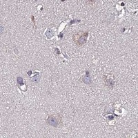


Comprehensive Human Protein Atlas IHC scoring per tissue (reliability: Uncertain — Low consistency between antibody staining and RNA expression data. At least one protein variant secreted, tissue location of RNA and protein might differ and correlation is complex. Pending external verification.). Rows are taken directly from the HPA tissue chart — click any row's HPA link to view the source.
| Tissue | Cell type | Level | Evidence | Source |
|---|---|---|---|---|
| Adrenal gland | Glandular cells | High | Protein (IHC) | HPA → |
| Bronchus | Respiratory epithelial cells | High | Protein (IHC) | HPA → |
| Fallopian tube | Glandular cells | High | Protein (IHC) | HPA → |
| Liver | Hepatocytes | High | Protein (IHC) | HPA → |
| Nasopharynx | Respiratory epithelial cells | High | Protein (IHC) | HPA → |
| Tissue | Cell type | Level | Evidence | Source |
|---|---|---|---|---|
| Adipose tissue | Adipocytes | Not detected | Protein (IHC) | HPA → |
| Bone marrow | Hematopoietic cells | Not detected | Protein (IHC) | HPA → |
| Oral mucosa | Squamous epithelial cells | Not detected | Protein (IHC) | HPA → |
| Parathyroid gland | Glandular cells | Not detected | Protein (IHC) | HPA → |
| Soft tissue | Fibroblasts | Not detected | Protein (IHC) | HPA → |
Troubleshoot CDNF staining in paraffin sections by checking retrieval, controls, cellular distribution and scoring before interpreting chromogenic signal.
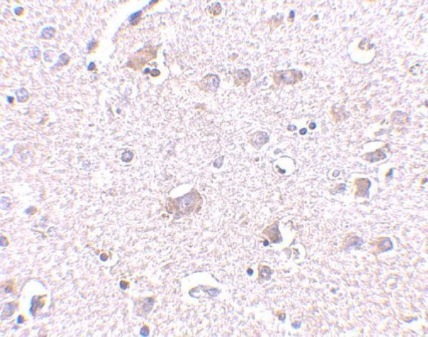
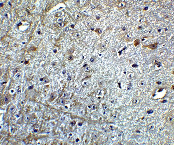
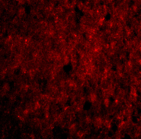
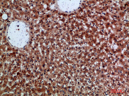
Anti-CDNF antibodies have IHC images from human brain, mouse brain, and paraffin-embedded human liver (catalog image captions). IF images cover human brain cells and mouse brain tissue (A10069-1 image captions).
A10069-1 has IHC-P images from human and mouse brain and IF images from human brain cells and mouse brain tissue (A10069-1 applications and image captions). A10069 has an IHC image from paraffin-embedded human liver; IF is listed as an application, but no IF image is supplied (A10069 applications and image captions).
Which to pick: For brain tissue IHC, choose A10069-1 based on its human and mouse brain IHC images; for paraffin-embedded human liver, choose A10069 based on its own IHC caption (catalog image captions). For IF/ICC, A10069-1 has an image from human brain cells, while A10069 lists IF without an image (catalog applications and image captions). Both list human, mouse, and rat reactivity and a rabbit host, but neither provides a clone designation; the captions do not report the fixative (catalog reactivity, host, clone fields, and image captions).