CDR2 / Cerebellar degeneration-related protein 2 · IHC design guide

Design Immunohistochemistry for CDR2

Plan chromogenic IHC on paraffin sections with the IHC-validated antibody at 0.5–1 μg/mL (datasheet A02973-2). Use the reported nuclear pattern as a tentative reference because tissue IHC reliability is uncertain (HPA tissue IHC).

Evidence assembled Oct 2026 · For research use; verify linked source records and product datasheet before use
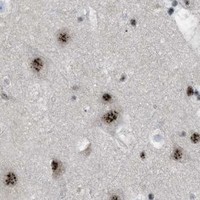
Immunohistochemistry protocol sheet for CDR2 (IHC for CDR2): expected localisation Nuclear staining in tissue sections (HPA tissue IHC), antibody A02973-2, validated IHC image, and IHC protocol steps
Printable CDR2 IHC protocol sheet — expected localisation Nuclear staining in tissue sections (HPA tissue IHC), antibody A02973-2, controls and protocol steps. Open the full CDR2 IHC guide →

CDR2 Immunohistochemistry Experimental Design Guide

Expected localisation, validated protocols, controls and antibodies — the at-a-glance facts below, then the full design guide.

Must know before staining
Expected localisation Nuclear staining in tissue sections (HPA tissue IHC)
Staining pattern General nuclear staining across tissue cell types (HPA tissue IHC)
Antigen retrieval EDTA pH 8.0 HIER, heat-mediated (datasheet A02973-2)
Positive control ⓘ Adrenal gland+4 more · see all
Negative control ⓘ None in HPA (detected in all 45 tissues); use no-primary + isotype controls
Important caveats
Reasons your staining may differ from the expected pattern.
Fixation Keep fixation consistent across sections. (standard IHC practice; not target-specific)
Caveat Tissue staining awaits external verification (HPA tissue IHC)
Regulation Regulation not established (UniProt)
Isoform / epitope No annotated isoforms; one 1–454 chain (UniProt)
Section 1

Recommended CDR2 IHC & IF Protocols

The catalog antibody’s IHC-P protocol uses heat-mediated EDTA retrieval at pH 8.0 (datasheet A02973-2). The published CDR2 staining excerpt below reports samples and a retrieval attempt, but no chromogenic protocol details (PMC3688866).

Recommended immunohistochemistry (IHC-P) protocol parameters
SampleParaffin-embedded human lung cancer tissue; fixative not specified (datasheet A02973-2)
FixationImage fixative and duration unreported (datasheet A02973-2); verify before use.
Sectioning4–5 µm sections on charged slides (standard)
DeparaffinisationXylene, graded ethanol series to water (standard)
Antigen retrievalHeat retrieval: EDTA pH 8.0 (datasheet A02973-2); 20 min, 95–100 °C (standard)
Peroxidase block3% H2O2, 10 min, room temperature (standard)
Blocking10% goat serum (datasheet A02973-2)
Primary antibodyRabbit anti-CDR2, 0.5-1μg/ml (datasheet A02973-2)
Primary incubationOvernight at 4 °C (datasheet A02973-2)
DetectionStreptavidin-biotin complex (SABC), DAB chromogen (datasheet A02973-2)
CounterstainHematoxylin, blue, dehydrate and mount (standard)
Expected resultCDR2-positive staining in glandular cells of adrenal gland (HPA tissue IHC: High). HPA tissue profile: General nuclear expression. No signal in the no-primary control.
💡Decision noteStart with heat-mediated EDTA retrieval at pH 8.0 for the catalog antibody (datasheet A02973-2); the published rat-section retrieval attempt does not specify conditions (PMC3688866).
Section 2

What Is the Expected CDR2 Staining Pattern?

CDR2 shows a general nuclear IHC pattern, with high staining reported in glandular, respiratory epithelial, squamous epithelial, and neuronal cells in the listed tissues (HPA: tissue IHC). ICC-IF places it in the nucleoplasm (HPA: approved nucleoplasm). It has no annotated transmembrane segment (UniProt Q01850 topology). Interpret the IHC pattern cautiously: its reliability is Uncertain, pending external verification (HPA: tissue IHC).

What am I looking at on my slide?
Nuclear staining in glandular cells of colon or duodenum, or neuronal cells of cerebral cortex.This fits reported high staining in those cells (HPA: tissue IHC) and the nucleoplasmic location seen by ICC-IF (HPA: approved nucleoplasm). Record intensity and the fraction of stained cells; neither source establishes a required cutoff.
Predominantly cytoplasmic or membrane staining, with little nuclear signal.This conflicts with the general nuclear IHC profile (HPA: tissue IHC) and approved nucleoplasmic ICC-IF location (HPA: subcellular). Check morphology and controls before interpreting it as CDR2; an artefact or off-target signal is possible (general IHC practice).
Staining confined to a cell population other than the reported positive population within a listed tissue.Compare cell identity carefully: high staining is assigned to specific cell types, such as colon glandular cells and bronchial respiratory epithelial cells (HPA: tissue IHC). Cross-reactivity or endogenous detection activity is possible, but the Uncertain IHC reliability prevents a definitive call from this mismatch alone (HPA: IHC reliability; general IHC practice).
Diffuse stain across nuclei, cytoplasm, and empty tissue spaces.This is hard to reconcile with the reported nuclear profile (HPA: tissue IHC). Uneven detection, residual reagent, or endogenous activity can produce background (general IHC practice). Assess the no-primary control and whether the stain follows recognizable cells before scoring.
No nuclear signal in a section containing an expected positive cell population.Absence in, for example, cerebral cortex neuronal cells or adrenal gland glandular cells contrasts with reported high staining (HPA: tissue IHC). Review the positive control and assay conditions before calling the sample negative; HPA rates its IHC evidence Uncertain (HPA: IHC reliability).
💡Expected CDR2 appearanceA plausible positive is predominantly nuclear staining in the reported glandular, epithelial, or neuronal cells, potentially high in the listed tissues (HPA: tissue IHC); widespread noncellular or predominantly membrane stain is suspect against that profile (HPA: tissue IHC; general IHC practice).
How each factor affects the staining
Compartment evidenceHPA describes general nuclear staining by tissue IHC and an approved nucleoplasmic location by ICC-IF (HPA: tissue IHC; HPA: subcellular). The ICC-IF result helps assess location, but it does not establish IHC assay reliability (HPA: IHC reliability).
Cell and tissue contextHigh IHC staining is reported in named populations across adrenal gland, appendix, bronchus, cerebral cortex, cervix, colon, duodenum, and epididymis (HPA: tissue IHC). The supplied record lists no negative tissue or cell population, so these examples cannot define a specific negative control (HPA: tissue IHC).
IHC antibody validationBoth listed antibodies, HPA018151 and HPA023870, have Uncertain IHC status; HPA023870 has Approved ICC status (HPA: antibodies). Agreement with an ICC location therefore supports a compartment check, while independent IHC confirmation remains valuable (HPA: antibodies; general IHC practice).
Protein topologyUniProt annotates no transmembrane segment or signal peptide for CDR2 (UniProt Q01850 topology and processing). Strong membrane-only staining warrants scrutiny; these annotations do not predict antigen retrieval conditions or fixation sensitivity (UniProt Q01850 topology; general IHC practice).
Why is my staining missing, weak or wrong?
SituationLikely causeNext action
Nuclei are unstained in a listed high-staining tissue.The assay may have failed, or this section may differ from the reported pattern; HPA IHC reliability is Uncertain (HPA: tissue IHC).Check a same-run positive control, section integrity, reagent steps, and detection before interpreting absence (general IHC practice).
Stain is strongest at membranes or throughout cytoplasm.The location conflicts with nuclear tissue IHC and nucleoplasmic ICC-IF findings (HPA: tissue IHC; HPA: subcellular).Compare cellular morphology and control sections; reassess specificity before scoring it as CDR2 (general IHC practice).
Unexpected cells stain while the reported population does not.Cell assignment may be mistaken, or the signal may reflect cross-reactivity or endogenous activity (general IHC practice).Identify cells from morphology and compare the reported cell-specific pattern and no-primary control (HPA: tissue IHC; general IHC practice).
Brown signal is diffuse or extends into empty spaces.Nonspecific background or detection activity can obscure the nuclear pattern (general IHC practice; HPA: tissue IHC).Inspect the no-primary control; review blocking, washing, and chromogen development as general IHC checks (general IHC practice).
The nuclear signal is weak but background is low.The supplied sources provide no CDR2-specific retrieval or dilution setting; weak signal alone does not identify its cause (HPA: tissue IHC; UniProt Q01850).Use the selected antibody's documented IHC-P conditions and a positive control when assessing assay performance (general IHC practice).
Does ICC-IF show the same compartment?HPA assigns an approved nucleoplasmic ICC-IF location, while its tissue IHC reliability remains Uncertain (HPA: subcellular; HPA: tissue IHC).Use the ICC-IF finding as a location cross-check only; interpret the paraffin-section result with its IHC controls (HPA: subcellular; general IHC practice).

Sample controls for CDR2 IHC & IF

🧪Run cerebral cortex first and score its neuronal cells, which show High CDR2 staining (HPA: cerebral cortex neuronal cells High). HPA detects CDR2 in all 45 scored tissues, so there is no validated negative tissue; use no-primary and isotype controls, and expect any genuinely negative cells within the section to show only background staining without treating neighboring non-neuronal cells as proven negatives (HPA: no negative tissue; standard IHC practice).
Positive control tissue: Adrenal gland (Glandular cells, HPA High)
Negative control tissue: None in HPA: CDR2 is detected in all 45 scored tissues. Use a no-primary (secondary-only) and an isotype control instead.
ICC-IF cell lines (HPA subcellular resource): HPA ICC-IF images show CDR2 in A-431, U-251MG, U2OS, with annotated localisation: Nucleoplasm (approved) (HPA subcellular).
Technical controls: Include a no-primary, secondary-only slide and a rabbit host- and class-matched isotype control in the appropriate monoclonal or polyclonal format (selected-SKU caption: rabbit primary; standard IHC practice). Use a matched CDR2 knockout sample or a validated peptide-blocking control where available, and check cerebral cortex for pigment that could resemble DAB staining and for endogenous peroxidase or biotin background with the planned detection system (standard IHC practice; selected-SKU caption: biotin–SABC/DAB detection).
⚠️Feasibility: A target-specific fixation window or fixation effect is unreported, and the selected-SKU paraffin-section caption does not state a fixative (selected-SKU caption: fixative unreported). The caption uses heat retrieval in EDTA at pH 8.0, providing a starting condition without establishing that retrieval is required (selected-SKU caption: EDTA pH 8.0). There is insufficient matched evidence to say frozen sections or IF are easier; assess cerebral cortex pigment against the no-primary slide when interpreting DAB signal (HPA: ICC-IF images available; standard IHC practice).

HPA tissue IHC evidence for CDR2

Comprehensive Human Protein Atlas IHC scoring per tissue (reliability: Uncertain — Pending external verification.). Rows are taken directly from the HPA tissue chart — click any row's HPA link to view the source.

Positive expression · recommended positive controls

TissueCell typeLevelEvidenceSource
Adrenal gland Glandular cells High Protein (IHC) HPA →
Appendix Glandular cells High Protein (IHC) HPA →
Bronchus Respiratory epithelial cells High Protein (IHC) HPA →
Cerebral cortex Neuronal cells High Protein (IHC) HPA →
Cervix Squamous epithelial cells High Protein (IHC) HPA →

Undetected expression · recommended negative controls

TissueCell typeLevelEvidenceSource
None in HPA: CDR2 is detected in all 45 scored tissues. Use a no-primary (secondary-only) and an isotype control instead.
Section 3

Advanced CDR2 IHC Tips

Troubleshoot CDR2 staining in paraffin sections by checking retrieval, compartment, tissue controls, and detection artefacts before comparing scores.

Which retrieval conditions should I try when CDR2 staining is weak?
Start with heat-mediated retrieval in EDTA at pH 8.0 for paraffin sections (datasheet A02973-2). The selected tissue image used that retrieval before an overnight incubation at 4°C with 1 μg/mL primary antibody (caption A02973-2). If staining remains weak, compare a second retrieval condition on adjacent sections while holding antibody concentration, incubation, and detection constant (standard IHC practice). Check whether stronger retrieval also increases background or damages tissue, and choose the condition that preserves interpretable cellular detail (standard IHC practice). Record heating and cooling conditions for each run so retrieval differences can be traced (standard IHC practice).
Could fixation explain variable CDR2 staining between paraffin blocks?
CDR2-specific sensitivity to fixation is unknown from the supplied evidence; the selected paraffin-section caption does not state a fixative (caption A02973-2). Record each block’s fixative and fixation duration, then compare blocks with matched retrieval and detection conditions (standard IHC practice). Include a previously staining control section in the same run to distinguish a block-related problem from a reagent or detection failure (standard IHC practice). If morphology and several antigens are weak, review processing records before changing the CDR2 antibody concentration (standard IHC practice). Do not infer a fixation effect from tissue staining patterns or CDR2’s reported sequence features (HPA: tissue IHC; UniProt Q01850).
Where should I expect CDR2 signal in a paraffin section?
Prioritise interpretable nuclear staining: the tissue IHC profile reports general nuclear expression, and the subcellular profile places CDR2 in the nucleoplasm (HPA: tissue IHC; HPA: nucleoplasm, approved). UniProt does not independently annotate a subcellular location, so treat that pattern as a reference rather than proof of specificity (UniProt Q01850; HPA: tissue IHC reliability uncertain). Compare stained cells with a nuclear counterstain and score only cells whose compartment can be resolved (standard IHC practice). Diffuse cytoplasmic colour without convincing nuclear signal warrants review of background, detection controls, and antibody conditions (HPA: nucleoplasm; standard IHC practice). Confirm that the same pattern persists away from damaged tissue edges (standard IHC practice).
How should I assess epitope-related loss of CDR2 staining?
The supplied record lists a single 1–454 chain, no annotated isoforms, and no signal peptide or transmembrane segment (UniProt Q01850). It also reports a phosphoserine at residue 311, but supplies no antibody epitope or evidence that phosphorylation changes binding (UniProt Q01850; datasheet A02973-2). If staining differs between blocks, first compare section processing and the documented EDTA pH 8.0 retrieval under otherwise matched conditions (datasheet A02973-2; standard IHC practice). Use a separately validated antibody with a documented, distinct epitope if epitope masking remains a concern (standard IHC practice). Do not assign a negative region to an unreported splice isoform (UniProt Q01850).
How can I check a CDR2 IHC pattern by multiplex immunofluorescence?
Use IF as a separate validation experiment, since the supplied product image documents chromogenic staining of paraffin sections rather than an IF protocol (caption A02973-2). Pair CDR2 with a marker validated for the expected cell population and a nuclear counterstain, then assess nuclear overlap within identified cells (HPA: nucleoplasm; standard IF practice). Choose a fluorophore channel with low tissue autofluorescence and include single-colour controls to check bleed-through (standard IF practice). For an intracellular, nucleoplasmic target, optimise permeabilisation after fixation so antibody can reach the nuclear compartment (HPA: nucleoplasm; standard IF practice). Establish IF fixation and antibody conditions directly; the IHC caption does not report fixation (caption A02973-2).
What should I change when CDR2 DAB staining is widespread?
First inspect a no-primary control and a known staining control under the same detection conditions to locate nonspecific signal (standard IHC practice). The selected image used 10% goat serum blocking, biotinylated secondary antibody, a streptavidin–biotin complex, and DAB (caption A02973-2). Apply a peroxidase block and check whether tissue-dependent endogenous biotin contributes to colour in that detection system (standard IHC practice). If controls indicate antibody-related background, titrate the primary below the caption’s 1 μg/mL condition and reassess nuclear contrast (caption A02973-2; standard IHC practice). Review wash conditions and exclude damaged edges or necrotic areas from interpretation (standard IHC practice).
How should I quantify CDR2 staining across tissue samples? ⚠ ANSWER MARKED FOR VERIFICATION
Define a nuclear scoring rule before reviewing sample groups, consistent with the reported nucleoplasmic location (HPA: nucleoplasm; standard IHC practice). For each annotated cell population, report the percentage of positive nuclei and an H-score using intensity categories 0–3; document the threshold and reader agreement (standard IHC practice). Normalise positive counts to the number of eligible intact nuclei, or express counts per mm² of analysable tissue when cell density is the endpoint (standard IHC practice). Keep imaging, counterstaining, and DAB development consistent across comparisons (standard IHC practice). Report excluded necrotic and edge regions, and analyse distinct cell populations separately (standard IHC practice).
How do I distinguish true CDR2 staining from tissue artefact?
Look for reproducible nuclear staining in intact cells, consistent with the reported nucleoplasmic location, while recognising that the tissue IHC profile is rated uncertain (HPA: nucleoplasm; HPA: tissue IHC reliability uncertain). The HPA tissue list reports high staining in several glandular, epithelial, and neuronal cell populations, so identify the cells present before interpreting a positive region (HPA: tissue IHC). Question isolated cytoplasmic colour, sharp edge-only staining, and signal confined to necrotic tissue (HPA: nucleoplasm; standard IHC practice). Compare no-primary and detection controls to assess endogenous enzyme or detection-system colour (standard IHC practice). Require agreement across well-preserved regions and replicate sections before calling a sample positive (standard IHC practice).
Boster reagents

Best CDR2 / Cerebellar degeneration-related protein 2 IHC Antibodies

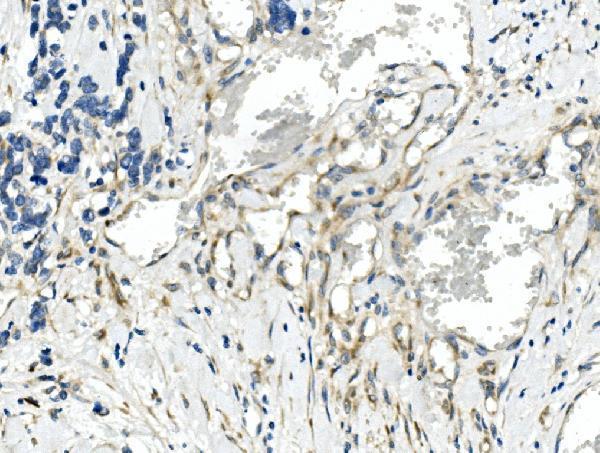
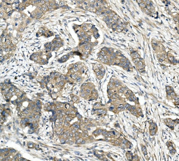
The catalog antibody A02973-2 has human IHC data from paraffin sections of lung, mammary and rectal cancer tissue (catalog IHC image captions); no IF data are supplied (catalog applications and images).

Real IHC data IHC analysis of CDR2 using anti-CDR2 antibody (A02973-2). CDR2 was detected in paraffin-embedded section of human lung cancer tissue. Heat mediated antigen retrieval was performed in EDTA buffer (pH8.0, epitope retrieval solution). The tissue section was blocked with 10% goat serum. The tissue section was then incubated with 1μg/ml rabbit anti-CDR2 Antibody (A02973-2) overnight at 4°C. Biotinylated goat anti-rabbit IgG was used as secondary antibody and incubated for 30 minutes at 37°C. The tissue section was developed using Strepavidin-Biotin-Complex (SABC) (Catalog # SA1022) with DAB as the chromogen.
Anti-CDR2 Antibody ®
Cat # A02973-2

A02973-2 is listed for human IHC (catalog applications and reactivity). Its IHC captions show paraffin sections of human lung, mammary and rectal cancer tissue (catalog IHC image captions).

Which to pick: Choose A02973-2 for human paraffin-section IHC; its caption documents EDTA retrieval at pH 8.0 and 1 μg/ml primary antibody (A02973-2 IHC image caption). The fixative is unreported (A02973-2 IHC image caption). No IF/ICC or other-species choice is supported by this catalog entry (catalog applications, reactivity and images).

Each figure is that product's own IHC / IF validation image from its datasheet.

References

  1. UniProt Consortium. UniProt entry Q01850 (CDR2_HUMAN, Cerebellar degeneration-related protein 2).
  2. Human Protein Atlas. CDR2 tissue IHC expression (reliability: Uncertain).
  3. Human Protein Atlas. CDR2 subcellular location (ICC-IF): Localized to the nucleoplasm..
  4. Human Protein Atlas. CDR2 antibody validation summary (2 antibodies).
  5. CDR2L Antibodies: A New Player in Paraneoplastic Cerebellar Degeneration. PloS one 2013 — PMC3688866.
  6. CDR2 antigen and Yo antibodies. Cancer immunology, immunotherapy : CII 2011 — PMC3024499.
  7. Proteolytic degradation and potential role of onconeural protein cdr2 in neurodegeneration. Cell death & disease 2016 — PMC5143381.
  8. T cells specific for post-translational modifications escape intrathymic tolerance induction. Nature communications 2018 — PMC5783942.
  9. PubMed PMID:2014264 — UniProt-cited evidence.
  10. PubMed PMID:14702039 — UniProt-cited evidence.
  11. PubMed PMID:15489334 — UniProt-cited evidence.