This website uses cookies to ensure you get the best experience on our website.


- Table of Contents
Plan CEACAM1 chromogenic IHC in paraffin sections using gut enterocyte microvilli as a positive staining pattern (HPA tissue IHC). Use the catalog antibody at 1:50 (datasheet M00923-2), and score membrane staining by cell type against negative controls (standard IHC practice).

Expected localisation, validated protocols, controls and antibodies — the at-a-glance facts below, then the full design guide.
| Expected localisation | Enterocyte microvilli and epithelial membranes (HPA tissue IHC) | |
| Staining pattern | Membranous staining, strongest at gut enterocyte microvilli (HPA tissue IHC) | |
| Antigen retrieval | EDTA pH 8.0 HIER, heat-mediated (datasheet M00923-2) | |
| Positive control | Appendix+4 more · see all | |
| Negative control | Adipose tissue+4 more · see all |
| Fixation | Keep paraffin-section fixation consistent (standard IHC practice; not target-specific) | |
| Caveat | Staining may include products of other genes (HPA tissue IHC) | |
| Regulation | No staining-linked induction specified (UniProt) | |
| Isoform / epitope | 11 isoforms; check extracellular versus cytoplasmic epitope (UniProt) |
The catalog antibody protocol is followed by published CEACAM1 IHC methods for lymphoma and myeloma tissue microarrays, lung cancer specimens, and liver graft sections (PMC13312280; PMC3728234; PMC6686173).
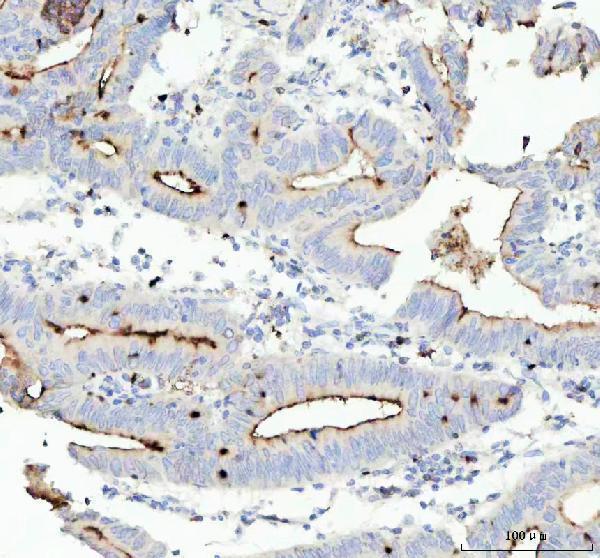
| Sample | Paraffin-embedded human colorectal adenocarcinoma tissue; fixative not specified (datasheet M00923-2) |
| Fixation | Image fixative and duration unreported (datasheet M00923-2); verify before use. |
| Sectioning | 4–5 µm sections on charged slides (standard) |
| Deparaffinisation | Xylene, graded ethanol series to water (standard) |
| Antigen retrieval | Heat retrieval: EDTA pH 8.0 (datasheet M00923-2); 20 min, 95–100 °C (standard) |
| Peroxidase block | 3% H2O2, 10 min, room temperature (standard) |
| Blocking | 10% goat serum (datasheet M00923-2) |
| Primary antibody | Rabbit monoclonal (clone 19C63) anti-CEACAM1, 1:50 (datasheet M00923-2) |
| Primary incubation | Overnight at 4 °C (datasheet M00923-2) |
| Detection | HRP-conjugated secondary, DAB chromogen (datasheet M00923-2) |
| Counterstain | Hematoxylin, blue, dehydrate and mount (standard) |
| Expected result | CEACAM1-positive staining in enterocytes - Microvilli of appendix (HPA tissue IHC: High). HPA tissue profile: Membranous expression in a few tissues, most abundant in glandular epithelia of the gastrointestinal tract, bile canaliculi of liver and intercalated ducts of salivary gland. No signal in the no-primary control. |
CEACAM1 is predominantly at lateral cell membranes, with apical, basal, junctional and microvillus locations also reported; it has one transmembrane segment and an extracellular domain (UniProt P13688 topology and subcellular location). In tissue IHC, expect the clearest staining along gastrointestinal enterocyte microvilli (HPA: High in appendix, colon and rectum). HPA rates tissue staining reliability Enhanced, while noting medium consistency with RNA data and possible detection of proteins from more than one gene (HPA: tissue IHC reliability).
| High staining outlines enterocyte microvilli in appendix, colon or rectum (HPA: High in these cells). | This matches the strongest supplied tissue pattern. A crisp surface distribution is consistent with CEACAM1 membrane and microvillus localization; assess the stained structure as well as its intensity (HPA: tissue IHC; UniProt P13688 subcellular location). |
| Strong nuclear staining dominates, with little discernible epithelial surface staining. | Nuclear dominance does not match the reported CEACAM1 locations (UniProt P13688 subcellular location). Treat it as a possible staining artifact and reassess the slide and controls before calling it CEACAM1 positive (standard IHC interpretation). |
| Strong staining appears in cells reported as undetected, such as adipocytes in adipose tissue or respiratory epithelial cells in bronchus (HPA: Not detected in those cells). | This is discordant with the supplied tissue observations. Cross-reactivity or endogenous detection activity is possible; the HPA tissue summary also cautions that staining may detect proteins from more than one gene (HPA: tissue IHC reliability; standard IHC interpretation). |
| Broad, diffuse color covers tissue and background without a clear cell boundary or microvillus pattern. | That distribution is hard to reconcile with the reported membrane predominance (UniProt P13688 subcellular location). Review background and detection controls before scoring cells; diffuse color alone does not establish a positive CEACAM1 result (standard IHC interpretation). |
| No signal appears in appendix, colon or rectum enterocyte microvilli (HPA: High in those cells). | The expected positive structure is missing, so this run cannot support a negative interpretation on its own. Check tissue preservation, retrieval, reagent performance and detection controls using the validated IHC workflow (standard IHC practice). |
| Membrane topology (UniProt P13688 topology) | CEACAM1 has an extracellular region at residues 35–428, one transmembrane segment at 429–452 and a cytoplasmic region at 453–526. Interpret staining against the expected membrane distribution; the supplied record does not locate the antibody epitope (UniProt P13688 topology). |
| Tissue and cell context (HPA: tissue IHC) | Appendix, colon and rectum enterocyte microvilli are High; duodenum and small intestine enterocyte microvilli are Medium (HPA: tissue IHC). A weaker result in a Medium site should be judged against that site's reference level, not the strongest gastrointestinal fields. |
| Restricted structures outside the intestine (HPA: tissue IHC) | The HPA profile describes bile canaliculi in liver and intercalated ducts in salivary gland, while listing hepatocytes and salivary gland glandular cells as Low (HPA: tissue IHC). Examine the named structures before interpreting a broadly stained liver or salivary section. |
| Antibody and profile confidence (HPA: tissue IHC and antibody validation) | HPA lists IHC Supported for HPA011041 and IHC Enhanced for CAB002146 (HPA: antibody validation). Its overall tissue reliability is Enhanced, with medium staining–RNA consistency and a caution about proteins from more than one gene; a plausible pattern still needs controls (HPA: tissue IHC reliability). |
| Isoforms and processing (UniProt P13688 isoforms and processing) | UniProt lists 11 isoforms, a signal peptide at residues 1–34, and a mature chain beginning at residue 35 (UniProt P13688). Without an epitope assignment, the supplied evidence cannot establish which isoforms the IHC-validated antibody detects. |
| Additional reported locations (UniProt P13688 subcellular location) | UniProt also reports secreted CEACAM1 and secretory-vesicle membrane localization (UniProt P13688 subcellular location). Such annotations broaden the biological context, but they do not make uniform cytoplasmic color a sufficient positive call when the tissue reference is membranous (HPA: tissue IHC). |
| Situation | Likely cause | Next action |
|---|---|---|
| Known-positive gastrointestinal tissue is blank. | A technical run failure is possible when HPA-High enterocyte microvilli have no signal (HPA: appendix, colon and rectum; standard IHC practice). | Confirm the expected cells are present, then review the validated antigen-retrieval and detection steps and a run control (standard IHC practice). No target-specific fixation sensitivity or retrieval condition is established by the supplied sources. |
| Diffuse chromogen obscures cell borders. | Background can arise from detection chemistry, insufficient blocking or an overly strong reagent condition (standard IHC practice). | Inspect a no-primary control, review blocking and detection, and use the validated antibody conditions before scoring. Seek a resolved membrane or microvillus pattern (standard IHC practice; HPA: tissue IHC). |
| Nuclear staining is the dominant finding. | A nuclear pattern conflicts with the supplied membrane, junction, vesicle and secreted locations (UniProt P13688 subcellular location). | Check counterstain and detection controls, then compare the same run with HPA-High enterocyte microvilli. Do not score nuclei alone as CEACAM1 positive (standard IHC interpretation; HPA: tissue IHC). |
| A reportedly negative cell population stains strongly. | Off-target binding or endogenous detection activity may explain staining that conflicts with an HPA Not detected entry; HPA also cautions about detection of proteins from more than one gene (HPA: tissue IHC reliability; standard IHC practice). | Verify the cell identity and compare no-primary and known-positive controls. Interpret the discordant result cautiously, especially if the expected membrane pattern is absent (standard IHC interpretation; UniProt P13688 subcellular location). |
| Liver or salivary gland looks uniformly strong. | Uniform intensity exceeds the Low cell-level entries and loses the canalicular or duct emphasis in the HPA profile (HPA: liver and salivary gland tissue IHC). | Inspect bile canaliculi or intercalated ducts at higher magnification and compare background controls before assigning a tissue-wide score (HPA: tissue IHC; standard IHC interpretation). |
| IF/ICC question: should a bright intracellular signal be accepted as the expected pattern? | HPA gives a Membrane subcellular summary but supplies no ICC-IF image-bearing cell lines or detailed main location; UniProt also reports secretory-vesicle membrane localization (HPA: subcellular ICC-IF; UniProt P13688 subcellular location). | Use membrane localization as the main comparison and interpret intracellular signal with controls and the separate IF/ICC guide. The supplied HPA ICC-IF record does not establish a cell-line-specific pattern (HPA: subcellular ICC-IF; standard IF interpretation). |


Comprehensive Human Protein Atlas IHC scoring per tissue (reliability: Enhanced — Medium consistency between antibody staining and RNA expression data. Caution, targets protein from more than one gene.). Rows are taken directly from the HPA tissue chart — click any row's HPA link to view the source.
| Tissue | Cell type | Level | Evidence | Source |
|---|---|---|---|---|
| Appendix | Enterocytes - Microvilli | High | Protein (IHC) | HPA → |
| Colon | Enterocytes - Microvilli | High | Protein (IHC) | HPA → |
| Rectum | Enterocytes - Microvilli | High | Protein (IHC) | HPA → |
| Duodenum | Enterocytes - Microvilli | Medium | Protein (IHC) | HPA → |
| Kidney | Cells in glomeruli | Medium | Protein (IHC) | HPA → |
| Tissue | Cell type | Level | Evidence | Source |
|---|---|---|---|---|
| Adipose tissue | Adipocytes | Not detected | Protein (IHC) | HPA → |
| Adrenal gland | Glandular cells | Not detected | Protein (IHC) | HPA → |
| Breast | Adipocytes | Not detected | Protein (IHC) | HPA → |
| Bronchus | Respiratory epithelial cells | Not detected | Protein (IHC) | HPA → |
| Caudate | Glial cells | Not detected | Protein (IHC) | HPA → |
Troubleshoot CEACAM1 staining in paraffin sections by checking retrieval, membrane localisation, antibody specificity and cell level scoring before interpreting chromogenic signal.
M00923-2 has IHC images from human paraffin sections (catalog IHC captions) and is listed for IF/ICC (catalog applications); no IF image is supplied (catalog IF images).
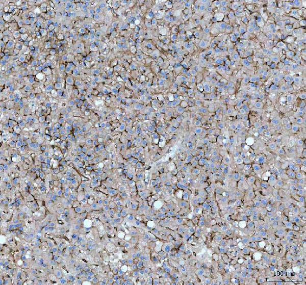
M00923-2 will render with its human colorectal adenocarcinoma paraffin-section IHC figure (catalog IHC caption). The catalog also shows IHC in human liver cancer paraffin sections and lists IF/ICC applications, without an IF image (catalog IHC captions; catalog applications; catalog IF images).
Which to pick: Choose M00923-2 for human paraffin-section IHC: its own figure uses EDTA retrieval at pH 8.0, antibody at 1:50, and DAB detection; the fixative is unreported (M00923-2 IHC caption). For IF/ICC, M00923-2 is listed at 1:50, though no IF image is supplied (catalog applications; catalog IF dilution; catalog IF images). No cross-species choice is supported because M00923-2 lists human reactivity only (catalog reactivity).